Secondary prevention of type 1 diabetes
mellitus: stopping immune destruction
and promoting ß-cell regeneration
1Divisão de Endocrinologia and Metabologia,
2Divisão de Imunologia Clínica e Unidade de Transplante de Medula Óssea,
Departamento de Clínica Médica, Faculdade de Medicina de Ribeirão Preto, Universidade de São Paulo, Ribeirão Preto, SP, Brasil
C.E.B. Couri1,
M.C. Foss1
and J.C. Voltarelli2
Abstract
Type 1 diabetes mellitus results from a cell-mediated autoimmune attack against pancreatic ß-cells. Traditional treatments involve nu-merous daily insulin dosages/injections and rigorous glucose control. Many efforts toward the identification of ß-cell precursors have been made not only with the aim of understanding the physiology of islet regeneration, but also as an alternative way to produce ß-cells to be used in protocols of islet transplantation. In this review, we summarize the most recent studies related to precursor cells implicated in the regeneration process. These include embryonic stem cells, pancreas-derived multipotent precursors, pancreatic ductal cells, hematopoietic stem cells, mesenchymal stem cells, hepatic oval cells, and mature ß-cells. There is controversial evidence of the potential of these cell sources to regenerate ß-cell mass in diabetic patients. However, clinical trials using embryonic stem cells, umbilical cord blood or adult bone marrow stem cells are under way. The results of various immunosuppressive regimens aiming at blocking autoimmunity against pancreatic ß-cells and promoting ß-cell preservation are also ana-lyzed. Most of these regimens provide transient and partial effect on insulin requirements, but new regimens are beginning to be tested. Our own clinical trial combines a high dose immunosuppression with mobilized peripheral blood hematopoietic stem cell transplantation in early-onset type 1 diabetes mellitus.
Correspondence
J.C. Voltarelli Hemocentro Regional de Ribeirão Preto Campus da USP
14051-140 Ribeirão Preto, SP Brasil
Fax: +55-16-2101-9309 E-mail: jcvoltar@fmrp.usp.br
Research supported by FAEPA-HCRP, FUNDHERP, FAPESP, CNPq, and FINEP.
Received September 30, 2005 Accepted July 11, 2006
Key words
•Diabetes mellitus •ß-cell regeneration •ß-cell preservation •ß-cell precursors •Immune intervention •Stem cell
Introduction
Diabetes mellitus is a metabolic syndrome characterizedby hyperglycemia resulting from defects in insulin secretion,insulin ac-tion, or both. Chronic hyperglycemia is as-sociated with long-term damage, dysfunc-tion and failureof various organs, including retinopathy withpotential loss of vision; ne-phropathy leading to renal failure;peripheral
neuropathy with risk of foot ulcers, amputa-tionsand Charcot joints; autonomic neuro-pathy causing gastrointestinal,genitourinary, cardiovascular symptoms, and sexual dys-function.Patients with diabetes have an in-creased incidence of atherosclerotic cardio-vascular disease (1).
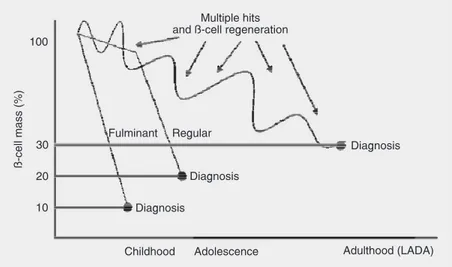
pan-creatic ß-cells. Autoimmune destruction of ß-cells is due to multiple genetic predisposi-tions and is also related to environmental factors that are still poorly defined (1). When clinical symptoms are observed the autoim-mune process is markedly advanced (60-80% of the ß-cell mass have been destroyed at the time of diagnosis (2)). The rate of ß-cell destruction is variable, being rapid in children and slow in adults (3) (Figure 1). In the later stage of disease there is little or no insulin secretion, as indicated by low or undetectable plasma levels of peptide. C-peptide is consecrated with insulin by the ß-cells as a by-product of the enzymatic cleav-age of proinsulin to insulin. Measurement of C-peptide provides a fully validated means of quantifying endogenous insulin secretion, being closely related to the amount of ß-cell mass (4). Patients with type 1 diabetes de-pend on exogenous insulin administration for survival. The best classical treatment is based on 3-4 subcutaneous injections of in-sulin per day, i.e., intensive inin-sulin therapy (5). This treatment is responsible for a 35-90% reduction of the risk of retinopathy, nephropathy and neuropathy compared with conventional therapy with 1-2 injections per day (6).
Stopping the destruction - a way to preserve ß-cell mass
Subgroup analysis of the Diabetes Con-trol and Complication Trial (DCCT) showed an important aspect related to long-term com-plications of diabetes, i.e., patients with ini-tially higher serum levels of C-peptide pre-sented a slower decline of these levels dur-ing the study and suffered less microvascu-lar complications than patients with low or undetected levels of C-peptide. In other words, ß-cell preservation is another impor-tant target in the management of type 1 diabetes and its related complications (7).
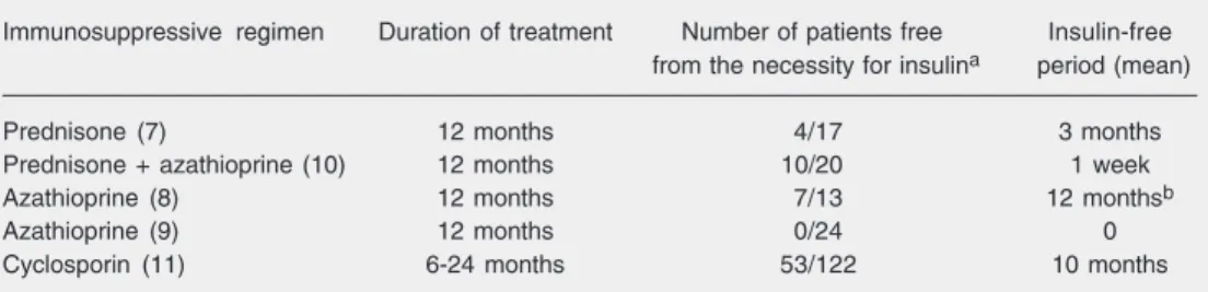
Preservation of ß-cell mass, however, was attempted with immunosuppression long time before the DCCT. In 1981, Eliot and colleagues (8) treated recently diagnosed children with prednisone with the aim of stopping pancreatic ß-cell destruction by the autoimmune process. Urinary C-peptide lev-els in the group treated with corticosteroid were significantly higher than control for one year after therapy was initiated. Subse-quently, short-term studies were conducted using azathioprine (9,10), azathioprine plus prednisone (11), and cyclosporine (12,13) and demonstrated a slower decline (or even some improvement) in plasma C-peptide lev-els. In those studies, some patients experi-enced short periods (<1 year) during which they were free from insulin treatment (Table 1). The chronic toxicity of immunosuppres-sion and the loss of the metabolic benefits after the withdrawal of the immunosuppres-sive agents limited the routine use of these therapies.
Recently, two studies were reported us-ing acute immunosuppressive treatments with humanized antibodies against a T-cell anti-gen (CD3) for recently diagnosed type 1 diabetic patients. The first study showed that 12 patients who were treated with the anti-body had better ß-cell function (measured by C-peptide levels) and lower exogenous insulin requirements after 1 year than
tients in the placebo group (14). A subse-quent study on a larger sample of patients (40 in each group) showed that the effects on ß-cell function and insulin requirements could be maintained for up to 18 months (15). However, very few patients became insulin-free in these trials.
Studies using immunosuppressive agents should be done shortly after or even before the clinical diagnosis of type 1 diabetes, since a larger ß-cell mass can be preserved. In 2002, Shapiro et al. (16) showed that low doses of sirolimus (0.1 mg/kg) and tacroli-mus (0.1 mg/kg) were able to reduce insulitis, preserving pancreatic insulin content and preventing diabetes in female non-obese dia-betic (NOD) mice (8% diabetes incidence at 35 weeks vs 66% in vehicle-treated mice). The rationale of the immunosuppressive treatment is focused on stopping autoim-mune aggression to the endocrine pancreas, facilitating its natural reconstitution and maintaining the residual functional capacity of the ß-cell mass (see below). The same goal can be reached by a different mechan-ism at least transiently in early onset type 1 diabetes mellitus (DM1) treated with inten-sive insulin therapy (17), somatostatin (18) or diazoxide (19). These therapies preserve ß-cell reserve, inhibiting insulin secretion and providing a rest to ß-cell activity.
As overt type 1 diabetes is preceded by a chronic phase in which T-cells and other mononuclear cells infiltrate the islets, some
trials have analyzed the impact of certain drugs on the prevention of ß-cell loss early in the preclinical phase. In these trials, drugs such as oral and parenteral insulin and oral nicotinamide were unable to delay the onset of diabetes and to prevent insulin use (20-22). In 2004, a Finnish birth-cohort study was conducted to analyze the effect of di-etary vitamin D supplementation on the inci-dence of type 1 diabetes. Children who regu-larly took the recommended dose of vitamin D (2000 IU daily) during the first year of life had a risk reduction of 0.22 (0.05-0.89) com-pared with those who regularly received less than the recommended amount. Thus, vita-min D seems to promote a protective action against pancreatic ß-cell immune-mediated destruction (23). Immunosuppressive treat-ments have not yet been employed in pri-mary prevention trials.
In late 2003, our research group started a phase I/II study of high dose immunosup-pression with intravenous cyclophosphamide plus antithymocyte globulin followed by autologous hematopoietic stem cell trans-plantation for patients with new-onset type 1 diabetes. The aim of this treatment was to stop the autoimmune destruction of ß-cells with immunosuppressive drugs and to “re-set” the deleterious immunologic system to a normally reconstituted one with autolog-ous hematopoietic stem cells (24). This thera-py is being successfully used in other au-toimmune diseases such as systemic lupus
Table 1. Effect of different immunosuppressive treatments on insulin-free time in patients with newly diag-nosed type 1 diabetes mellitus.
Immunosuppressive regimen Duration of treatment Number of patients free Insulin-free
from the necessity for insulina period (mean)
Prednisone (7) 12 months 4/17 3 months
Prednisone + azathioprine (10) 12 months 10/20 1 week
Azathioprine (8) 12 months 7/13 12 monthsb
Azathioprine (9) 12 months 0/24 0
Cyclosporin (11) 6-24 months 53/122 10 months
aNumber of patients free from the necessity for insulin/number of treated patients. bOne patient remained in
erythematous and multiple sclerosis (25). The exact mechanism of action of autolog-ous hematopoietic stem cell transplantation is unclear, but it may shift the balance be-tween destructive immunity and tolerance through yet undefined mechanisms such as clonal exhaustion, suppressor cells, immune indifference, cytokine alterations, changes in T- or B-cell clonality or changes in immu-nodominant autoantigens (25). The results of transplantation are monitored by endo-crine (HbA1c, C-peptide levels and dose of insulin kg-1 day-1) and immunologic
param-eters (lymphocyte subsets, cytokine produc-tion, T-cell receptor diversity, and humoral and cellular autoreactivity against pancre-atic ß-cells). Severe adverse effects and mor-tality are expected to be very low in this population of young patients with no previ-ous morbidity except for DM1 (24). Prelimi-nary results obtained with 13 patients are encouraging but they have not yet been pub-lished in full (26).
New clinical trials using various forms of immunosuppression (mycophenolate mofe-til, anti-IL-2 receptor antibodies, anti-CD20 antibody, or anti-thymocyte globulin) for DM1, without infusion of stem cells, started recently or are prepared to start soon.
ß-cell regeneration: is it possible?
Differentiated ß-cells first appear around the 13th embryonic day (at the onset of the secondary transition), a phase of pancreatic organogenesis during which endocrine cells detach from the exocrine matrix, increase in number and reorganize to form mature is-lets. Islet growth continues after birth result-ing from a combination of hypertrophy and hyperplasia (27).
The maintenance of ß-cell mass is the result of a dynamic state consisting of neo-genesis, proliferation and apoptosis (28). This is a physiological process whose objective is to guarantee the harmony of glucose homeo-stasis in the presence of challenges
experi-enced after birth such as obesity and other states of insulin resistance (29,30).
The vast majority of studies analyzing pancreatic regeneration were conducted on rodents (see below). Thus, in 1983, Bonner-Weir and colleagues demonstrated that young near-totally pancreatectomized rats presented an eight-week recuperation of 27% of creas weight and 42% of the endocrine pan-creas. Quantitative measurements estimated ß-cell turnover in adult rat islets to be as high as 3% per day (31). Some sporadic cases in humans are impressive in this regard: for example, a 39-year-old type 1 diabetic pa-tient submitted to simultaneous pancreas-kidney transplantation was referred for treat-ment of an abdominal incision herniation 2 years after the initial procedure. A regimen with tacrolimus, prednisolone and myco-phenolate was used post-transplantation and good glycemic control was achieved with-out exogenous insulin. At the time of the corrective surgery, a native pancreas biopsy showed that the percent of ß-cells was 4-fold higher than that observed in long-term type 1 diabetic patients conventionally treated with insulin (32).
In light of recent discoveries demonstrat-ing the regenerative potential of the pan-creas, which cells could be the precursors of adult ß-cells?
Probable precursors of adult ß-cells
potential than its parents (33,34). Eventually a stem cell becomes a “progenitor” or “pre-cursor” cell, committed to produce one or a few terminally differentiated cells such as neurons or muscle cells (35) (Table 2). The clonal isolation of putative pancreatic pre-cursors has been an elusive objective of researchers who look for a more complete knowledge of ß-cell physiology and for new replacement strategies for DM1.
Pancreas-derived stem cells
Many studies have looked for ß-cell pre-cursors in pancreatic tissue. Georgia and Bhushan (36) demonstrated that cyclin D2
expression in the endocrine pancreas coin-cides with a massive increase in islet mass, with ß-cell replication being the main mech-anism for the maintenance of ß-cell mass. Using cyclin D2-/- mice, ß-cell replication
was reduced 4-fold and cyclin D2-/- mice
were glucose intolerant. These results sug-gested that cyclin D2 plays a key role in regulating the transition of ß-cells from a state of quiescence to replication. In addi-tion, many other investigators support the hypothesis of self-replication as the main source of ß-cell mass. Their analysis shows
that pre-existing ß-cells, rather than pluripo-tent stem cells, are the major source of new ß-cells during adult life and after pancreate-ctomy in mice (37,38). On the other hand, in other studies, the increase in ß-cell mass in adult mice is considered to result from dif-ferentiation of adult multipotent precursors. Thus, Seaberg and colleagues (35) reported the isolation of pancreas-derived multipotent precursors from pancreatic islets and ducts. These cells proliferate in vitro to form indi-vidual clonal colonies of neurons and glial cells, pancreatic endocrine α-, ß-, δ-cells
and pancreatic exocrine cells. The in vitro
proliferated ß-cells were shown to be able to produce insulin. The pancreas-derived multipotent precursors did not express mark-ers of embryonic stem cells or genes sugges-tive of mesodermal or neural crest origin. Therefore, they could represent a previously unidentified adult intrinsic pancreatic pre-cursor proposed by other investigators using flow-cytometric cell sorting (39).
Lechner et al. (40) reported that cells expressing the intermediate filament protein nestin, a marker of neural stem cells, can be isolated from human and rodent islets and expanded extensively in vitro. Insulin, glu-cagon, and pancreatic duodenal
homeobox-Table 2. Stem cell nomenclature.
Cell line Homogeneous population of cells capable of self renewal.
Cloned cell line Population of cells that derives from replication of a single cell.
Stem cell Cell that can divide indefinitely to produce differentiated tissues and of identical stem cells.
Zygote Diploid cell resulting from the fusion of male and female gametes.
Morula Spheroidal mass of cells resulting from early cleavage divisions of the zygote.
Blastocyst Four- to five-day-old embryo formed prior to implantation in the uterus; consisted of only a few undifferentiated stem cells.
Embryoid body Cluster of embryonic cells presented in cultures of stem cells or in tumors.
Totipotent cell Cell committed to a specific lineage that is capable of giving rise to all cells and tissues, including extraembryonic tissues. Example: blastomeres from the morula.
Pluripotent cell Cell not committed to a specific lineage that may differentiate into all tissues, with exception of extraembryonic tissues.
Example: cells from the inner cell mass of the blastocyst.
Multipotent cell Progenitor cell that can give rise to diverse cell types in response to appropriate environmental cues.
Example: adult bone marrow mesenchymal stem cells.
Progenitor cell Parent cell that is committed to dividing and multiplying in order to form a specific cell type.
1 protein/insulin promoter factor-1 (IPF-1/ IPF-1) expression, as well as low-level insu-lin secretion, can be detected in cultures of nestin-positive islet-derived stem cells after the addition of differentiating cytokines and growth factors. These cells also form insu-lin-producing islet-like clusters in vitro, a process that is markedly enhanced by the addition of the insulinotropic hormone glu-cagon-like peptide-1.
Few studies, however, have been con-ducted on human pancreatic tissues. In 2000, Bonner-Weir et al. (41), using fractions of digested human pancreas enriched for duc-tal tissue, generated in vitro insulin-produc-ing islet-like clusters. When plastic adherent cells from these preparations were overlaid with an inert matrix (Matrigel), they formed cysts and clusters (cultured human islet buds). Most cells in these aggregates were positive for the ductal marker cytokeratin 19 and others showed immunoreactivity for insulin and other islet hormones. The insulin con-tent of the cultures increased over time, and a low level of glucose-responsive insulin secretion was observed in vitro. However, the capacity to expand the cultivated tissue was limited.
Bone marrow and umbilical cord blood
Although the existence of stem cells in adult organs appears to be relatively well accepted, the intriguing question is how did these cells actually get there? The first hypo-thesis is that they represent a population of cells that are preserved throughout develop-ment in every single organ. These cells ap-pear to have maintained many properties of pluripotential blastocyst cells and could par-ticipate in local tissue repair throughout the lifespan of the organism. A second hypo-thesis is that a distant source such as bone marrow may contain a self-renewing popu-lation of multipotent stem cells that are con-tinuously released into the circulation. It is
tempting to speculate that stem cells are an essential component of this circulating bone marrow surveillance system and that they are prepared to home to areas of injured tissues to participate in tissue repair and regeneration (42).
Ianus and colleagues (43) demonstrated an extra-pancreatic source of ß-cells that may play a role in the adaptation of ß-cell mass in response to physiological and envi-ronmental stimuli. In this study, bone mar-row cells that selectively expressed the en-hanced green fluorescent protein (EGFP) if the insulin gene was actively transcribed were transplanted into lethally irradiated re-cipient mice and gave rise to EGFP-positive insulin-producing cells in pancreatic islets. Through a genetic approach, the authors ruled out cell fusion as the mechanism for EGFP-positive cells with islet-like charac-teristics. Moreover, these cells seemed to be functional since glucose and incretins stimu-lated insulin secretion.
secre-tion in vitro and, when transplanted into streptozotocin-induced diabetic rats, could down-regulate blood glucose levels.
Umbilical cord blood (UCB) is another source of stem cells with the potential to promote in vivo ß-cell regeneration. In fact, in a xenogenic model of stem cell transplan-tation, human mononuclear UCB cells were able to reduce blood glucose levels and in-crease survival in mouse models of DM1 (49) and DM2 (50). In DM2 animals, UCB stem cell infusion also improved renal ab-normalities caused by diabetes, suggesting a regenerative action on the renal parenchyma. However, there is no indication of the type of stem cells or the mechanism responsible for the clinical effect.
The possible role of adult stem cells de-rived either from UCB or bone marrow on ß-cell regeneration led to the organization of some clinical trials to use those cells to treat human DM. In the San Nicolas Clinic, Ar-gentina, adult bone marrow mononuclear cells are being injected into the pancreas of DM1 and DM2 patients by arteriography without any additional treatment. The re-sults have not been fully published but have shown improvement of metabolic param-eters and a reduced use of antidiabetic drugs more pronounced in DM2 than in DM1 pa-tients. Proposed mechanisms are stimula-tion of angiogenesis and ß-cell regenerastimula-tion by stem cells (Fernandez-Viña R, personal communication, and Ref. 51). On the other hand, at the University of South Florida, USA, autologous UCB cells cryopreserved at birth from infants that become DM1 pa-tients later in life are injected back into these children intravenously without previous im-munosuppression. The expectation is that UCB cells would migrate preferentially to the damaged pancreas, either inducing the proliferation of insulin-producing islets, pro-viding protection to existing islets by modu-lating the autoimmune process, or possibly differentiating into islet cells. Eight patients have been treated in this manner, apparently
with positive responses (Haller M and Wingard J, personal communication).
Hepatic oval cells
Pancreas and liver have a close anatomic association starting from the primitive fo-regut during embryogenesis and this fact has prompted attempts to isolate pancreatic pro-genitor cells from the liver, especially the hepatic oval cells. Oval cells are considered to be hepatic stem cells that can give rise to hepatocytes and bile duct cells (52). Yang et al. (53), using in vitro proliferation of these cells, obtained islet-like clusters that could express several endocrine hormones includ-ing insulin. In the same publication, a pre-liminary in vivo study described the success-ful reversal of diabetes in streptozotocin-treated NOD mice with severe combined immunodeficiency (NOD-SCID) using he-patic stem cells.
Embryonic stem cells
Embryonic stem cells (ESC) are pluripo-tent cell lines derived from the inner cell mass of blastocyst stage embryos and their differentiation in culture may reproduce char-acteristics of early embryonic development. Pancreatic and islet cell replacement are cur-rently considered to be the only curative therapies for type 1 diabetes. However, the shortage of human donations is a primary obstacle for these approaches, making ESC a promising source of ß-cell generation in the near future in countries where their use is permitted.
and glucokinase genes, Pdx-1/Ipf-1 and neurogenin-3 transcription factors. Func-tional analyses indicated secretion of insulin into the medium.
Other studies have used cell-trapping systems or various growth factors to obtain insulin-secreting clones from undifferenti-ated ESC of mice. Soria et al. (55) developed a gene construction that allows the expres-sion of a neomycin selection system under the control of regulatory regions of the hu-man insulin gene. The chimeric gene also contained a hygromycin resistance gene used to select transfected cells. A resulting clone displayed regulated hormone secretion in vitro in the presence of various secretagogues. After the in vitro isolation of clusters of differentiated ß-cells from ESC,these cells were implanted in the spleen of streptozoto-cin-induced diabetic animals. Normoglyce-mia and normal body weight were restored after 1 and 4 weeks, respectively.
Based on several recent reports claiming the generation of insulin-producing cells from ESC, Hanssonet al. (56) investigated the prop-erties of these insulin-containing progenitors. In their study they found that, although differ-entiated cells containing immunoreactive in-sulin were isolated, they did not contain proin-sulin-derived C-peptide. Furthermore, in spite of variable insulin release from these cells upon glucose addition, C-peptide release was never detected. They suggested that C-peptide biosynthesis and secretion should be demon-strated in order to claim insulin production
from an ESC progeny.
The potential of ESC to regenerate pan-creatic ß-cells attracted the attention of bio-technological companies interested in de-veloping embryonic cell lines to be used by diabetic patients (57) and of for-profit clin-ics that are injecting embryonic stem cells from 4-8-week embryos into type 2 diabetic patients. Favorable responses were reported in abstract form (58,59).
Much effort for the identification of ß-cell precursors has been made not only with the aim of understanding the physiology of islet regeneration but also as an alternative way to produce ß-cells to be used in proto-cols of islet transplantation. Precursor cells implicated in the regeneration process in-clude ESC, pancreas-derived multipotent precursors, ductal cells, hematopoietic stem cells, mesenchymal stem cells, hepatic oval cells, and mature ß-cells. Whatever the source of ß-cells, immunosuppressive regimens as-sociated or not with stem cell infusion seem to facilitate these endogenous mechanisms of ß-cell regeneration in early-onset disease. For patients with a longer time of diagnosis of type 1 diabetes, inactivation of the au-toimmune response should be associated with strategies of ß-cell regeneration, since a much larger ß-cell mass has been destroyed in these patients. For this purpose, clinical tri-als using stem cells derived from embryonic tissues, umbilical cord blood or adult bone marrow are underway.
References
1. American Diabetes Association. Diagnosis and classification of dia-betes mellitus. Diabetes Care 2004; 27 (Suppl 1): S5-S10. 2. Notkins AL, Lernmark A. Autoimmune type 1 diabetes: resolved and
unresolved issues. J Clin Invest 2001; 108: 1247-1252.
3. Pozzilli P, Di Mario U. Autoimmune diabetes not requiring insulin at diagnosis (latent autoimmune diabetes of the adult): definition, char-acterization, and potential prevention. Diabetes Care 2001; 24: 1460-1467.
4. Palmer JP, Fleming GA, Greenbaum CJ, Herold KC, Jansa LD, Kolb H, et al. C-peptide is the appropriate outcome measure for type 1
diabetes clinical trials to preserve beta-cell function: report of an
ADA workshop, 21-22 October 2001. Diabetes 2004; 53: 250-264.
5. Writing Team for the Diabetes Control and Complications Trial/ Epidemiology of Diabetes Interventions and Complications Re-search Group. Effect of intensive therapy on the microvascular
complications of type 1 diabetes mellitus. JAMA 2002; 287:
2563-2569.
diabe-tes mellitus. N Engl J Med 1993; 389: 977-986.
7. The Diabetes Control and Complications Trial Research Group. Effect of intensive therapy on residual beta-cell function in patients with type 1 diabetes in the diabetes control and complications trial. A randomized, controlled trial. Ann Intern Med 1998; 128: 517-523. 8. Eliot RB, Berryman CC, Crossley JR, James AG. Partial
preserva-tion of pancreatic ß-cell funcpreserva-tion in children with diabetes. Lancet
1981; 19: 631-632.
9. Harrison LC, Colman PG, Dean B, Baxter R, Martin FI. Increase in remission rate in newly diagnosed type I diabetic subjects treated with azathioprine. Diabetes 1985; 34: 1306-1308.
10. Cook JJ, Hudson I, Harrison LC, Dean B, Colman PG, Werther GA, et al. Double-blind controlled trial of azathioprine in children with newly diagnosed type I diabetes. Diabetes 1989; 38: 779-783. 11. Silverstein J, Maclaren N, Riley W, Spillar R, Radjenovic D, Johnson
S. Immunosuppression with azathioprine and prednisone in recent-onset insulin-dependent diabetes mellitus. N Engl J Med 1988; 319: 599-604.
12. Assan R, Feutren G, Sirmai J, Laborie C, Boitard C, Vexiau P, et al. Plasma C-peptide levels and clinical remissions in recent-onset type I diabetic patients treated with cyclosporin A and insulin. Diabe-tes 1990; 39: 768-774.
13. Bougneres PF, Landais P, Boisson C, Carel JC, Frament N, Boitard C, et al. Limited duration of remission of insulin dependency in children with recent overt type I diabetes treated with low-dose cyclosporin. Diabetes 1990; 39: 1264-1272.
14. Herold KC, Hagopian W, Auger JA, Poumian-Ruiz E, Taylor L, Donaldson D, et al. Anti-CD3 monoclonal antibody in new-onset type 1 diabetes mellitus. N Engl J Med 2002; 346: 1692-1698. 15. Keymeulen B, Vandemeulebroucke E, Ziegler AG, Mathieu C,
Kaufman L, Hale G, et al. Insulin needs after CD3-antibody therapy
in new-onset type 1 diabetes. N Engl J Med 2005; 352: 2598-2608.
16. Shapiro AM, Suarez-Pinzon WL, Power R, Rabinovitch A. Combina-tion therapy with low dose sirolimus and tacrolimus is synergistic in preventing spontaneous and recurrent autoimmune diabetes in non-obese diabetic mice. Diabetologia 2002; 45: 224-230.
17. Shah SC, Malone JI, Simpson NE. A randomized trial of intensive insulin therapy in newly diagnosed insulin-dependent diabetes mel-litus. N Engl J Med 1989; 320: 550-554.
18. Grunt JA, al-Hakim H, Willoughby L, Howard CP. A randomized trial of a somatostatin analog for preserving beta cell function in children with insulin dependent diabetes mellitus. J Pediatr Endocrinol 1994; 7: 331-334.
19. Ortqvist E, Bjork E, Wallensteen M, Ludvigsson J, Aman J, Johansson C, et al. Temporary preservation of beta-cell function by
diazoxide treatment in childhood type 1 diabetes. Diabetes Care
2004; 27: 2191-2197.
20. Gale EA, Bingley PJ, Emmett CL, Collier T. European Nicotinamide Diabetes Intervention Trial (ENDIT): a randomised controlled trial of intervention before the onset of type 1 diabetes. Lancet 2004; 363: 925-931.
21. Chaillous L, Lefevre H, Thivolet C, Boitard C, Lahlou N, tlan-Gepner C, et al. Oral insulin administration and residual beta-cell function in recent-onset type 1 diabetes: a multicentre randomised controlled trial. Diabete Insuline Orale group. Lancet 2000; 356: 545-549. 22. Diabetes Prevention Trial - Type 1 Diabetes Study Group. Effects of
insulin in relatives of patients with type 1 diabetes mellitus. N Engl J
Med 2002; 346: 1685-1691.
23. Hypponen E, Laara E, Reunanen A, Jarvelin MR, Virtanen SM. Intake of vitamin D and risk of type 1 diabetes: a birth-cohort study.
Lancet 2001; 358: 1500-1503.
24. Voltarelli JC, Burt RK, Kenyon N, Kaufman DB, Squiers E. Hemato-poietic stem cell transplantation as treatment for type 1 diabetes. In:
Burt R, Marmont A (Editors), Stem cell therapy for autoimmmune
diseases. Georgetown: Landes Bioscience; 2004.
25. Burt RK, Slavin S, Burns WH, Marmont AM. Induction of tolerance in autoimmune diseases by hematopoietic stem cell transplantation: getting closer to a cure? Blood 2002; 99: 768-784.
26. Voltarelli J, Couri C, Oliveira M, Stracieri A, Moraes D, Coutinho M, et al. Autologous hematopoietic stem cell transplantation for type I diabetes mellitus. Bone Marrow Transplant 2006; 37 (Suppl 1): S16. 27. Lee VM, Stoffel M. Bone marrow: an extra-pancreatic hideout for the
elusive pancreatic stem cell? J Clin Invest 2003; 111: 799-801.
28. Bonner-Weir S, Sharma A. Pancreatic stem cells. J Pathol 2002;
197: 519-526.
29. Swenne I. Pancreatic beta-cell growth and diabetes mellitus. Diabe-tologia 1992; 35: 193-201.
30. Fernandes A, King LC, Guz Y, Stein R, Wright CV, Teitelman G. Differentiation of new insulin-producing cells is induced by injury in adult pancreatic islets. Endocrinology 1997; 138: 1750-1762. 31. Finegood DT, Scaglia L, Bonner-Weir S. Dynamics of beta-cell
mass in the growing rat pancreas. Estimation with a simple math-ematical model. Diabetes 1995; 44: 249-256.
32. Kuroda A, Yamasaki Y, Imagawa A. Beta-cell regeneration in a patient with type 1 diabetes mellitus who was receiving
immunosup-pressive therapy. Ann Intern Med 2003; 139: W81.
33. Fischbach GD, Fischbach RL. Stem cells: science, policy, and ethics. J Clin Invest 2004; 114: 1364-1370.
34. Wagers AJ, Weissman IL. Plasticity of adult stem cells. Cell 2004; 116: 639-648.
35. Seaberg RM, Smukler SR, Kieffer TJ, Enikolopov G, Asghar Z, Wheeler MB, et al. Clonal identification of multipotent precursors from adult mouse pancreas that generate neural and pancreatic lineages. Nat Biotechnol 2004; 22: 1115-1124.
36. Georgia S, Bhushan A. Beta cell replication is the primary mechan-ism for maintaining postnatal beta cell mass. J Clin Invest 2004; 114: 963-968.
37. Dor Y, Brown J, Martinez OI, Melton DA. Adult pancreatic beta-cells are formed by self-duplication rather than stem-cell differentiation.
Nature 2004; 429: 41-46.
38. Levine F, Mercola M. No pancreatic endocrine stem cells? N Engl J
Med 2004; 351: 1024-1026.
39. Suzuki A, Nakauchi H, Taniguchi H. Prospective isolation of multi-potent pancreatic progenitors using flow-cytometric cell sorting. Dia-betes 2004; 53: 2143-2152.
40. Lechner A, Leech CA, Abraham EJ, Nolan AL, Habener JF. Nestin-positive progenitor cells derived from adult human pancreatic islets of Langerhans contain side population (SP) cells defined by expres-sion of the ABCG2 (BCRP1) ATP-binding cassette transporter.
Biochem Biophys Res Commun 2002; 293: 670-674.
41. Bonner-Weir S, Taneja M, Weir GC, Tatarkiewicz K, Song KH, Sharma A, et al. In vitro cultivation of human islets from expanded ductal tissue. Proc Natl Acad Sci U S A 2000; 97: 7999-8004. 42. Lechner A, Habener JF. Stem/progenitor cells derived from adult
tissues: potential for the treatment of diabetes mellitus. Am J Physiol Endocrinol Metab 2003; 284: E259-E266.
43. Ianus A, Holz GG, Theise ND, Hussain MA. In vivo derivation of
glucose-competent pancreatic endocrine cells from bone marrow without evidence of cell fusion. J Clin Invest 2003; 111: 843-850. 44. Lagasse E, Connors H, Al-Dhalimy M, Reitsma M, Dohse M,
45. Theise ND, Badve S, Saxena R, Henegariu O, Sell S, Crawford JM, et al. Derivation of hepatocytes from bone marrow cells in mice after radiation-induced myeloablation. Hepatology 2000; 31: 235-240. 46. Kang EM, Zickler PP, Burns S, Langemeijer SM, Brenner S, Phang
OA, et al. Hematopoietic stem cell transplantation prevents diabetes in NOD mice but does not contribute to significant islet cell
regen-eration once disease is established. Exp Hematol 2005; 33:
699-705.
47. Jiang Y, Jahagirdar BN, Reinhardt RL, Schwartz RE, Keene CD, Ortiz-Gonzalez XR, et al. Pluripotency of mesenchymal stem cells derived from adult marrow. Nature 2002; 418: 41-49.
48. Chen LB, Jiang XB, Yang L. Differentiation of rat marrow mesenchy-mal stem cells into pancreatic islet beta-cells. World J Gastroenterol
2004; 10: 3016-3020.
49. Ende N, Chen R, Reddi AS. Effect of human umbilical cord blood
cells on glycemia and insulitis in type 1 diabetic mice. Biochem
Biophys Res Commun 2004; 325: 665-669.
50. Ende N, Chen R, Reddi AS. Transplantation of human umbilical cord blood cells improves glycemia and glomerular hypertrophy in type 2
diabetic mice. Biochem Biophys Res Commun 2004; 321: 168-171.
51. Fernandez-Viña R, Saslavsky J, Andrin O, Sosa N, Swirido P, Vrsalovick F, et al. Feasibility of implant autologous stem cells with
endovascular technique in diabetes mellitus. Cytotherapy 2004; 7
(Suppl 1): Abstract #37.
52. Petersen BE, Goff JP, Greenberger JS, Michalopoulos GK. Hepatic oval cells express the hematopoietic stem cell marker Thy-1 in the rat. Hepatology 1998; 27: 433-445.
53. Yang L, Li S, Hatch H, Ahrens K, Cornelius JG, Petersen BE, et al. In vitro trans-differentiation of adult hepatic stem cells into pancreatic endocrine hormone-producing cells. Proc Natl Acad Sci U S A 2002; 99: 8078-8083.
54. Assady S, Maor G, Amit M, Itskovitz-Eldor J, Skorecki KL,
Tzuker-man M. Insulin production by huTzuker-man embryonic stem cells. Diabetes
2001; 50: 1691-1697.
55. Soria B, Roche E, Berna G, Leon-Quinto T, Reig JA, Martin F. Insulin-secreting cells derived from embryonic stem cells normalize
glycemia in streptozotocin-induced diabetic mice. Diabetes 2000;
49: 157-162.
56. Hansson M, Tonning A, Frandsen U, Petri A, Rajagopal J, Englund MC, et al. Artifactual insulin release from differentiated embryonic stem cells. Diabetes 2004; 53: 2603-2609.
57. Sinden JD. ReNeuron Group plc. Regenerative Med 2006; 1:
143-147.
58. Smikodub OI, Noritskaya AV. Embryonic stem cells in new onset type 2 diabetes mellitus. Cytotherapy 2004; 6: 402 (Abstract). 59. Smikodub OI, Noritskaya AV. Embryonic stem cells in pernicious