Internet Journal of Medical Update
Journal home page: http://www.akspublication.com/ijmu
Review
Neonatal Sepsis: past, present and future; a review article
Dr. Shalini Tripathi
*ΨMD and Professor G. K. Malik
**MD
*
Lecturer, Department of Pediatrics, Era’s Lucknow Medical College, Lucknow, India
**
Director, Department of Pediatrics, RML Institute of Medical Sciences, Lucknow,
India
(Received 27 October 2009 and accepted 15 February 2010)
ABSTRACT: Sepsis is the most common cause of neonatal mortality. As per National Neonatal Perinatal Database (NNPD) 2002-2003, the incidence of neonatal sepsis in India was 30 per 1000 live birth. It is 3% among intramural babies and 39.7% among extramural admissions. The early manifestations of neonatal sepsis are vague and ill-defined. Novel approaches in the diagnosis of neonatal sepsis include heart rate analysis on ECG or colorimetric analysis of skin color. Although blood culture is the gold standard for the diagnosis of sepsis, culture reports would be available only after 48-72 hours. In this era of multidrug resistance, it is mandatory to avoid unnecessary use of antibiotics to treat non-infected infants. Thus, rapid diagnostic test(s) that differentiate non-infected from non-non-infected infants, particularly in the early newborn period, that include Interleukien-6 (IL-6), neutrophil CD64 index, procalcitonin and nucleated RBC count, have the potential to make a significant impact on neonatal care. The aim of this review is to specify the diagnostic criteria, treatment guidelines and a summary of the newer diagnostic tests of sepsis. KEY WORDS: Sepsis; Intramural; Extramural; Multidrug resistance
INTRODUCTIONΨ
Sepsis is the commonest cause of neonatal mortality and is responsible for 30-50% of total neonatal deaths each year in developing countries.
1-3
It is estimated that up to 20% of neonates develop sepsis and approximately 1% die of sepsis related causes.2 The term neonatal sepsis, refers to systemic infection of neonates including septicemia, pneumonia, meningitis, arthritis, osteomyelitis, and urinary tract infection. As per National Neonatal Perinatal Database (NNPD) 2002-2003, the incidence of systemic infection is 3% among intramural babies in tertiary care institutions in India, with septicemia being present in three fourth of the cases and pneumonia in one third of neonates. Infection is the primary cause of mortality in 18.6% of intramural neonates in which
Klebsiella pneumoniae is the most frequent bacterial isolate (32.5%), followed by
Staphylococcus aureus (13.6%).
Ψ Correspondence at: House No. 255/394, In front of Tikona Park, Kundri, Rakabganj, Lucknow 226003, India; Email: [email protected] Phone no: +919235237435
Sepsis is 12 times more common in extramural admissions (39.7%). In extramural admissions,
Klebsiella is the commonest bacteria responsible (27.5%), the next common being S. aureus
(14.9%). Sepsis is responsible for death in 38.0% of these extramural babies.3, 4
In a north Indian tertiary care center Sundaram et al studied the incidence of neonatal sepsis between the two time periods(epoch-1-1991-1996, and epoch-2-2001-2006) and it was found that that incidence of early onset sepsis did not change between the epochs but the incidence of late onset sepsis increased from 12-16.5 per 1000 live births(p<0.001).5
Definition
National Neonatal Forum of India has defined neonatal sepsis as follows:3
Probable (Clinical) Sepsis: In an infant having clinical picture suggestive of septicemia, if there is the presence of any one of the following criteria:
• Positive septic screen - presence of two of the four parameters namely, TLC (< 5000/mm), band to total polymorphonuclear cells ratio of >0.2, absolute neutrophil count < 1800/cumm, C-reactive protein (CRP) >1mg/dl and micro ESR > 10 mm-first hour.
• Radiological evidence of pneumonia.
Culture Positive Sepsis: In an infant having clinical picture suggestive of septicemia, pneumonia or meningitis, if there is presence of either of the following:
• Isolation of pathogens from blood or CSF or urine or abscess (es)
• Pathological evidence of sepsis on autopsy.
Classification
Neonatal sepsis is of two types:
Early onset Sepsis (EOS: Early onset sepsis presents within first 72 hours of life. In severe cases the neonate may be symptomatic in utero
(fetal tachycardia, poor beat to beat variability). Clinically, the neonate usually presents as respiratory distress and pneumonia. Presence of the following risk factors has been associated with an increased risk of EOS:6, 7
• Low birth weight (<2500gms) or preterm baby
• Febrile illness in the mother with in 2 weeks prior to delivery
• Foul smelling and/or meconium stained liquor amnii.
• Prolonged rupture of membrane (>24 hours).
• More than 3 vaginal examinations during labor.
• Prolonged and difficult delivery with instrumentation.
• Perinatal asphyxia (Apgar score <4 at 1 minute of age) or difficult resuscitation.
Neonates with presence of foul smelling liquor or three of above mentioned risk factors should be considered to have EOS & treated with antibiotics. Presence of ≥2 risk factors should be investigated with sepsis screen and treated accordingly.8
Late onset Sepsis (LOS): Late onset sepsis usually presents after 72 hours of age. The source of infection is either nosocomial or community acquired and neonates usually present with septicemia, pneumonia or meningitis.9,10 Risk factors for development of LOS include:
• NICU admission
• Poor hygiene
• Low birth weight (LBW)
• Poor cord care
• Prematurity
• Bottle feeding
• Invasive procedure
• Superficial infection (pyoderma, umbilical sepsis)
• Prelacteal feeding
• Ventilation
• Aspiration of feeds
CLINICAL FEATURES
Manifestations of neonatal sepsis are vague and ill-defined. Alteration in established feeding behavior is common and early, but is a nonspecific symptom. Other features are hypothermia or fever (former is more common in LBW babies), lethargy, poor cry, poor perfusion i.e. prolonged capillary refill time (>2 seconds), hypotonic or absent neonatal reflexes, bradycardia or tachycardia, respiratory distress i.e. apnea or gasping respiration, hypoglycemia or hyperglycemia and metabolic acidosis. System wise specific features are:
Central nervous system: These are bulging anterior fontanel, blank look, high-pitched cry, excessive irritability, coma, seizures, and neck retraction. Presence of these signs should raise clinical suspicion of meningitis
Cardiac: The cardiac signs are mainly hypotension and poor perfusion. A recent study emphasized the value of early diagnosis of sepsis using analysis of heart rate characteristics on ECG monitoring. Griffin et al found that abnormal heart rate characteristics such as reduced variability and transient decelerations occurred 24 hours prior to onset of symptoms in sepsis and sepsis like illness.11 Another group found that sample asymmetry of RR interval increased in the 3-4 days preceding sepsis with the steepest increase in the last 24 hours. These tests may prove helpful in starting the therapy long before the baby shows signs of deterioration.12
Gastrointestinal: These are feed intolerance, vomiting, diarrhea, abdominal distension, paralytic ileus, and necrotising enterocolitis.
Hepatic: The common hepatic signs are hepatomegaly and direct hyperbilirubinemia (Infants with the onset of jaundice after 8 days of age or with direct hyperbilirubinemia were more likely to have urinary tract infection.)13
Renal: There may be acute renal failure.
Hematological: Hematological signs are bleeding and petechiae, purpura.
INVESTIGATIONS
Blood culture
Blood culture is the gold standard for diagnosis of septicemia. It should be done in all cases before starting antibiotics. One-mL sample of blood should be adequate for a blood culture bottle containing 5-10 mL of culture media. Blood culture should be observed for 72 hours before labelling it sterile. It is now possible to detect growth in 12-24 hours by BACTEC or BACT/ALERT culture system which can detect bacteria at a concentration of 1-2 cfu per ml. Blood samples collected from indwelling catheters or lines are likely to be contaminated.
Sepsis screen15,16
This is a panel of tests consisting of: Component Abnormal value
TLC < 5000/mm3
ANC < as per Manroe chart for term11 and Mouzinho’s chart for very LBL (VLBW) infants12
Immature/ total neutrophil
> 0.2
Micro-ESR > 15mm in 1st hr
CRP > 1mg/dl
All neonates suspected to have sepsis should have a septic screen to corroborate the diagnosis. However, the decision to start antibiotics need not be conditional to the sepsis screen result, if there is a strong clinical suspicion of sepsis. Sepsis screen is considered positive if two of these are positive. If the screen is negative but clinical suspicion persists, it should be repeated within 12 hours. If the screen is still negative, sepsis can be excluded with reasonable certainty. The absolute neutrophil count varies considerably in the immediate neonatal period and normal reference ranges are available from Manroe’s chart.17 For very low birth weight infants, the reference ranges are available from Mouzinho’s charts.18 Presence of two abnormal parameters in screen is associated with sensitivity 93-100%, specificity 83%, positive and negative predictive value of 27% and 100%, respectively in detecting sepsis. Rosenberg et al validated the only existingfeasible score introduced by Singh et al. in 2003 and to createan improved score. In their five-sign model requiring at least one clinical sign of infection(apnea, hepatomegaly, jaundice, lethargy and pallor) had anarea under the receiver operating characteristic of 0.70, sensitivity of 77.1%, and PPV of 64.9.19
In a recently published paper, the authors have evaluated the SNAP II score for the assessment of their illness severity which consists of 6 physiological parameters, namely lowest mean
arterial pressure (MAP), worst ratio of partial pressure of oxygen (PaO2) to fraction of inspired oxygen (FiO2), lowest temperature (in ºF), lowest serum pH, occurrence of multiple seizures, and urine output (<1mL/kg/hr). They found that SNAP II can predict mortality as well as organ dysfunction in severely septic neonates; individual components of the score do not have equal predictive ability.20
Lumbar puncture (LP)
The incidence of meningitis in neonatal sepsis has varied from 0.3-3% in various studies3,9. In EOS, lumbar puncture is indicated in the presence of a positive blood culture or if the clinical picture is consistent with septicemia. In situations of late onset sepsis, LP should be done in all infants prior to starting antibiotics. LP should not be done in following cases:21
• Asymptomatic babies being investigated for maternal risk factors. However, LP should be preformed in these cases as well if blood culture becomes positive subsequently.
• Premature neonates presenting with respiratory distress syndrome.
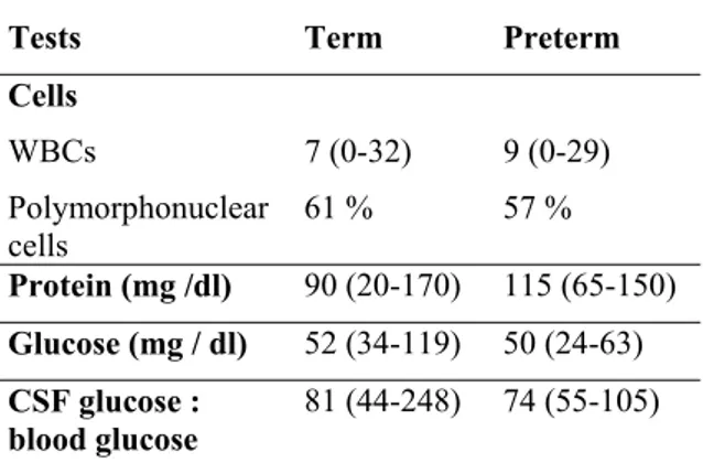
It should be postponed in critically ill and haemodynamically unstable baby; if traumatic it should be repeated with in 12-72 hours. The cerebrospinal fluid characteristics are unique in the newborn period and normal values are given in Table 121.
Table 1: Normal values of CSF in newborn period
Tests Term Preterm
Cells
WBCs 7 (0-32) 9 (0-29)
Polymorphonuclear cells
61 % 57 %
Protein (mg /dl) 90 (20-170) 115 (65-150) Glucose (mg / dl) 52 (34-119) 50 (24-63) CSF glucose :
blood glucose
81 (44-248) 74 (55-105)
Urine culture
urine examination done to exclude urinary tract infection (UTI). UTI may be diagnosed in the presence of one of the following: (a) >10 WBC/mm in a 10 mL centrifuged sample (b) >10 organisms /mL in urine obtained by catheterization and (c) any organism in urine obtained by suprapubic aspiration.
Radiology
Chest X-Ray is done in cases of respiratory distress or apnea. Abdominal X-ray should be done for diagnosis of necrotizing enterocolitis.
NEWER DIAGNOSTIC TESTS FOR DIAGNOSIS OF NEONATAL SEPSIS
Isolation of bacteria from blood is a standard and most specific method used to diagnose neonatal sepsis. Positive cultures ranged from 8% to 73% in the diagnosis of potential neonatal sepsis.23 An additional drawback of culture based diagnosis is the 24–48 hour assay time. Newer diagnostic tests can be grouped into:
1. Acute phase reactants 2. Cell surface markers
3. Granulocyte colony stimulating factor 4. Cytokines
5. Molecular genetics 6. Mol cell proteomics Acute phase reactants
These groups of endogenous peptides are produced by the liver as part of an immediate response to infection or tissue injury. These reactants are C-reactive protein, procalcitonin, fibronectin, haptoglobin, lactoferrin, neopterin and
oromucosoid
C-reactive protein: CRP is synthesized within six to eight hours of exposure to an infective process or tissue damage, with a half life of 19 hours, and can increase more than 1000-fold during an acute phase response. In a study it was concluded that CRP, IL-6 and IgM are helpful in the early diagnosis of Gram-negative neonatal sepsis and CRP continues to be the best single test. A CRP value of 5 mg/l was the best among the three parameters with 95% sensitivity and 98% negative predictive value. The best combination was CRP > or = 5 mg/l and/or IgM of > or =20 mg/dl. The use of both CRP and IgM in combination was the most helpful in predicting Gram-negative neonatal sepsis which has a significant role in making decisions regarding antibiotics treatment.24
Another recent study was done to compare the efficiency of Serum Amyloid A (SAA) with that of C-reactive protein (CRP) and procalcitonin (PCT) in diagnosis and follow-up of neonatal sepsis in preterm infants showed that SAA is an accurate and
reliable marker for diagnosis and follow-up of neonatal sepsis. It is especially useful at the onset of inflammation for rapid diagnosis of neonatal sepsis and can be safely and accurately used in combination with other sepsis markers such as CRP and PCT in diagnosis and follow-up of neonatal sepsis in preterm infants.25
Procalcitonin: Procalcitonin (PCT) is produced by monocytes and hepatocytes which begins to rise four hours after exposure to bacterial endotoxin, peaking at six to eight hours, and remaining raised for at least 24 hours with a half life of 25–30 hours. Several studies have shown that serum procalcitonin concentrations increase appreciably in systemic bacterial infection. In a study done by Cetinkaya M et al median procalcitonin levels were higher in the septic group compared with the non-septic group at the time of the sepsis workup (2.7 vs 0.5 ng/ml, p=0.003), at 1-24 h after the sepsis workup (4.6 vs 0.6 ng/ml, p=0.003), and at 25-48 h (6.9 vs 2.0 ng/ml, p=0.016). Using high cut-off levels, both procalcitonin (2.3 ng/ml) and CRP (30 mg/l) had high specificity and positive predictive value (97%, 91% and 96%, 87%, respectively) but low sensitivity (48% and 41%, respectively) to detect sepsis. The conclusion was procalcitonin >2.3 ng/ml or CRP >30 mg/l indicates a high likelihood for neonatal sepsis, and antibiotic therapy should be continued even in the presence of sterile cultures. However, it is not a readily available diagnostic assay in most institutions.26 Cell surface markers
Neutrophil CD11b and CD64 appear to be promising markers for diagnosis of early and late onset infections.
For culture-positive sepsis episodes, the CD64 index had the highest area under the curve (0.852) of all hematological variables, with a sensitivity of 80% and a specificity of 79%, with a cutoff value of 4.02. So, neutrophil CD64 is a highly sensitive marker for neonatal sepsis. Prospective studies incorporating CD64 into a sepsis scoring system are warranted.27
inflammatory response. Therefore, CD11b is not a good marker for neonatal sepsis28.
Granulocyte colony stimulating factor
Granulocyte colony stimulating factor (GCSF), a mediator produced by bone marrow, facilitates proliferation and differentiation of neutrophils, and has been proposed to be a reliable infection marker for early diagnosis of neonatal sepsis. A concentration ≥ 200 pg/ml has a high sensitivity (95%) and negative predictive value (99%) for predicting early onset neonatal bacterial and fungal infections.29
Cytokines
As antigen specific immunity develops later e.g. at 2 years of age in the case for encapsulated bacteria, neonates initially depend on natural (innate) immunity. This includes phagocytes (by monocytes, tissue macrophages, and neutrophils), natural killer cells, and humoral mediators (CRP, complements, and transplacentally acquired maternal antibodies). In response to antigens such as bacterial endotoxins, activated tissue macrophages produce tumor necrosis factor (TNF) and interleukin (IL). These proinflammatory cytokines stimulate endothelial cells to express receptors for intercellular adhesion molecule on white blood cells. This initiates the cytokine cascade towards increased production of IL6, IL8, and chemokines. Newborn infants display a higher percentage of IL6 and IL8 positive cells than do adults. There is sharp rise in IL6 concentration on exposure to bacterial products, which precedes the increase in CRP. Umbilical cord blood IL6 has consistently been shown to be a sensitive marker for diagnosing early onset neonatal sepsis at the onset of infection compared with other biochemical markers, including CRP, IL1ß, TNFα, and E-selectin, but sensitivity is reduced at 24 and 48 hours because IL6 concentrations fall rapidly and become undetectable after 24 hours. The combined measurement of IL6 (early and sensitive) with CRP (late and specific) in the first 48 hours of presumed septic episodes improves the sensitivity compared with either marker alone.30 Median values of IL-6 (pg/ml) were higher in infants with sepsis vs. those with no sepsis on days 0 [40 vs. 13] (p = 0.03) and 1 [24 vs. 9] (p < 0.001). IL-8 levels were not significantly different. IL-6 levels may be useful in the initiation as well as early termination of antibiotic therapy in late-onset neonatal sepsis.31
IL8 is a proinflammatory cytokine that is predominantly produced by monocytes, macrophages, and endothelial cells, with similar kinetics to IL6. IL8 is considered to be a highly accurate marker with sensitivities ranging from 80% to 91% and specificities from 76% to 100%. A
recent multicentric randomised controlled trial of 1291 clinically stable infants with clinical signs or obstetric risk factors suggesting early onset neonatal sepsis revealed that the combination of IL8 >70 pg/ml and/or CRP >10 mg/l significantly reduced antibiotic therapy from 49.6% to 36.1% (p<0.0001) without missing infections; sensitivity was 80%, specificity 87%, positive predictive value 68%, and negative predictive value 93%.32 In the model of assessing concentration suggested by Ng PC et al., they obtained 100% sensitivity and 97% specificity of IL-10, IL-6 and RANTES for identifying infected preterm infants, who developed DIC. It would be particularly valuable to broaden the panel of studies by the chemokines helpful in diagnosing sepsis in adults such as GRO-α, MIP-1α or ENA-78.33
TNFα is a proinflammatory cytokine that stimulates IL6 production and has a broad spectrum of biological actions on several types of target cell, both immune and non-immune. Newborns developing early onset infection are born with higher TNFα concentrations than non-infected infants.
Other markers studied over the last few years include adhesion molecules (intercellular adhesion molecule 1, vascular cell adhesion molecule 1, E-selectin, L-selectin) and complement activation products (C3a-desArg, C3bBbP, sC5b-9), IL-1alpha, IL-1beta, IL receptor antagonist (IL1ra) which have been found to be significantly increased during sepsis, but require further evaluation for clinical application in the diagnosis of newborn infection.
It has been demonstrated that median IL-6 and TNF-α levels were significantly higher in groups with patients with a diagnosis of clinical sepsis than in controls. The optimal cut-off point was 32 pg /ml for IL-6 and 12 pg /ml for TNF-α. The combination of TNF-α and IL-6 provided a sensitivity of 98.5% and it is a highly sensitive marker of sepsis in the immediate postnatal period.34
In a recent study it was demonstrated that cytokines released in sepsis have an important role in stimulating Nucleated RBC (NRBC) production independent of hypoxia. In this study significantly elevated NRBC demonstrated in EOS (no EONS (n=49)) 1330 cells/ cmm (665-2630), EONS (n=19)3020 cells/cmm (1388-4558), p=0.011) along with significantly elevated IL-6 in EONS, but no increase in level of umbilical cortisol or erythropoietin. Increase NRBC count immediately after birth could be an interesting marker of EONS in absence of hypoxia and awaits further evaluation.35
Molecular genetics36-39
highly conserved in all bacterial genomes, is a useful method for identification of bacteria in clinical samples. Amplification targeting of this 16S rRNA gene is a potentially valuable clinical tool in samples with low copy numbers of bacterial DNA, as this gene is present at 1 to more than 10 copies in all bacterial genomes. The gene also has a number of divergent regions nested within it, so PCR can be targeted for species specific detection of bacteria in clinical samples. This technology has also been reported to be a very sensitive (100%) and rapid method for detecting potential pathogens in amniotic fluid commonly involved in the pathogenesis of preterm labour and adverse neonatal outcome.36
However, the performance of broad range PCR analysis at a level of high analytical sensitivity is complex and remains one of the most challenging PCR applications in the diagnostic laboratory. For example, as 16S rRNA gene amplification targets all bacterial species, small amounts of inherent residual DNA present in reagents may be co-amplified, resulting in false positivity. Methods for the removal of potential background contamination include long wave UV light gamma irradiation DNAse, restriction endonuclease digestion, ultrafiltration, and low DNA polymerases. However, many of these methods result in a reduced sensitivity in detecting target DNA, with a detection limit range of 103–104 copies/ml, which is not ideal for diagnosing sepsis in clinical settings. It was found that a combination of pre-PCR culture with the use of AmpliTaq Low DNA achieves an acceptable level of sensitivity (5–50 copies/ml in a turnaround time of eight hours) for the real time amplification of bacteria in blood samples, without the need to remove any inherent DNA contamination. Detection by PCR does not yield the antimicrobial sensitivity pattern of the pathogen. Early exclusion of bacterial infection could help to reduce overuse of antibiotics. It is predicted that eventually real time PCR combined with DNA Micro Array technology will allow not only identification of the organism but also the antimicrobial sensitivity pattern, which is so critical to clinical care.
It has been revealed from an Indian study that PCR is useful and superior to blood culture for early diagnosis of sepsis in neonates with 100% sensitivity and 95.6% specificity, and once available in most tertiary centers can help in early and accurate diagnosis.37
Role of proteomics for diagnosis of neonatal infection40, 41
In a study, it was found significant alterations in levels of eight serum proteins in infected preterm neonates (specifically P- and E-selectins, interleukin 2, soluble receptor α, interleukin 18,
neutrophil elastase, urokinase plasminogen activator and its cognate receptor, and C-reactive protein) were observed at statistically significant increased levels. 40
Molecular tools (16S-rRNA) demonstrate that the diversity of microbial agents of intra-amniotic infection exceeds what is suspected clinically or is documented by cultures. The resulting inflammatory process has the potential to damage the fetus in utero. Stepwise algorithms (mass restricted score) have been developed to extract proteomic profiles characteristic of amniotic fluid inflammation. The mass restricted score includes four proteomic biomarkers: defensin-2, defensin-1, S100A12, and S100A8 proteins. Other amniotic fluid biomarkers relevant for preterm birth are S100A9 and insulin-like growth factor-binding protein 1. S100A12 - ligand for the receptor of advanced glycation end products - has the strongest association with histological chorioamnionitis and funisitis. So the conclusion was - Presence of S100A12 and S100A8 in amniotic fluid is predictive of early-onset neonatal sepsis and poor neurodevelopmental outcome.41
MANAGEMENT
Supportive Treatment
Attention should be given to basic supportive care in a sick child. The infant should be nursed in a thermoneutral environment taking care to avoid hypothermia and hyperthermia. Oxygen saturation should be maintained in the normal range and ventilation should be initiated as required. The infant should be regularly monitored for hypoglycemia/ hyperglycemia. Colloids and inotropes are used for maintaining normal blood pressure and tissue perfusion. Enteral feeds should be avoided till the baby is haemodynamically stable. Packed cells and fresh frozen plasma should be used appropriately for the management of anemia and bleeding diathesis.
Antimicrobial Treatment
When infection is hospital acquired and there is high probability of resistant strain, cefotaxime in combination with an aminoglycoside should be used for septicemia, pneumonia as well as meningitis. 3rd generation cephalosporins have very good CSF penetration and are traditionally thought to have excellent antimicrobial activity against gram-negative organisms. Hence they were considered to be a good choice for the treatment of nosocomial infections and meningitis. However, recent reports suggest that at least 60-70% of the gram-negative organisms are resistant to them. Moreover, routine use of these antibiotics might increase the risk of infections with extended spectrum beta-lactamase (ESBL) positive organisms. Therefore it is preferable to use antibiotics such as piperacillin-tazobactam or methicillin/vancomycin in units with high incidence of resistant strains. A combination of piperacillin-tazobactam with amikacin should be considered if pseudomonas sepsis is suspected. Penicillin resistant staphylococcus aureus should be treated with cloxacillin, nafcillin or methicillin. Addition of an aminoglycoside is useful in therapy against staphylococcus. Methicillin resistant
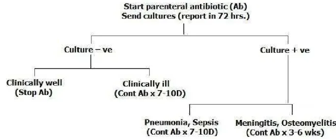
staphylococcus aureus (MRSA) should be treated with a combination of ciprofloxacin or vancomycin with amikacin. Ciprofloxacin has excellent activity against gram-negative organisms also; however, it does not have good CSF penetration. It may be used for the treatment of resistant gram-negative bacteremia after excluding meningitis. For sepsis due to enterococcus, a combination of ampicillin and gentamicin is a good choice for initial therapy. Vancomycin should be used for the treatment of enterococcus resistant to the first line of therapy. Reserve antibiotics: Newer antibiotics like aztreonam, meropenem and imipenem are also now available in the market. Aztreonam has excellent activity against gram-negative organisms while meropenem is effective against most bacterial pathogens except methicillin resistant staphylococcus aureus (MRSA) and enterococcus. Imipenem is generally avoided in neonates because of the reported increase in the incidence of seizures following its use. Empirical use of these antibiotics should be avoided; they should be reserved for situations where sensitivity of the isolated organism warrants its use. A simple approach for the management of sepsis is given in Figure 1.
Figure 1: Suspected Neonatal Sepsis
Duration of antibiotic therapy:
• Clinical sepsis (Based on clinical suspicion and/or sepsis screen positivity) -7-10 days
• Culture positive sepsis (not meningitis), UTI -14 days
• Meningitis-2 weeks after sterilization of CSF culture or for a minimum of 2 weeks for gram positive meningitis and 3 weeks for gram negative meningitis, whichever is longer.
• Bone and joint infection-4-6 weeks
Adjunctive therapy
Intravenous Immune Globulin (IVIG): According to Cochrane database systemic review there is
insufficient evidence to support the routine administration of IVIG preparations investigated to date to prevent mortality in infants with suspected or subsequently proved neonatal infection.42
growth factors. Future studies should focus on investigating other abnormalities of neonatal host defence and/or combined immunotherapy approaches in an attempt to circumvent the immaturity of host defense and potentially reduce both the incidence and severity of neonatal sepsis.44 Granulocyte colony stimulating factor (G-CSF
45-47
: Carr and colleagues reported a randomized trial (PROGRAMS) of GM-CSF for the prevention of sepsis in small for gestational age preterm neonates. This increased the neutrophil count, but had no effect on the primary end point of sepsis free survival to 14 days from trial entry.45
According to the Cochrane database systemic review there is currently insufficient evidence to support the introduction of either G-CSF or Granulocyte monocyte colony stimulating factor (GM-CSF) into neonatal practice, either as treatment of established systemic infection to reduce resulting mortality, or as prophylaxis to prevent systemic infection in high risk neonates. The limited data suggesting that G-CSF treatment may reduce mortality when systemic infection is accompanied by severe neutropenia should be
investigated further in adequately powered trials which recruit sufficient infants infected with organisms associated with a significant mortality risk.46
Exchange transfusion: Exchange transfusion in neonatal sepsis has not been extensively studied. It may be used with caution in neonatal sepsis associated with neutropenia, sclerema, earliest evidence of disseminated intravascular coagulation and metabolic acidosis (pH <7.2).48
Pentoxifylline: Pentoxifylline is a methylxanthine that has been postulated to improve outcomes in sepsis through modulating the activity of the reticuloendothelial system and decreasing the neutrophil activation that contributes to acute tissue injury. Large scale clinical trials have not yet been performed.49
ACCURACY OF DIAGNOSTIC TESTS
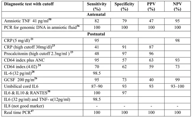
The accuracy of diagnostic tests or combinations of tests for early Onset neonatal sepsis has been summarized in Table 2.
Table 2 Accuracy of Diagnostic Tests or Combinations of Tests for Early Onset Neonatal Sepsis
Diagnostic test with cutoff Sensitivity (%)
Specificity (%)
PPV (%)
NPV (%) Antenatal
Amniotic TNF 41 pg/ml50 82 79 47 95
PCR for genomic DNA in amniotic fluid36 100 100 100 100
Postnatal
CRP(5 mg/dl)21 95 98
CRP (high cutoff 30mg/dl)23 41 91 87
Procalcitonin (high cutoff 2.3ng/ml ) 23 48 97 96
CD64 index plus ANC 95 57 63 93
CD64 index(4.02)24 70 62 59 73
IL-6(32 pg/ml)28 98.5
GCSF 200 pg/m26 95 73 40 99
Umbilical cord IL6 87–90 93 93 93–100
IL6 &IL10 & RANTES30 100 97
IL6 (32 pg/ml) and TNF- α(12pg/ml) 98.5
IL8 (not good marker) - - - -
Real time PCR47 100 100 100 100
CONCLUSION
Clinical diagnosis of sepsis in new born infants is not easy because symptoms and signs are non-specific. There is no laboratory test with 100% specificity and sensitivity, and hence, the search has continued for a reliable test. Blood culture has been the gold standard for confirmation of diagnosis but the results of the test are available only after 48-72 hours. The neonates with "risk
Array chip technology, should allow rapid identification of potential pathogens together with antimicrobial resistance markers.
REFERENCES
1. Bang AT, Bang RA, Baitule SB, et al. Effect of home-based neonatal care and management of sepsis on neonatal mortality: field trial in rural India. Lancet 1999 Dec;354(9194):1955-61.
2. Stoll BJ. The global impact of neonatal infection. Clin Perinatol. 1997 Mar;24(1):1-21.
3. Report of the National Neonatal Perinatal Database. Report 2002-2003. NNPD Network. 2005 Jan; http://www.newbornwhocc.org/pdf/nnpd_repo rt_2002-03.PDF
4. Sankar MJ, Agarwal A, Deorari AK, et al. Sepsis in the newborn. Indian J Pediatr. 2008 Mar;75(3):261-6.
5. Sudaram V, Kumar P, Dutta S, et al. Blood culture confirmed bacterial sepsis in neonates in a north Indian tertiary care center: changes over the last decade. Jpn J Infect Dis. 2009 Jan;62(1):46-50.
6. Kaftan H, Kinney JS. Early onset neonatal bacterial infections. Semin Perinatol. 1998 Feb;22(1):15-24.
7. Belady PH, Farkouh LJ, Gibbs RS. Intra-amniotic infections and premature rupture of membranes. Clin Perinatol. 1997 Mar;24(1):43-57.
8. Aggarwal R, Sarkar N, Deorari AK, et al. Sepsis in the newborn. Indian J Pediatr. 2001 Dec;68(12):1143-7.
9. Baltimore RS. Neonatal nosocomial infection.
Semin Perinatol 1998 Feb;22(1):25-32. 10. Wolach B. Neonatal sepsis: pathogenesis and
supportive therapy. Semin Perinatol. 1997 Feb;21(1):28-38.
11. Griffin MP, Lake DE, O'Shea TM, et al. Heart rate characteristics and clinical signs in neonatal sepsis. Pediatr Res. 2007 Feb;61(2):222-7.
12. Kovatchev BP, Farhy LS, Cao H, et al. Sample asymmetry of heart rate characteristics with application to neonatal sepsis and systemic inflammatory response syndrome. Pediatr Res. 2003 Dec;54(6):892-8.
13. Garcia FG, Nager AL. Jaundice as an early diagnostic sign of urinary tract infection in infancy. Pediatrics. 2002 May;109(5):846-51. 14. De Felice C, Flori ML, Pellegrino M, et al.
Predictive value of skin color for illness severity in the high risk newborn. Pediatr Res. 2002 Jan;51:100-5.
15. Polinski C. The value of the white blood cell count and differential count in the prediction of
neonatal sepsis. Neonatal Netw. 1996 Oct;15(7):13-23.
16. Da Silva O, Ohlossan A, Kenyon C. Accuracy of leucocyte indices and C- reactive protein for diagnosis of neonatal sepsis: a critical review.
Pediatr Infect Dis J. 1995;14:362-6.
17. Manroe BL, Weinberg AG, Rosenfeld CR, et al. The neonatal blood count in health and disease. I. Refernce values for neutrophilic cells. J Pediatr. 1979 Jul;95(1):89-98.
18. Mouzinho A, Rosenfeld CR, Sanchez PJ, et al. Revised reference ranges for circulating neutrophils in very-low-birth-weight neonates.
Pediatrics. 1994 Jul;94(1):76-82.
19. Rosenberg AE, Ahmed AS, Saha SK, et al. Nosocomial sepsis risk score for preterm infants in low-resource settings. J Trop Pediatr. 2010 Apr;56(2):82-9.
20. Sundaram V, Dutta S, Ahluwalia J, et al. Score for neonatal acute physiology II predicts mortality and persistent organ dysfunction in neonates with severe septicemia. Indian Pediatr. 2009 Sep;46(9):775-80.
21. Paul V, Agrawal R. Neonatal sepsis. In: NNF Manual of Neonatal Care. 1st ed, Prism Books Pvt. Ltd, Bangalore, 121-34.
22. Richard A, Polin, Elvira P, et al. Bacterial sepsis and meningitis. William T, Roberta A, Christine A Avery’s Disease of the Newborn, 8th ed 2005;551-77.
23. Chiesa C, Pellegrini G, Panero A. C-reactive protein, interleukin-6, and procalcitonin in the immediate postnatal period: influence of illness severity, risk status, antenatal and perinatal complications, and infection. Clin Chem. 2003 Jan;49(1):60-8.
24. Khassawneh M, Hayajneh WA, Kofahi H, et al. Diagnostic markers for neonatal sepsis: comparing C-reactive protein, interleukin-6 and immunoglobulin M. Scand J Immunol. 2007 Feb;65(2):171-5.
25. Cetinkaya M, Ozkan H, Köksal N, et al. Comparison of serum amyloid A concentrations with those of C-reactive protein and procalcitonin in diagnosis and follow-up of neonatal sepsis in premature infants. J Perinatol. 2009 Mar;29(3):225-31.
26. Turner D, Hammerman C, Rudensky B, et al. The role of procalcitonin as a predictor of nosocomial sepsis in preterm infants. Acta Paediatr. 2006 Dec;95(12):1571-6.
27. Bhandari V, Wang C, Rinder C, et al. Hematologic profile of sepsis in neonates: neutrophil CD64 as a diagnostic marker.
Pediatrics. 2008 Jan;121(1):129-34.
29. Kennon C, Overturf G, Bessman S, et al. Granulocyte colony-stimulating factor as a marker for bacterial infection in neonates. J Pediatr. 1996 Jan;128:765-9.
30. Mehr S, Doyle LW. Cytokines as markers of bacterial sepsis in newborn infants: a review.
Pediatr Infect Dis J. 2000 Sep;19(9):879-87. 31. Gonzalez BE, Mercado CK, Johnson L, et al.
Early markers of late-onset sepsis in premature neonates: clinical, hematological and cytokine profile. J Perinat Med. 2003;31(1):60-8. 32. Franz AR, Bauer K, Schalk A. Measurement
of interleukin 8 in combination with C-reactive protein reduced unnecessary antibiotic therapy in newborn infants: a multicenter, randomized, controlled trial. Pediatrics 2004 Jul;114:1–8. 33. Ng PC, Li K, Leung TF, et al. Early prediction
of sepsis-induced disseminated intravascular coagulation with interleukin-10, interleukin-6, and RANTES in preterm infants. Clin Chem. 2006 Jan;52(6):1181-9.
34. Silveria RC, Procianoy RS. Evaluation of interleukin-6, tumor necrosis factor-alpha and interleukin-1beta for early diagnosis of neonatal sepsis. Acta Paediatr. 1999 Jan;88(6):647-50.
35. Dulay AT, Buhimschi IA, Zhao G, et al. Nucleated red blood cells are a direct response to mediators of inflammation in newborns with early-onset neonatal sepsis. Am J Obstet Gynecol. 2008 Apr;198(4):426.e1- e9.
36. Maiwald M. Broad-range PCR for the detection and identification of bacteria. Molecular microbiology: diagnostic principles and practice. 2nd ed. Washington DC: American Society of Microbiology, 2004;379-90.
37. Yadav AK, Wilson CG, Prasad PL, et al. Polymerase chain reaction in rapid diagnosis of neonatal sepsis. Indian Pediatr. 2005 Jul;42(7):681-5.
38. Frayha HH, Kalloghlian, A. Gram-specific quantitative polymerase chain reaction for diagnosis of neonatal sepsis: implications for clinical practice. Crit Care Med. 2009 Aug;37(8):2487-8.
39. Straka M, Dela Cruz W, Blackmon C. Rapid detection of group B streptococcus and Escherichia coli in amniotic fluid using
real-time fluorescent PCR. Infect Dis Obstet Gynecol. 2004 Sep-Dec;12(3-4):109-14.
40. Kingsmore SF, Kennedy N, Halliday HL, et al. Identification of diagnostic biomarkers for Infection in premature neonates. Mol Cell Proteomics. 2008 Oct;7(10):1863-75.
41. Buhimschi CS, Bhandari V, Han YW, et al. Using proteomics in perinatal and neonatal sepsis: hopes and challenges for the future.
Curr Opin Infect Dis. 2009 Jun;22(3):235-43. 42. Ohlsson A, Lacy J. Intravenous
immunoglobulin for suspected or subsequently proven infection in neonates. Cochrane Database Syst Rev. 2004;(1):CD0011239. 43. Cohen-Wolkowiez M, Benjamin DK Jr,
Capparelli E. Immunotherapy in neonatal sepsis: advances in treatment and prophylaxis.
Curr Opin Pediatr. 2009 Apr; 21(2):177-81. 44. Shaw CK, Thapalial A, Shaw P, et al
Intravenous immunoglobulins and haematopoietic growth factors in the prevention and treatment of neonatal sepsis: ground reality or glorified myths? Int J Clin Pract. 2007 Mar;61(3):482-7.
45. Carr R, Brocklehurst P, Dore C, et al. Granulocyte-macrophage colony stimulating factor administered as prophylaxis for reduction of sepsis in extremely preterm, small for gestational age neonates (the PROGRAMS trial): a single-blind, multicentre, randomised controlled trial. Lancet. 2009 Jan; 373(9659):226-33.
46. Carr R, Modi N, Dore C. G-CSF and GM-CSF for treating or preventing neonatal infection.
Cochrane database syst Rev. 2003;(3):CD003066.
47. Gathwala G, Bala H. Colony stimulating factors as adjunctive therapy in neonatal sepsis. Indian J Pediatr. 2006 May;73(5):393-4.
48. Mathur NB. Neonatal sepsis. Indian Pediatr.
1996;33:663-76.
49. Hasque E, Mohan P. Pentoxiphylline for neonatal sepsis. Cochrane database syst rev. 2003;(4):CD004205.