Late-onset sepsis in preterm children in a neonatal
intensive care unit: a three-year analysis
Sepse tardia em pré-termos de uma unidade de terapia intensiva
neonatal: análise de três anos
INTRODUCTION
he incidence of prematurity has increased in several countries and has become a relevant public health concern.(1) In this context, the increased number of preterm newborns with lower birth weights and gestational ages has resulted in longer durations in neonatal intensive care units (NICUs). Concomitantly, the incidence of late-onset neonatal sepsis has also trended upward, with an incidence ranging from 16% to 50%.(2-6)
Brunnella Alcantara Chagas de Freitas1,2, Mirene Peloso1, Lilyane Damasceno Manella3, Sylvia do Carmo Castro Franceschini4, Giana Zarbato Longo4, Andréia Patrícia Gomes1, Rodrigo Siqueira-Batista1
1. Department of Medicine and Nursing, Universidade Federal de Viçosa - UFV - Viçosa (MG), Brazil.
2. Postgraduate Program, Department of Nutrition and Health, Universidade Federal de Viçosa - UFV - Viçosa (MG), Brazil.
3. Residency Program in Pediatrics, Universidade Federal de Viçosa - UFV - Viçosa (MG), Brazil; Hospital São Sebastião - HSS - Viçosa (MG), Brazil. 4. Department of Nutrition and Health, Universidade Federal de Viçosa - UFV - Viçosa (MG), Brazil.
ABSTRACT
Objective: To evaluate the prevalence factors and etiologies associated with late neonatal sepsis in preterm neonates in a neonatal intensive care unit.
Methods: his was a cross-sectional
study of secondary data pertaining to preterm neonates admitted to the neonatal intensive care unit between 2008 and 2010 and was gathered from medical charts. he outcome variable, late neonatal sepsis, was characterized using the Brazilian national health surveillance agency criteria. Pearson’s Chi-squared test, Fisher’s exact test and the linear trend Chi-squared test were used to assess the qualitative variables for linear trends. he statistical signiicance level was set at p < 0.05. Bivariate and multivariate analyses of the independent and dependent variables were conducted to obtain a measure of the efect and prevalence ratios, considering a p-value of less than 0.20 to indicate statistical signiicance.
Results: his study included 267 preterm neonates. Of the participants, 28.5% were characterized as having late-onset sepsis. Positive blood cultures were recorded for 17.1% of the neonates. Death occurred in 8.2% of the total cases, and of these deaths, 68.2% occurred within the sepsis group.
hree deaths were associated with positive blood cultures, all of which grew Gram-negative bacteria. he bivariate analysis demonstrated that as the gestational age and birth weight decreased, the prevalence of late-onset sepsis trended upward. Ten or more days on mechanical ventilation was associated with late-onset neonatal sepsis in 80.8% of cases. Peripherally inserted central catheters left in place for 11 or more days were associated with late-onset neonatal sepsis in 76.2% of cases. he multivariate analysis demonstrated that a peripherally inserted catheter left in place for less than 11 days was associated with late-onset neonatal sepsis. Gram-negative bacteria, including Klebsiella pneumoniae
and Escherichia coli, were the most frequent causative agents.
Conclusions: Late sepsis remains a
concern because of its prevalence in intensive care units and because it increases the number of invasive procedures that preterm children usually undergo in these units. he authors emphasize the expanding role of Gram-negative bacteria in late-onset neonatal sepsis and the need for more eicient methods to identify conirmed sepsis.
Keywords: Sepsis; Intensive care units, neonatal; Infant, premature; Microbiology
his study was conducted at the Department of Nutrition and Health and Department of Medicine and Nursing, Universidade Federal de Viçosa - UFV - Viçosa (MG), Brazil.
Conlicts of interest: None.
Submitted on November 18, 2011 Accepted on March 1, 2012
Corresponding author:
Brunnella Alcantara Chagas de Freitas Universidade Federal de Viçosa Departamento de Medicina e Enfermagem (DEM)
Avenida P. H. Rolfs s/n - Campus Universitário
Zip Code: 36571-000 – Viçosa (MG), Brazil.
As the survival of more premature children increases, the spectrum of infectious diseases changes in response to current medical practices. Current medical practices are responsible for the increased survival of and the selective pressure on pathogenic organisms. Very-low-birth-weight survivors have a signiicant risk for infection, and the organisms that were once considered harmless and non-pathogenic are now commonly found to be non-pathogenic. he newborn’s gestational age and maturity, as well as the intensity of the required care, should be considered.(5)
he spectrum of etiologies, which now include an expanding role of Gram-negative bacteria, is changing. Gram-negative bacteria are associated with higher mortality rates, especially among children with very low birth weight,(5,7-9) which range from 19 to 24%.(7,8,10,11)
To understand the changes in late-onset neonatal sepsis epidemiology, historical changes in NICUs should be considered. Identifying risk factors is mandatory, as is establishing strategies to reduce late-onset sepsis, which should be continuously reassessed to further reduce colonization.(12) herefore, hand hygiene is critical, but potentially efective practices that pertain to nutrition, skin care, respiratory and venous access care, minimal handling and accurate diagnoses should also be considered.(13-15)
his study aims to assess the prevalence factors and associated etiologies for late-onset sepsis in preterm neonates in a neonatal intensive care unit.
METHODS
Study characteristics
his was a cross-sectional study conducted using secondary data gathered from medical charts. he study participants included preterm neonates admitted to the neonatal intensive care unit (NICU) of the Hospital São Sebastião (HSS) from January 1, 2008 to December 31, 2010. he study protocol was approved by the Universidade Federal de Viçosa (UFV) ethics committee under protocol number 063/2011.
Cases
he HSS is a hospital located in Viçosa, Minas Gerais, Brazil and has become a referral center for treatment of high-risk pregnancies since 2009. he NICU opened in March 2004 and is responsible for patients from the hospital itself and those referred from other institutions. he unit has nine beds, and 1,059 children have been admitted from the opening until December 2010. his study included premature neonates, independent of their place of birth, who stayed in the hospital beyond the irst
two days of life and who were followed until discharge from the NICU or death. Patients who were discharged or who died before reaching two days of age were excluded.
Assessed variables
he outcome variables were categorized into the following two groups: the patients who developed late-onset neonatal sepsis (the “sepsis group” - SG) and the patients who did not develop sepsis (“non-sepsis group” - NSG).
Late-onset neonatal sepsis was deined as occurring after the irst 48 hours of life, as indicated by the Brazilian health surveillance agency (ANVISA).(3,16) he unit’s criteria were adopted during the study. herefore, clinical sepsis was diagnosed if at least one of the clinical criteria (apnea, bradycardia, unstable temperature, food intolerance, worsened respiratory distress, glucose intolerance, hemodynamic instability, hypoactivity/lethargy) was present in association with the following: (a) a blood count with > 3 parameter changes and/or increased quantitative C-reactive protein was recorded; (b) a blood culture was not performed or was negative; c) there was no evidence of infection at any other sites; (d) antimicrobial therapy was started by the treating physician.(16) Bacteriologically conirmed sepsis was deined by a positive blood culture. his culture must have been obtained from one single blood draw of at least 1 mL.(17)
Brain heart infusion (BHI) broth was used for the cultures. When positive growth was detected after 24 hours of incubation, Gram staining and seeding in conventional microbiological media (agar-chocolate and agar-agar) were performed, and subsequently, the bacteria were identiied.
Gestational age (GA) was best estimated using early gestational ultrasonography (less than 20 weeks gestation), the date of the last period, obstetric notes and clinical examinations.(7) Gestational age was categorized as less than 28 weeks (extremely premature), 28-31 weeks (very premature) and 32 or more weeks (moderately premature).(18)
Birth weight was categorized as extremely low birth weight (ELB; birth weight less than 1,000 g); very low birth weight (VLB; birth weight between 1,000 and 1,499 g); low birth weight (LB; or birth weight between 1,500 and 2,499 g); and newborns with birth weights of 2,500 g or more.(18)
on mechanical ventilation (MV), and maintenance of peripherally inserted central venous access (CVA) for 11 or more days. Any surgical procedures performed were recorded. he gender (male or female) of the infant was also noted.
he receiver operating characteristic (ROC) curve was used to choose the best cutof points for MV and CVA times for correlation with late-onset sepsis.
Statistical analysis
he data were retrieved from the medical charts using a semi-structured form speciically designed for this study. he sample size was calculated with Stat Calc Epi Info 7.0 software, using a prevalence of 24%, a conidence level of 95% and a sample error of 4%. Given these parameters, a sample of 265 patients was required.
Pearson’s Chi-squared test, Fisher’s exact test, and a linear trend Chi-squared test were used to assess the qualitative variables. Results associated with p-values less than 0.05 were considered statistically signiicant. Bivariate and multivariate analyses of the independent and dependent variables were conducted to obtain the prevalence ratio (PR) using the Poisson regression and considering a p-value less than 0.20.(19) he SPPS version 17.0 and Stata version 9 software packages were used for the statistical analysis.
RESULTS
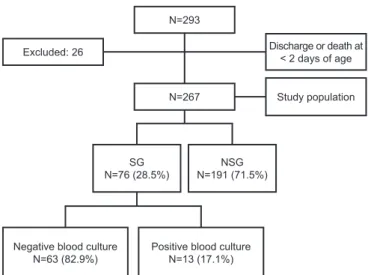
During the study’s time span, 502 patients were admitted to the unit (47.4% of the total population admitted since the opening), and of the admitted patients, 336 were preterm (66.9%). A total of 293 medical charts were reviewed. Of these, 267 preterm neonates met the inclusion criteria and were included in this study.
Late-onset neonatal sepsis was observed in 28.5% (76/267) of the neonates, and 17.1% (13/76) of these neonates had positive blood culture (Figure 1). A total of 22 children died (8.2%), and 68.2% were part of the SG. Gram-negative bacteria were isolated from three of the patients who died by ive days of age (E. coli), by eight days of age (Pseudomonas spp.), and by nine days of age (Klebsiella pneumoniae). Two of the three children were very premature, and one was extremely premature.
A total of 63.1% of the preterm deliveries were performed by cesarean section, 17.6% were born at other institutions, 54.5% were male, and 9.5% had ive-minute Apgar scores of less than seven. Of the included neonates, 11.2% were extremely premature, 31.1% were very premature, and 57.7% were moderately premature.
A total of 12.4% had ELB, 32.2% had VLB, and 41.9% had LB; the remaining 13.5% had birth weights of 2,500 g or more. Of the included neonates, 25.1% had central venous access in place for 11 days or longer, and 17.6% were on MV for 10 days or longer. Nine patients underwent surgical procedures (3.4%).
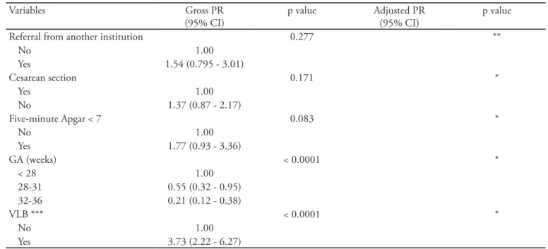
Table 1 illustrates the association between late-onset neonatal sepsis and population characteristics. No diferences were observed between the groups (SG and NSG) in regard to the following characteristics: referral from another institution, cesarean section, and gender (p > 0.05). However, the analysis revealed that as GA and birth weight decreased, the incidence of late-onset sepsis trended upward (p < 0.0001).
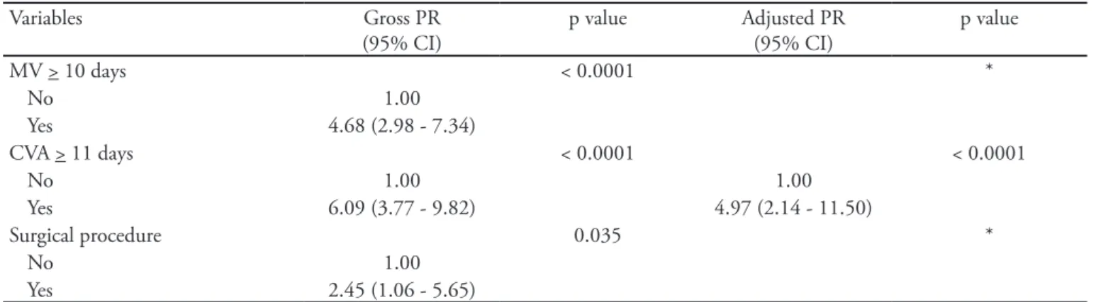
MV for 10 or more days and CVA for 11 or more days were associated with late-onset neonatal sepsis in 80.8% and 76.2% of the preterm newborns, respectively (p < 0.0001). In the SG, 66.7% of the newborns underwent surgical procedures; the diference was statistically signiicant (p = 0.018). Similarly, the occurrence of ive-minute Apgar scores of less than seven was diferent between the groups (p = 0.037).
Bivariate and multivariate analyses were conducted for the late-onset sepsis outcome variables, resulting in a p-value of less than 0.20. he calculation model is shown in table 2. he prevalence of late-onset sepsis was 397% higher in premature neonates with CVA for 11 or more days than in those with CVA for less than 11 days.
he data in Figure 2 data indicate that among the 76 patients who developed late-onset neonatal sepsis (n = 76; 28.5%), 13 (17.1%) had a positive blood culture; Gram-negative bacteria were commonly identiied (n = 8; 61.5%),
Figure 1 - Preterm neonates included in the study.
SG - sepsis group; NSG - non-sepsis group. N=293
N=267
Negative blood culture N=63 (82.9%)
Positive blood culture N=13 (17.1%)
Excluded: 26 Discharge or death at
< 2 days of age
Study population
SG N=76 (28.5%)
Table 2 - Bivariate and multivariate analysis of the variables included in the late-onset neonatal sepsis – preterm model
Variables Gross PR
(95% CI)
p value Adjusted PR
(95% CI)
p value
Referral from another institution 0.277 **
No 1.00
Yes 1.54 (0.795 - 3.01)
Cesarean section 0.171 *
Yes 1.00
No 1.37 (0.87 - 2.17)
Five-minute Apgar < 7 0.083 *
No 1.00
Yes 1.77 (0.93 - 3.36)
GA (weeks) < 0.0001 *
< 28 1.00
28-31 0.55 (0.32 - 0.95)
32-36 0.21 (0.12 - 0.38)
VLB *** < 0.0001 *
No 1.00
Yes 3.73 (2.22 - 6.27)
Continued...
Table 1 - Prevalence of late-onset neonatal sepsis according to population characteristics
Late-onset neonatal sepsis
Variables Yes (N=76), %
(SG)
No (N=191), % (NSG)
p value
Referral from another institution 36.2 63.8 0.197 *
Cesarean section 24.7 75.3 0.105 *
Five-minute Apgar < 7 47.8 52.2 0.037 *
Gestational age (weeks) < 0.0001 **
< 28 70.0 30.0
28-31 38.5 61.5
32-36 14.9 85.1
Birth weight (g) < 0.0001 **
< 1000 57.6 42.4
1001-1499 44.2 55.8
1500-2499 15.2 84.8
> 2500 5.6 94.4
Gender 0.606 *
Male 26.9 73.1
Female 29.7 70.3
MV > 10 days a 80.8 19.2 0.0001 *
CVA > 11 days a 76.2 23.8 < 0.0001 *
Surgical procedure b 66.7 33.3 0.018 ***
SG - sepsis group; NSG - non-sepsis group; MV - mechanical ventilation; CVA - peripherally inserted central venous access. he percentage is relative to the total number of valid responses; missing data were not considered. Signiicance test, p < 0.05.
*Pearson’s Chi-squared; **linear trend Chi-squared tests; *** Fisher’s exact test.
a Calculated based on the ROC curve.
b Surgical procedures (n=9): ileostomy (n=2), enterorraphy (n=1), exploratory laparotomy (n=1), tracheotomy (n=1), herniorraphy (n=1), chest drain
Table 2 - Continuation
Variables Gross PR
(95% CI)
p value Adjusted PR
(95% CI)
p value
MV > 10 days < 0.0001 *
No 1.00
Yes 4.68 (2.98 - 7.34)
CVA > 11 days < 0.0001 < 0.0001
No 1.00 1.00
Yes 6.09 (3.77 - 9.82) 4.97 (2.14 - 11.50)
Surgical procedure 0.035 *
No 1.00
Yes 2.45 (1.06 - 5.65)
PR - prevalence ratio; CI - conidence interval; GA - gestational age; VLB - very low birth weight; MV - mechanical ventilation; CVA - peripherally inserted central venous access.
*Noncontinuous variables in the multivariate model. **Not included in the multivariate analysis because p>0.20. ***VLB established as cutof point due to low incidence in the exposed group when categorized into four categories. Note: there was no interaction between the variables GA and VLB.
and the strains included Klebsiella pneumoniae (n=3), Escherichia coli (n=2), Klebsiella spp. (n=1), Enterobacter spp. (n=1) and Pseudomonas spp. (n=1). he Gram-positive bacteria that were identiied included oxacillin-sensitive Staphylococcus epidermidis (n=2), oxacillin-resistant Staphylococcus epidermidis (n=1), Staphylococcus aureus (n=1) and Staphylococcus saprophyticus (n=1). No fungi were isolated.
DISCUSSION
The incidence of late-onset neonatal sepsis (28.5%) in this study was comparable to that reported in the medical literature (16% to 50%).(2-6) However, the proportion of positive blood cultures among the patients with sepsis (17.1%) was lower than the figures reported in the literature (18% to 65%).(6,7,20,21)
The blood cultures from the three patients who died revealed Gram-negative bacteria, raising the possibility that these agents are associated with increased mortality. This finding agrees with Cohen-Wolkowiez et al. and Gordon et al., who found that increased mortality ranging from 19% to 24% was related to Gram-negative bacteria.(7,8) Kayange et al. also found that increased mortality was associated with positive blood cultures.(11)
In our results, the duration of central venous access was an independent factor associated with late-onset neonatal sepsis. This finding is supported by other authors who found an association between late infection and invasive procedures.(22-24)
The prevalence of Gram-negative bacteria in late-onset sepsis has also been documented in other reports. Graham et al. reported a higher prevalence of Gram-negative bacteria, which are likely to be associated other risk factors and preventive measures compared with Gram-positive bacteria and Candida spp.(22) Furthermore, other authors have reported that the maintenance of peripherally inserted central venous access and other invasive procedures, such as mechanical ventilation, are risk factors for
Gram-Figure 2 - Prevalence of late-onset neonatal sepsis, positive blood culture and isolated bacteria - preterm.
Gram-negative (n=8): Klebsiella pneumoniae (n=3), Escherichia coli (n=2),
Klebsiella spp. (n=1), Enterobacter spp. (n=1) and Pseudomonas spp. (n=1). Gram-positive (n=5): oxacillin-sensitive Staphylococcus epidermidis
(n=2), oxacillin-resistant Staphylococcus epidermidis (n=1), Staphylococcus
aureus (n=1) and Staphylococcus saprophyticus (n=1).
SG - sepsis group; NSG - non-sepsis group; BC - blood culture; pos - positive; neg - negative.
NSG n=191 (71.5%)
BC neg.; n=63 (82.9%) SG
n=76 (28.5%)
BC pos.; n=13 (17.1%)
Gram-neg.; n=8; (61.5%) Gram-pos.;
negative sepsis.(10,22,25) The study by Nambiar et al. demonstrates a higher prevalence of Gram-negative bacteria (43%), predominantly Enterobacter spp.(26)
A high proportion of Klebsiella pneumoniae among isolated Gram-negative bacteria was also documented by Tragante et al. and Meireles et al.(4,21) However, some studies have reported a predominance of Gram-positive bacteria, including coagulase-negative Staphylococcus. Among cultured Gram-negative bacteria, Pseudomonas spp. and Enterobacter spp., were isolated most frequently.(3,6,12,27-29) An American study assessing only late-premature children has demonstrated that S. aureus and E. coli are often present in blood cultures.(7)
The cross-sectional and retrospective nature of this study is a limitation. A causative relationship cannot be identified from this type of study. This study only allows for an analysis of associations. In addition, this study is subject to information bias.
CONCLUSIONS
Late-onset sepsis remains a concern due to its prevalence in neonatal intensive care units and its association with invasive procedures commonly performed on preterm neonates. The increasing participation of Gram-negative bacteria in late-onset neonatal sepsis should be emphasized, as should the need for more efficient methods to identify cases of proven sepsis.
RESUMO
Objetivo: Avaliar a prevalência, os fatores e os agentes etiológicos associados à sepse neonatal tardia em pré-termos de uma unidade de terapia intensiva neonatal.
Métodos: Estudo transversal, de dados secundários
de prontuários de pré-termos admitidos em uma unida-de unida-de terapia intensiva neonatal, no triênio 2008-2010. Caracterizou-se a variável desfecho sepse neonatal tardia pelos critérios da Agência Nacional de Vigilância Sanitá-ria. Empregaram-se os testes do Qui-quadrado de Pearson, exato de Fisher ou Qui-quadrado de tendência linear para as variáveis qualitativas. Considerou-se significante p<0,05. Realizaram-se análises bivariadas e multivariadas entre as variáveis independentes e a dependente, obtendo-se como medida de efeito as razões de prevalências, considerando-se p<0,20.
Resultados: Participaram do estudo 267 prematuros.
Destes, 28,5% evoluíram com sepse tardia, com positivida-de positivida-de hemocultura em 17,1%. Evoluíram a óbito 8,2% dos pré-termos e, destes, 68,2% eram do grupo sepse. Associa-ram-se à hemocultura positiva três óbitos, todos com a par-ticipação de Gram-negativos. Na análise bivariada para o desfecho sepse tardia observou-se que, à medida que decres-ceram a idade gestacional e o peso ao nascer, houve aumen-to de sua prevalência. A duração de ventilação mecânica e de cateter central de inserção periférica por períodos iguais ou superiores respectivamente a 10 e 11 dias se associaram ao desfecho sepse neonatal tardia em 80,8% e 76,2% dos pré-termos. Na análise multivariada, permaneceu como fa-tor associado à sepse tardia o tempo de cateter central de inserção periférica igual ou superior a 11 dias. Houve maior participação dos Gram-negativos como agentes etiológicos, sendo mais frequentes a Klebsiella pneumoniae e a Escheri-chia coli.
Conclusões: A sepse tardia mantém-se uma preocupação
por sua prevalência nas unidades de terapia intensiva e pela associação a procedimentos invasivos a que são submetidos os pré-termos. Ressaltam-se a tendência à emergência dos Gram-negativos na participação da sepse neonatal tardia e a necessidade de melhores e mais eficientes métodos para identificar os quadros de sepse comprovada.
Descritores: Sepse; Unidades de terapia intensiva
neo-natal; Prematuro; Microbiologia
REFERENCES
1. Barros FC, Victora CG, Barros AJ, Santos IS, Albernaz E, Matijasevich A, et al. he challenge of reducing neonatal mortality in middle-income countries: indings from three Brazilian birth cohorts in 1982, 1993, and 2004. Lancet. 2005;365(9462):847-54.
2. Couto RC, Carvalho EA, Pedrosa TM, Pedroso ER, Neto MC, Biscione FM. A 10-year prospective surveillance of nosocomial infections in neonatal intensive care units. Am J Infect Control. 2007;35(3):183-9.
3. Pessoa-Silva CL, Richtmann R, Calil R, Santos RM, Costa ML, Frota AC, Wey SB. Healthcare-associated infections among neonates in Brazil. Infect Control Hosp Epidemiol. 2004;25(9):772-7.
4. Tragante CR, Ceccon MEJR, Falcão MC, Seiti M, Sakita N, Vieira RA. Prevalência de sepse por bactérias Gram negativas produtoras de beta-lactamase de espectro estendido em Unidade de Cuidados Intensivos Neonatal. Rev Paul Pediatr. 2008;26(1):59-63.
on the rate of late-onset bloodstream infections in very low-birth-weight infants. Am J Perinatol. 2011;28(3):227-32. 6. Pinheiro MSB, Nicoletti C, Boszczowsk I, Puccini DMT,
Ramos SR. Infecção hospitalar em Unidade de Terapia Intensiva Neonatal: há inluência do local de nascimento? Rev Paul Pediatr. 2009;27(1):6-14.
7. Cohen-Wolkowiez M, Moran C, Benjamin DK, Cotten CM, Clark RH, Benjamin DK Jr, Smith PB. Early and late onset sepsis in late preterm infants. Pediatr Infect Dis J. 2009;28(12):1052-6.
8. Gordon A, Isaacs D. Late onset neonatal Gram-negative bacillary infection in Australia and New Zealand: 1992-2002. Pediatr Infect Dis J. 2006;25(1):25-9.
9. Alfaleh KM. Incidence of Late Onset Neonatal Sepsis in Very Low Birth Weight Infants in a Tertiary Hospital: An ongoing challenge. Sultan Qaboos Univ Med J. 2010;10(2):227-30.
10. Hervas JA, Ballesteros F, Alomar A, Gil J, Benedi VJ, Alberti S. Increase of Enterobacter in neonatal sepsis: a twenty-two-year study. Pediatr Infect Dis J. 2001;20(2):134-40. 11. Kayange N, Kamugisha E, Mwizamholya DL, Jeremiah
S, Mshana SE. Predictors of positive blood culture and deaths among neonates with suspected neonatal sepsis in a tertiary hospital, Mwanza-Tanzania. BMC Pediatr. 2010;10:39.
12. Stoll BJ, Hansen N, Fanarof AA, Wright LL, Carlo WA, Ehrenkranz RA, et al. Late-onset sepsis in very low birth weight neonates: the experience of the NICHD Neonatal Research Network. Pediatrics. 2002;110(2 Pt 1):285-91.
13. Mussi-Pinhata MM, Rego MA. Immunological
peculiarities of extremely preterm infants: a challenge for the prevention of nosocomial sepsis. J Pediatr (Rio J). 2005;81(1 Suppl):S59-68.
14. Mussi-Pinhata MM, Nascimento SD. Neonatal nosocomial infections. J Pediatr (Rio J). 2001;77 (Suppl 1):S81-96. 15. Kilbride HW, Powers R, Wirtschafter DD, Sheehan
MB, Charsha DS, LaCorte M, et al. Evaluation and development of potentially better practices to prevent neonatal nosocomial bacteremia. Pediatrics. 2003;111(4 Pt 2):e504-18.
16. Infecções relacionadas à assistência à saúde em neonatologia. In: Agência Nacional de Vigilância Sanitária. Neonatologia: critérios nacionais de infecção relacionadas à assistência à saúde. Brasília: ANVISA; 2008.
17. Sarkar S, Bhagat I, DeCristofaro JD, Wiswell TE,
Spitzer AR. A study of the role of multiple site blood cultures in the evaluation of neonatal sepsis. J Perinatol. 2006;26(1):18-22.
18. Behrman RE, Butler AS, editors. Preterm birth: causes, consequences, and prevention. Washington (DC): National Academies Press (US); 2007.
19. Barros AJ, Hirakata VN. Alternatives for logistic regression in cross-sectional studies: an empirical comparison of models that directly estimate the prevalence ratio. BMC Med Res Methodol. 2003;3:21.
20. Shin YJ, Ki M, Foxman B. Epidemiology of neonatal sepsis in South Korea. Pediatr Int. 2009;51(2):225-32.
21. Meireles LA, Vieira AA, Costa CR. Avaliação do diagnóstico da sepse neonatal: uso de parâmetros laboratoriais e clínicos como fatores diagnósticos. Rev Esc Enferm USP. 2011;45(1):33-9.
22. Graham PL 3rd, Begg MD, Larson E, Della-Latta P, Allen A, Saiman L. Risk factors for late onset gram-negative sepsis in low birth weight infants hospitalized in the neonatal intensive care unit. Pediatr Infect Dis J. 2006;25(2):113-7. 23. Herrmann DMML, Amaral LMB, Almeida SC. Fatores de
risco para o desenvolvimento de sepse neonatal tardia em uma unidade de terapia intensiva. Pediatria (São Paulo). 2008;30(4):228-36.
24. Pereira SM, de Almeida Cardoso MH, Figuexeds AL, Mattos H, Rozembaum R, Ferreira VI, et al. Sepsis-Related Mortality of Very Low Birth Weight Brazilian Infants: he Role of Pseudomonas aeruginosa. Int J Pediatr. 2009;2009:427682.
25. Aly H, Hammad TA, Ozen M, Sandhu I, Taylor C, Olaode A, et al. Nasal colonization among premature infants treated with nasal continuous positive airway pressure. Am J Perinatol. 2011;28(4):315-20.
26. Nambiar S, Singh N. Change in epidemiology of health care-associated infections in a neonatal intensive care unit. Pediatr Infect Dis J. 2002;21(9):839-42.
27. Sadeck ESR, Ceccon MEJR. Aspectos clínicos das infecções estailocócicas em unidade de terapia intensiva neonatal. Pediatria (São Paulo). 2006;28(4):234-41. 28. Freitas BAC, Leao RT, Gomes AP, Siqueira-Batista R.
Terapia nutricional e sepse neonatal. Rev Bras Ter Intensiva. 2011;23(4):492-8.