JSCS–4881 547.831.5:547.26’11
Original scientific paper
Synthesis, structural characterization and myorelaxant activity
of 4-naphthylhexahydroquinoline derivatives containing
different ester groups
MİYASE GÖZDE GÜNDÜZ1*, EMİNE ALBAYRAK1, FATMA İŞLİ2, GÖKÇE SEVİM ÖZTÜRK FİNCAN3, ŞENİZ YILDIRIM4, RAHİME ŞİMŞEK1, CİHAT ŞAFAK1,
YUSUF SARIOĞLU3, SEMA ÖZTÜRK YIDIRIM5 and RAY J. BUTCHER6 (Received 6 December 2015, revised 24 March, accepted 29 March 2016)
1Department of Pharmaceutical Chemistry, Faculty of Pharmacy, Hacettepe University, Ankara 06100, Turkey, 2Department of Rational Drug Use and Drug Supply Management,
Turkish Medicines and Medical Devices Agency, Ankara 06520, Turkey, 3Department of Pharmacology, Faculty of Medicine, Gazi University, Ankara 06500, Turkey, 4Department of Pharmacology, Faculty of Medicine, Kırıkkale University, Kırıkkale 71450, Turkey, 5
Depart-ment of Physics, Faculty of Sciences, Erciyes University, Kayseri 38039, Turkey and 6Department of Chemistry, Howard University, Washington DC 20059, USA
Abstract: The present study reports the synthesis, structural characterization
and myorelaxant activity evaluation of a series of 16 novel 4-naphthylhexa-hydroquinoline derivatives. The compounds were achieved by one-pot micro-wave-assisted method via a modified Hantzsch reaction. The structures of the
compounds were confirmed by various spectral methods, such as IR, 1D and 2D NMR techniques and mass analysis. X-Ray studies of compound 10 pro-vided further evidence for the proposed structure. To evaluate their myorelax-ant activities, the Emax and pD2 values of the compounds and nifedipine were
determined on isolated rabbit gastric fundus smooth muscle strips. The obtained results indicated that the introduction of long chain alkyl groups, such as the 2-methoxyethyl or 2-(methacryloyloxy)ethyl moiety, to the ester group led to the most active compounds.
Keywords:1,4-dihydropyridine; synthesis; myorelaxant activity; crystal
struc-ture, structure elucidation.
INTRODUCTION
Dihydropyridines (DHPs) represent low molecular weight heterocyclic
com-pounds based on a pyridine core. Although theoretically five isomeric DHPs
could exist, the most recognized ones have the 1,4-dihydro structure.
11,4-Dihyd-ropyridines are one of the most important chemical classes introduced into
logical sciences and are common in many commercialized drugs. These
com-pounds mainly present a well-known capacity as calcium channel blockers, thus
acting as vital drugs against cardiovascular diseases, particularly hypertension
and angina pectoris.
2Although DHPs were primarily developed as
cardiovas-cular agents, medicinal chemists decorated the 1,4-DHP nucleus and achieved
diverse activities at several receptors, channels and enzymes with different
medi-cal applications, such as antitubercular, antioxidant, antitumor, antithrombotic,
antimicrobial, antidiabetic, antidyslipidemic and anticonvulsant.
3Since their introduction into clinical medicine, 1,4-DHPs have been one of
the most studied class of drugs and many modifications have been performed on
the structure of nifedipine, the prototype of DHPs, in order to enhance calcium
modulating effects and obtain structure–activity relationships (SAR). According
to SAR studies, a 1,4-DHP ring with an unsubstituted nitrogen and pseudoaxial
oriented aryl ring substituent at C-4 are essential for activity.
4Ester
function-alities at C-3 and C-5 position are of utmost importance to modulate the activity
and tissue selectivity. It was proved that modification of the ester moiety plays a
key role in the ability of condensed 1,4-DHPs to block calcium currents.
5Fused
DHPs, such as hexahydroquinolines, that could be obtained by introducing the
DHP ring into condensed ring systems, are active derivatives exhibiting calcium
antagonistic effects.
6The classical method for the synthesis of 1,4-DHPs is a one-pot Hantzsch
reaction, which proceeds effectively and involves dehydrative coupling of an
aldehyde, two equivalents of a 1,3-dicarbonyl compound and ammonia.
7Dep-ending on the reagents and reaction conditions, long reaction times, unexpected
products or low yields can be obtained.
8Although most of the efforts focused on
improving the reaction using various catalysts, such as Fe
3O
4, cobalt and
ytter-bium,
9–11microwave (MW) irradiation has recently gained great popularity as an
energy source for Hantzsch reactions because of its ability to reduce reaction
times, to improve yields and to simplify the work-up processes.
12In the present study, sixteen DHP derivatives in which substituted
cyclohex-ane rings were fused to the DHP ring under microwave irradiation were
syn-thesized and how different ester groups and the naphthyl moiety attached to this
backbone affected the myorelaxant activities of these compounds was
inves-tigated to obtain additional information to enrich the classical SAR studies.
EXPERIMENTAL
General
Germany). Melting points were determined on a Thomas Hoover capillary melting point apparatus (Philadelphia, PA, USA) and are uncorrected. Infrared spectra were recorded on a Perkin–Elmer Spectrum BX FT-IR instrument (Beaconsfield, UK) and are reported in cm-1. The 1H-NMR spectra were obtained in dimethyl sulfoxide (DMSO) solutions on a Varian Mercury 400, 400 MHz high performance digital FT-NMR spectrometer (Palo Alto, CA, USA). 13C-NMR and COSY (2D-1H–1H homonuclear correlation spectrum) spectra were recorded on the same instrument. The chemical shifts are reported in parts per million (ppm) relative to tetramethylsilane. The X-ray crystallographic analysis was realized on an Agilent Xcalibur (Ruby, Gemini) diffractometer. The ESI-MS spectra were measured on a micromass ZQ-4000 single quadruple mass spectrometer. Elemental analyses were performed on a Leco CHNS-932 Elemental Analyzer (Philadelphia, PA, USA).
Analytical, physical and spectral data of the synthesized compounds are given in Sup-plementary material to this paper.
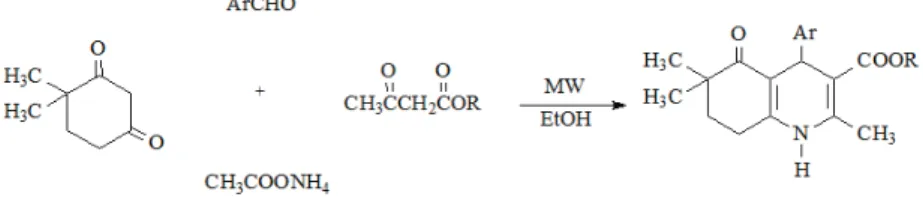
Chemistry
The general procedure for the preparation of alkyl 2,6,6-trimethyl-4-(1-naphthyl/2-naph-thyl)-5-oxo-1,4,5,6,7,8-hexahydroquinoline-3-carboxylates (compounds 1–16) was as follows: a one-pot four component mixture of 2 mmol 4,4-dimethyl-1,3-cyclohexanedione, 2 mmol of 1- or 2-naphthaldehyde, 2 mmol of an appropriate alkyl acetoacetate and 10 mmol of ammo-nium acetate was placed into a 35-mL microwave pressure vial and heated under microwave irradiation (power 50 W, maximum temperature 120 °C) for 5 min. in 5 mL ethanol. After completion of the reaction, monitored by TLC, the reaction mixture was poured into ice–water and the obtained precipitate was filtered and crystallized from ethanol–water. The synthetic route used to synthesize the target compounds is outlined in Fig. 1.
Fig. 1.Synthesis of compounds 1–16.
X-Ray crystallography
Computing details. Data collection: CrysAlis PRO,13 cell refinement: CrysAlis PRO,13;
data reduction: CrysAlis PRO.13 Program(s) used to solve and refine the structure: SHELXS97.14 Program(s) used for molecular graphics and to prepare the material for pub-lication: SHELXTL.14
Refinement. Carbon-bound H-atoms were placed in calculated positions (C–H, 0.93–0.98
Å) and were included in the refinement in the riding-model approximation, with Uiso(H) =
The N-bound H-atoms were located in a difference Fourier map but were refined with a distance restraint: N–H = 0.83 (1) Å with Uiso(H) = 1.2Ueq(N).
Pharmacology
New Zealand white rabbits, weighing 2.5–3 kg, were used in this study. The rabbits were sacrificed with i.v. injection of sodium pentobarbital (30–40 mg kg-1), followed by removal of the stomach through abdominal incision. The fundal part of the stomach was then dissected parallel to the longitudinal muscle wall. One muscle strip about 15–20 mm length and 2 mm width was obtained and allowed to equilibrate for 60 min in 20 mL organ baths filled with calcium (Ca2+) free Krebs–Henseleit solution (KHS). The composition of the Krebs solution was as follows (in mmol L-1): NaCl 118; KCl 4.7; NaHCO3 25; MgCl2 0.54; NaHPO4 0.9; glucose 11. The solution was gassed with 95 % O2 and 5 % CO2 during the study and tempe-rature was maintained at 37 °C by a thermoregulated water circuit. The pH of the saturated solution was 7.4. Each strip was connected to a force transducer (FDT 10-A, May IOBS 99, COMMAT Iletisim Co., Ankara, Turkey) for measurement of the isometric force, which was continuously displaced and recorded on an online computer via a four-channel transducer data
acquisition system (MP30B-CE, BIOPAC Systems Inc., Santa Barbara, CA) using software (BSL PRO v. 3.6.7, BIOPAC Systems Inc.) which also had the capacity to analyze the data. After mounting, each strip was allowed to equilibrate with a basal tension of 1 g for 60 min. Ca2+ free KHS was replaced with fresh solution every 15 min during this time. Nω-nitro-L- -arginine methyl ester hydrochloride (L-NAME, a nitric oxide synthase inhibitor, 10-4 M), indomethacin (COX inhibitor, 10-5 M), tetraethylammonium chloride (Ca2+-activated K+ channel blocker, 10-4 M), glibenclamide (ATP-sensitive K+ channel blocker, 10-6 M) and guanethidine (adrenergic nerve blocker, 10-6 M) were added into the organ bath 20 min before the compounds were added in order to eliminate the effects of nitric oxide, cyclooxygenase, Ca2+-activated K+ channel, ATP sensitive K+ channel and adrenergic pathways, respectively.
After the smooth muscle strips of rabbit gastric fundus had been placed in a high K+- -containing (80 mM) solution, 2.5 mM Ca2+ was added to the organ bath to develop con-traction. Concentration–relaxation responses for the compounds 1–16 (10-8–3×10⁻⁴ M) and nifedipine (10-9–10-6 M) were obtained by adding these into the bath in a cumulative manner. A cumulative concentration–response curve was constructed in a stepwise manner after the response to the previous concentration had reached a plateau. The relaxant effects of the com-pounds and nifedipine were expressed as percentage of the precontraction with 2.5 mM Ca2+ in the high K+ containing solution. DMSO, used in activity studies as solvent, was also tested. To evaluate the effects of the compounds, the maximum response (Emax) values of
com-pounds and nifedipine were established at 3×10⁻⁴ M and 10-6 M concentrations, respectively and pD2 values (the negative logarithm of the concentration for the half-maximal response
(EC50)) were calculated, as predicted from the Scatchard equation for drug–receptor
inter-action. Agonist pD2 values (apparent agonist affinity constants) were calculated from each
agonist concentration–response curve by linear regression of the linear part of the curve and taken as a measure of the sensitivity of the tissues to each agonist. While Emax is the parameter
for efficacy, pD2 is the parameter for potency. All data are expressed as mean ± standard
error. Statistical comparison between groups were performed using general linear models by the Scheffe F-test and p values less than 0.05 were considered to be statistically significant.
The study was approved by the Gazi University Ethics Committee. Procedures involving animals and their care were conducted in conformity with international laws and policies.
chloride and guanethidine were dissolved in distilled water, indomethacin, glibenclamide, nifedipine and the compounds were dissolved in DMSO.
RESULTS AND DISCUSSION
Chemistry
A series of condensed 4-naphthyl-1,4-DHP derivatives were prepared
via
a
modified Hantzsch reaction. In order to obtain the target compounds, 4,4-
-dimethyl-1,3-cyclohexanedione, 1-naphthaldehyde/2-naphthaldehyde, an
appro-priate alkyl acetoacetate were heated in the presence of excess ammonium
ace-tate under microwave irradiation in ethanol, which was classified as an excellent
microwave-absorbing solvent.
15,16The appearance of the products was monitored by TLC and the reaction time
was determined as 5 min., which is quite a short time compared to conventional
heating for the Hantzsch reaction.
17,18The structures and chemical characteristics of the synthesized compounds
are reported in Table S-I of the Supplementary material.
The structures of the synthesized compounds were elucidated by spectral
methods (IR,
1H-NMR,
13C-NMR, COSY, X-ray analysis and mass spectra) and
confirmed by elemental analysis. In the IR spectra, characteristic N–H, C=O
(ester) and C=O (ketone) stretching bonds were observed. In the
1H-NMR
spectra, the protons of the methyl substituents at the 6-position of the
hexahydro-quinoline ring were observed separately and as singlets at 0.73–0.99 ppm. The
methylene groups of the same ring were at 1.57–2.57 ppm. The methine protons
of the 1,4-DHP ring were seen as a singlet at 4.96–5.63 ppm.
The aromatic
protons of the naphthyl and phenyl rings were at 6.91–8.73 ppm, while the N–H
protons of the DHP ring were seen at 9.02–9.21 ppm. In the
13C-NMR spectra,
the number of the signals fitted exactly the number of carbon atoms. The
correl-ations between the interacting protons of compound
2 were determined by
COSY. The correlations between H-7 and H-8, CH
2and CH
3protons in the ester
side chain and H-4 and the aromatic protons were observed. These correlations
are demonstrated in Fig. 2 and the COSY spectrum is provided as Supplementary
material. Based on this information; the structure of compound
2 was
conclu-sively identified as ethyl
2,6,6-trimethyl-4-(1-naphthyl)-5-oxo-1,4,5,6,7,8-hexa-hydroquinoline-3-carboxylate.
The mass spectra of the compounds were recorded
via
the electrospray
ion-ization technique. The quasimolecular ions created by the addition of sodium ion
[M+Na]
+and also of a hydrogen cation [M+1+Na]
+were observed in the spectra
of all compounds. Cleavage of the ester group and the naphthyl ring from the
parent molecule were the next most observed fragmentations.
Fig. 2. COSY correlations of compound 2.
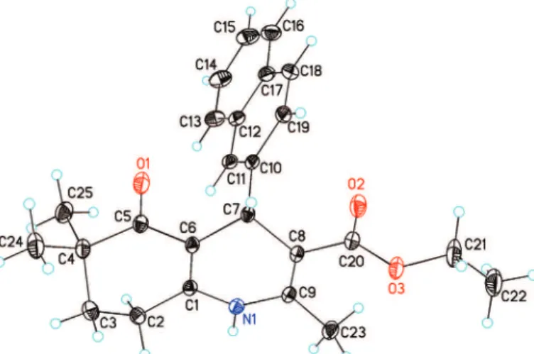
X-Ray analysis of compound
10
The three-dimensional structure of ethyl 2,6,6-trimethyl-4-(2-naphthyl)-5-
-oxo-1,4,5,6,7,8-hexahydroquinoline-3-carboxylate
(10) was evaluated by X-ray
crystallography (Fig. 3).
Fig. 3.X-Ray molecular structure of compound 10 with the atom-numbering scheme used in the crystallographic analysis.
The crystal data and a summary of intensity data collections and structural
refinements, selected bond lengths, bond angles and torsion angles are given in
the Supplementary material.
In compound 10 (Fig. 3), the naphthalene ring is almost planar with a
max-imum deviation from the mean plane of –0.029(2) Å for atom C(17). The
cyclo-hexene rings adopt a sofa conformation and are puckered with puckering
para-meters
19of
Q
T= 0.4383(17) Å,
θ
= 50.6(2)°,
φ
= 127.6(3)°. The values of the
bond lengths
20and angles in the title compound are within the normal ranges and
are comparable with those of related relationships.
21–23The X-ray
these dimers are connected by N–H…O hydrogen bonds, generating
one-dimen-sional chains along [011] (Supplementary material).
Pharmacology
The maximum relaxant effects (
E
max) and p
D
2values of compounds
1–16,
DMSO and nifedipine on isolated strips of rabbit gastric fundus smooth muscle
are given in Table I.
TABLE I. Maximum relaxant responses (Emax) and pD2 values of the compounds, nifedipine
and DMSO on strips of rabbit gastric fundus smooth muscle; the relaxation is expressed as the percentage of the precontraction induced by 2.5 mM Ca2+. The negative logarithm of the concentration for the half-maximal response (pD2) and Emax values represent mean value ± SEM; p < 0.05 compared with the control responses, n = 6
Compound Emax pD2
1 81.20±10.26 5.42±0.71
2 82.78±8.98 5.53±0.65
3 77.42±4.71 4.63±0.37
4 45.52±9.10 4.92±0.66
5 53.32±7.53 5.00±0.57
6 93.54±9.31 5.56±0.67
7 91.83±5.29 4.95±0.42
8 43.17±5.97 5.39±0.47
9 63.93±5.90 4.52±0.47
10 42.30±4.67 5.31±0.37
11 50.42±10.01 4.37±0.70
12 36.88±5.96 6.09±0.47
13 48.90±10.41 5.37±0.71
14 92.68±8.15 4.67±0.61
15 91.50±5.94 5.07±0.47
16 21.94±3.68 6.34±0.26
Nifedipine 100.00±2.18 8.33±0.04
DMSO 11.35±2.25 6.31±0.05
Tissues were pretreated with indomethacin, guanethidine,
L-NAME,
tetra-ethylammonium chloride and glibenclamide to investigate whether the relaxation
induced by the compounds occurred through cyclooxygenase, the adrenergic
system, nitric oxide pathways, Ca
2+-activated K
+channel and ATP-sensitive K
+channel, respectively. Pretreatment of the strips with indomethacin, guanethidine,
L
-NAME, tetraethylammonium chloride and glibenclamide confirmed that
cyclo-oxygenase, adrenergic and nitric oxide pathways, Ca
2+-activated K
+channel and
ATP-sensitive K
+channel played no roles on the relaxations evoked by these
substances.
The results of this study indicate that all of the compounds (10
–8–3×10
–4M)
and nifedipine (10
–9–10
–6M) produced concentration-dependent relaxation on
from the control relaxations produced by DMSO. The compounds and nifedipine
exerted concentration-dependent relaxation responses on the gastric fundus
smooth muscle strips precontracted with Ca
2+(2.5 mM) with the efficacy order:
nifedipine ≥ 6 ≥ 14 ≥ 7 = 15 > 2 ≥ 1 ≥ 3 > 9≥ 5 ≥ 11 ≥ 13 ≥ 4 ≥ 8 ≥ 10 ≥ 12 > 16.
The efficacy of compounds
6, 7,
14
and 15
were found to be the same as
nifedipine. The obtained results suggested that myorelaxant effects of the
com-pounds seem to exert their effects by blocking Ca
2+channels as does nifedipine.
Given that the main difference between these compounds is their ester
groups, it follows that the ester moiety plays a key role in the ability of these
compounds to block calcium channels.
Increasing the side chain length of the ester group mediated an increase
whereas introducing a ring structure at the same locus did not lead to a significant
improvement in blocking the activity. The introduction of a
2-(methacryloyl-oxy)ethyl or
2-methoxyethyl group as the side chain of the ester group resulted in
a series of highly active compounds.
Two methyl groups at the 6-position of the hexahydroquinoline ring are
pre-sent in all compounds and therefore they are not the most critical components for
the preferential activity. When the obtained results are analyzed in terms of the
substitution position of the naphthyl ring at the C-4 position of DHP, generally
the 1-naphthyl derivatives possessed better activities. Although all compounds
are potent myorelaxant agents on the gastric fundus smooth muscle strips,
intro-duction of a naphthalene substituent into the 4-position of the 1,4-DHP nucleus
decreased the myorelaxant effect of the compounds compared to nifedipine. This
could suggest that the substitution of an
o
-nitrophenyl ring by a naphthyl ring
increases the size of the molecules and may have a negative effect on the ability
of these compounds to show their effects. As a result, the naphthyl ring could be
a good choice as the aromatic substituent at the C-4 position of DHP only in
combination with long chain alkyl esters.
CONCLUSIONS
An easy, very rapid and convenient method for the preparation of condensed
1,4-DHPs under MW irradiation was reported. The target compounds were
achieved by the reaction of 4,4-dimethyl-1,3-cyclohexanedione,
1-naphthalde-hyde/2-naphthaldehyde, an appropriate alkyl acetoacetate and ammonium acetate
in ethanol. This method also offers a reduction of solvent use and reaction time in
addition to higher yields.
The obtained pharmacological results showed that all the synthesized
com-pounds had relaxing effects on isolated rabbit gastric fundus smooth muscle,
pos-sibly due to the blockade of the Ca
2+channels, similar to the action of nifedipine.
com-pounds, suggesting that two hydrogen bond acceptor groups might be required
for the calcium channel blocking activity. It was also proved that there is no
con-tribution of cyclooxygenase, adrenergic and nitric oxide pathways, ATP-sensitive
K
+channels and Ca
2+-activated K
+channels to the myorelaxant effects of the
compounds. As a result, further investigations are required to ascertain the Ca
2+channel blockage effects of the compounds.
SUPPLEMENTARY MATERIAL
Data on the characterization of the synthesized compounds are available electronically from http://www.shd.org.rs/JSCS/, or from the corresponding author on request.
Acknowledgements. The authors gratefully acknowledge the financial support provided
by the Scientific Research Fund of Hacettepe University, Turkey through Project 013.D03.301.001.
RJB wishes to acknowledge the NSF–MRI program (grant CHE-0619278) for funds to purchase the diffractometer and the Howard University Nanoscience Facility for access to liquid nitrogen.
И З В О Д
СИНТЕЗА, СТРУКТУРНАКАРАКТЕРИЗАЦИЈАИМИОРЕЛАКСНТНААКТИВНОСТ
ДЕРИВАТА 4-НАФТИЛХЕКСАХИДРОХИНОЛИНАКОЈИСАДРЖЕРАЗЛИЧИТЕ
ЕСТАРСКЕГРУПЕ
MIYASE GÖZDE GÜNDÜZ1, EMINE ALBAYRAK1, FATMA
İŞLI2, GÖKÇE SEVIM ÖZTÜRK FINCAN3,
ŞENIZ YILDIRIM4, RAHIME ŞIMŞEK1, CIHAT ŞAFAK1, YUSUF SARIOĞLU3, SEMA ÖZTÜRK YIDIRIM5 и RAY J. BUTCHER6
1
Department of Pharmaceutical Chemistry, Faculty of Pharmacy, Hacettepe University, Ankara 06100, Turkey, 2Department of Rational Drug Use and Drug Supply Management, Turkish Medicines and Medical Devices Agency, Ankara 06520, Turkey, 3Department of Pharmacology, Faculty of Medicine, Gazi University,
Ankara 06500, Turkey, 4Department of Pharmacology, Faculty of Medicine, Kırıkkale University, Kırıkkale 71450, Turkey, 5Department of Physics, Faculty of Sciences, Erciyes University, Kayseri 38039, Turkey и
6
Department of Chemistry, Howard University, Washington DC 20059, USA
Приказана је синтеза, структурна карактеризација и миорелаксантна активност
серијеод 16 новихдеривата 4-нафтилхексахидрохинолина. Једињењасусинтетисанау
једномреакциономкоракуподмикроталснимозрачивањем, модификованомХанчовом
реакцијом. СтруктуреједињењаодређенесунаосновуспектралнихподатакаИЦ, 1D и
2D NMR спектроскопије и масене спектрометрије. Анализом рендгенске структуре
монокристаладеривата10додатнојепотврђенапредложенаструктураједињења. Током
испитивањамиорелаксантнеактивностиодређенесуEmaxиpD2вредностииспитиваних
једињења и нифедипина, на исечцима глаткомишићних ћелија желудачног дна зеца.
Добијенирезултатиуказујунадоприносалкил-групадугогланца, каоштосу 2-метокси
-етилили 2-(метакрилоилокси)етил-естара, добројактивности.
(Примљено 6. децембра 2015, ревидирано 24. марта, прихваћено 29. марта 2016)
REFERENCES
1. N. Edraki, A. R. Mehdipour, M. Khoshneviszadeh, R. Miri, Drug Discovery Today 14
(2009) 1058
3. E. Carosati, P. Ioan, M. Micucci, F. Broccatelli, G. Cruciani, B. S. Zhorov, A. Chiarini, R. Budriesi, Curr. Med. Chem.19 (2012) 4306
4. P. Ioan, E. Carosati, M. Micucci, G. Cruciani, F. Broccatelli, B. S. Zhorov, A. Chiarini, R. Budriesi, Curr. Med. Chem.18 (2011) 4901
5. C. Bladen, M. G. Gunduz, R. Simsek, C. Safak, G. W. Zamponi, Pfluegers Arch. 466
(2014) 1355
6. R. Simsek, G. S. Ozturk, I. M. Vural, M. G. Gunduz, Y. Sarioglu, C. Safak, Arch. Pharm.
341 (2008) 55
7. V. G. Santos, M. N. Godoi, T. Regiani, F. H. S. Gama, M. B. Coelho, R. O. M. A. de Souza, M. N. Eberlin, S. J. Garden, Chem. Eur. J.20 (2014) 12808
8. K. A. Undale, Y. Park, K. Park, D. H. Dagade, D. M. Pore, Synlett (2011) 791
9. S. Sueki, R. Takei, J. Abe, I. Shimizu, Tetrahedron Lett. 52 (2011) 4473
10. M. Nasr-Esfahani, S. J. Hoseini, M. Montazerozohori, R. Mehrabi, H. Nasrabadi, J. Mol. Catal., A: Chem.382 (2014) 99
11. J. Safari, S. H. Banitaba, S. D. Khalili, Chin. J. Catal. 32 (2011) 1850
12. A. Debache, W. Ghalem, R. Boulcina, A. Belfaitah, S. Rhouati, B. Carboni, Tetrahedron Lett.50 (2009) 5248
13. Agilent. 2011. CrysAlis PRO and CrysAlis RED. Agilent Technologies Yarnton England. 14. G. M. Sheldrick, Acta Crystallogr., A64 (2008) 112
15. C. O. Kappe, Angew. Chem. Int. Ed. 43 (2004) 6250
16. A. Saini, S. Kumar, J. S. Sandhu, J. Sci. Ind. Res.67 (2008) 95
17. P. Lidstrom, J. Tierney, B. Wathey, J. Westman, Tetrahedron57 (2001) 9225
18. C. Safak, M. G. Gunduz, S. O. Ilhan, R. Simsek, F. Isli, S. Yildirim, G. S. O. Fincan, Y. Sarioglu, A. Linden, Drug Dev. Res. 73 (2012) 332
19. D. Cremer, J. A. Pople, J. Am. Chem. Soc. 97 (1975) 1354
20. F. H. Allen, Acta Crystallogr., B58 (2002) 380
21. M. G. Gunduz, R. J. Butcher, S. Ozturk Yildirim, A. El-Khouly, C. Safak, R. Simsek,
Acta Crystallogr., E68 (2012) o3404
22. A. El-Khouly, S. Ozturk Yildirim, R. J. Butcher, R. Simsek, C. Safak, Acta Crystallogr., E68 (2012) o3337
23. S. Ozturk Yildirim, R. J. Butcher, A. El-Khouly, C. Safak, R. Simsek, Acta Crystallogr., E68 (2012) o3365
24. J. Bernstein, R. E. Davis, L. Shimoni, N. L. Chang, Angew. Chem. Int. Ed. 34 (1995)
1555