Introduction
Biotransformations use living organisms or isolated enzimes to modify chemical structures and have some advantages over conventional chemical reactions [1]. This methodology have been showing a quick development along the time and represents a potential alternative for obtaining biologically active compounds of diffi-cult access by classic synthetic methods [2,3].
Kaurane diterpenes are widely distributed in nature [4-6] and present very important biologi-cal activities, such as antitumor [7] anti-HIV [8], trypanocidal [9-10] antimicrobial [11] and allelo-pathic [12-13], among others. Ent -kaur-16-en-19-oic and ent-kaur-9(11),16-dien-19-oic acids and their chemical transformations derivatives are very
used substrates for biotransformations [14-17]. Kaurane diterpenes are also intermediates in the biosynthesis of a number of plant and fungal metabolites, including gibberellins, a group of diterpene lactones, as GA3, used in agriculture to stimulate plant growth. Allelopathic activity of ent -kaur-16-en-19-oic and ent -kaur-9(11),16-dien-19-oic acids and derivatives obtained by chemical transformations of them were previously evaluated by our research group [12-13].
The fungus C. aphidicola promotes hydroxylations in different classes of organic compounds and has been widely used in bio-transformations of kaurane diterpenes [18-20].
In this work, we describe the biotransfor-mation of ent-kaur-16-en-19-ol (1) by C. aphidico-la. Two hydroxylated compounds, ent-kauran-16b,
www.scielo.br/eq www.ecletica.iq.unesp.br
Volume 34, número 1, 2009
Di-And Tri-Hydroxylated Kaurane Derivatives From
Microbial Transformation Of
Ent
-Kaur-16-En-19-Ol By
Cephalosporium Aphidicola
And Their Allelopathic Activity
On
Lactuca Sativa
(Lettuce)
D. Rocha, J. A. Takahashi, M. A. D. Boaventura*
Departamento de Química, ICEx, Universidade Federal de Minas Gerais, Belo Horizonte, MG, CEP 31270-901 , Brazil *[email protected]
Abstract: The use of microorganisms to induce chemical modifications in organic molecules is a very useful tool in organic synthesis, to obtain biologically active substances. The fungus Cephalosporium aphidicolais known by its ability to hydroxylate several skeleton positions of many classes of organ-ic compounds. In this work, the morgan-icrobial transformation of ent-kaur-16-en-19-ol (1) by C. aphidico-la, afforded two hydroxylated compounds, ent-kauran-16b,19-diol (2) and ent-kauran-16b,17,19-triol (3). Their structures were established by 1D and 2D-NMR studies. Both compounds were tested for their action on the growth of radical and shoot of Lactuca sativa.
Key words: Biotransformations, Cephalosporium aphidicola, ent-kaur-16-en-19-ol, ent-kauran-16b,
19-diol (2) and ent-kauran-16b,17,19-triol (3), were isolated (Scheme 1) and tested over their action on the growth of radical and shoot of
Lactuca sativa (lettuce). This is the first time these compounds were obtained from this substrate using
C. aphidicola. This is also the first report on their allelopathic activity by the best of our knowledge.
Materials And Methods General
Solvents (hexane, dichloromethane, ethyl acetate, methanol) from VETEC (Brazil), and were of PA grade. Silica gel Merck (Darmstadt, Germany) 70-240 and 230-400 mesh were used for chromatography column and silica gel Merck 60G was used for thin layer chromatography. Reagents to prepare the culture medium were pur-chased from Difco (Sparks, MD, USA). Melting points were determined with a Kofler hot plate apparatus and are uncorrected. Nuclear Magnetic Resonance (NMR) spectra (1D and 2D) were recorded in CDCl3, at room temperature, on a Bruker Avance DRX 400 MHz, from Bruker Analytic, Ettlingen, Germany. Ent- kaur-16-en-19-ol (1) was furnished by Dr. Henriete S. Vieira, Departamento de Química, ICEx, UFMG, Brazil.
Biotransformation
Stock culture of C. aphidicola (CCT 2163) was maintained on PDA under refrigera-tion and small secrefrigera-tions of this agar were trans-ferred to Erlenmeyer flasks containing a liquid medium (200 mL/flask) comprised of: glucose (50.0 g/L), NaH2PO4 (5.0 g/L), MnSO4.6H2O (2.0 g/L), KCl (1.0 g/L), glicine (2.0 g/L) and 2.0 mL of trace elements solution [CO(NO3)2,
0.10 g/L, FeSO4, 1.0 g/L, CuSO4, 0.15 g/L, ZnSO4, 1.61 g/L, MnSO4, 0.10 g/L, (NH4)6Mo7O24.4H2O, 1.0 g/L] of distilled water; pH was adjusted to 5 with 10% HCl solution and the culture was incubated over magnetic stirring for 3 days. Cells in suspension (15 mL portions) were transferred to new Erlenmeyer flasks and reached suitable growth after 24 h. Then, the sub-strate 1 (0.69 g) was added (0.5 mL/flask, 53 mg/mL). After 13 days, the flasks contents were extracted with ethyl acetate and the solvent was taken out under vacuum. Residue (1.37 g) was chromatographed over silica gel (21.5 g), using hexane, CH2Cl2, AcOEt and CH3OH, either neat or in mixtures of increasing polarity. A group of fractions (G-7, 81.5 mg), eluted from CH2Cl2/AcOEt 1:1, was rechromatographed over silica gel (3.2. g), using the same solvent system. G-7-2 (25.0 mg) gave an amorphous white solid [3.7 mg, ent-kauran-16b,19-diol, (2)], corresponding to a single spot by TLC (hexane/AcOEt 75:25), by another silica gel col-umn chromatography. G-7-3 (17.0 mg), after new purification by silica gel column chromatogra-phy, gave a crystalline white solid [12.8 mg, ent -kauran-16b,17,19-triol (3)], also pure by TLC (eluents: hexane/AcOEt 70:30).
Bioassay
Lactuca sativa (cv Grand Rapids) seeds were purchased from Isla Pak, RS, Brazil. All undersized and damaged seeds were discarded. Germination and growth were conducted in 100 mm Petri dishes containing a 9.0 cm sheet of Whatman no. 1 filter paper as suport. Then, 25 let-tuce seeds were placed per dish with 10 mL of a test (10-4, 10-6and 10-8M) or a control solution. All
solutions were prepared with deionized water and their pH values [buffered with 10 mM 2-(N -mor-pholino) ethanesulfonic acid, MES] were adjusted to 6.0 - 6.5 with NaOH solution. Concentrations lower than 10-4M were obtained by dilution series. All tests were triplicated. Dishes were covered with Parafilm to reduce evaporation and incubated in the dark at 25 oC, in a controlled-environment growth chamber, for 5 days. After this time, number of ger-minated seeds were counted (a seed was considered to be germinated when the radicle was at least 0.2 mm long), the lengths of radical and shoots were measured (using a pachymeter). During the meas-urement process, the dishes were kept at 4 oC to avoid subsequent growth. The osmotic pressure val-ues were measured on a microsmometer and ranged between 30 and 38 mOsmolar [15].
Data Analysis
The effects on germination and growth are given as percent differences from control, and con-sist of the differences (in cm) between mean values of seeds with tested compounds and mean values for control (seeds grown without addition of tested compounds)/ mean values for control x 100. Thus, zero represents the control, positive values repre-sent stimulation of the studied parameter and neg-ative values represent inhibition.
The data were evaluated using Student’s t -tests and the differences between the experiment and control were significant at a value of P ≤0.05.
The results obtained for compounds 2and
3are shown in Figure 2.
Results and Discussion
Compounds 2and 3were identified by 1D and 2D 1H and 13C- NMR as products of mono and di- hydroxylation, respectively, of the original C16-C17 double bond of substrate 1, since in the 1H- NMR spectrum of both products, the
unsatu-rated exocyclic methylene signals, present on starting material at dH 4.73 and 4.79 were not observed . Also, this spectrum showed the signals for C-19 hydroxymethyl AB system for both prod-ucts, those of 2at dH3.44 (1H, d, J=11.2 Hz) and 3.72 (1H, d, J=11.2 Hz). The 1H NMR of 2 exhib-ited signals for two methyl groups at dH0.95 (3H,
s, H-18) and 1.01 (3H, s, H-20) and for a new methyl group at dH1.35. 13C-NMR spectrum and DEPT experiment showed a chemical shift at dC
65.6, of the C-19 oxygenated methylene carbon, from the starting material. Additionally, signals for nine methylene carbons, three methine carbons, and four quaternary carbons (including a new oxy-genated carbon at dC79.3) were observed. In order to carry on a more detailed spectroscopic analysis of the products, bidimensional spectra (HSQC, HMBC and ROESY) were obtained for 2, since they are of special help for complete attribution of terpene compounds [21]. HSQC spectrum showed the correlations between the signal at dH1.82 with
dC49.0, attributed to C-13, by comparison to the spectroscopic data of the starting material; the sig-nals at dH1.43 and 1.48 were correlated to a signal at dC18.0 (C-11); the signals at dC1.57 and 1.88 to a signal at dC37.5 (C-14). A correlation of the signal at dH 0.99 with the signal atdC57.0 (C-9) was also observed. The signals corresponding to methine positions 5, 9 and 13 appeared at dH0.91 and dC56.8; at dH0.99 and dC57.0; and at dH 1.82 and dC 49.0, respectively. Also, the signal at dH
1.35 (3H) was correlated, in the HSQC spectrum, to a signal at dC24.4, and was associated with C-17, since this was the only option for the introduc-tion of an addiintroduc-tional methyl group in the molecule, and consequently the new oxygenated quaternary carbon signal observed at dC79.3 can be associat-ed to C-16.
In the HMBC spectrum, 3J correlations between H19b(dH3.72) with C-18 (dC27.0) and between H19b(dH3.44) with C-3 (dC35.7) were observed. Besides, correlations between hydro-gens of the methyl group at C17 (dH1.35) and C13 (dC49.0), C15 (dC57.9) and C16 (dC78.3) were further evidence of the presence of a methyl group at C-17. Therefore, 2was identified as the product of monohydroxylation of the original exocyclic double bond from starting material (Table 1).
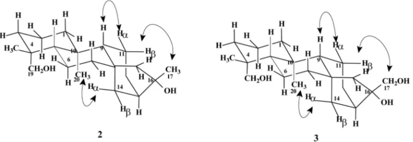
The configuration of C-16 was estab-lished by analysis of ROESY spectrum, where the correlation of CH3-20 (dH1.01) with H-14a, at dH 1.57, was observed. Thus, by using HSQC spectrum, the signal of H-14bwas located at dH
was found to be dH 1.43, due to its correlation with H-9 (dH0.99) therefore by HSQC spectrum H-11b (dH1.48) was located and, as this signal correlates with CH3-17 (dH1.35) in the ROESY spectrum, the later is unequivocally at the
a-position (Figure 1). Thus, 2was proposed to be
ent-kauran-16b,19-diol. This compound was already isolated as natural product from stems of
Aristolochia rodriguesii[22] and leaf and stems of Ozothamnus hookeri(Asteraceae) [23]; 2was also obtained by chemical modification of ent -kaur-16-en-19-oic acid [2].
NMR spectra of product 3 were not consis-tent with the presence of a methyl group at C-17, since only a new signal in 1H- NMR spectrum at dH4.03 (2H) was observed. Two new oxygenated carbon signals were detected in 13C NMR spec-trum, at dC78.7 and 71.8 (quaternary and methyl-ene, respectively, according with DEPT experi-ment). HSQC spectrum presented correlations between the signal at dH0.95 with dC57.1,
The effect of compounds 2and 3on radical and shoot growth of L. sativa, in three different concentrations, was tested and the results are shown in Figure 3. Both diol and triol inhibited radical growth in all three concentrations, and the best result was observed for compound 2at the concentration of 10-4 M. On shoot growth, the effect of the tested compounds was stimulatory at the concentrations of 10-4 and 10-6M. At 10-8M, both biotransformation products 2and 3inhibited shoot growth. The best growth stimulatory concen-tration was 10-6M. With kaurenol (1), the com-pound used to provide derivatives 2and 3, radical
Figure 1 – Correlations observed in the ROESY spectra of compounds 2and 3.
Figure 2- Effect of diol (2) and triol (3) on radical and shoot length of L. sativa. Values are presented as percentage differences from the control, zero repre-senting an observed value identical to the control, a positive value representing stimulation and a negative value representing inhibition.
and shoot growth were also inhibited at the higher concentration, while stimulation of both occurred at middle concentration [14].
Aknowledgements
The authors thank CNPq for ADR, JAT and MADB grants, to FAPEMIG and to IFS for financial help.
Received November 15 2008 Accepted December 20 2008
Referências Bibliográficas
[1] J. R. Hanson, An Introduction to Biotransformations in Organic Chemistry, W. H. Freeman/Spectrum and Co. Ltd, New York, 1995.
[2] J. R. Hanson, P. B. Hitchcock J. A. Takahashi, Phytochemistry 40 (1995) 797.
[3] H. S. Vieira, J. A. Takahashi, M. A. D. Boaventura, J. Agric Food Chem. 50 (2002) 3704.
[4] J. A. Takahashi, H. S. Vieira, J. R. Hanson, P. B. Hitchcock, A. B. Oliveira, M. A. D. Boaventura, Quim. Nova 24 (2001) 616.
[5] H. S. Vieira, J. A. Takahashi, M. A. D. Boaventura, Fitoterapia 72 (2001) 854.
[6] G. G. Harrigan, V. S. Bolzani, L. A. A. Gunatilaka, D. G. I. Kingston, Phytochemistry 36 (1994) 109.
[7] A. Morales, P. Pérez, R. Mendoza, R. Compagnone, A. Suarez, F. Arvelo, J. L. Ramírez, I. Galindo-Castro, Cancer Lett. 218 (2005) 109.
[8] Y. Wu, Y. Hung, F. R. Chang, M. Cosentino, H. K. Wang, K.- H. Lee, J. Nat. Products 59 (1996) 635.
[11] A. Urzúa, M. C. Rezende, C. Mascayano, L. Vásquez, Molecules 13 (2008) 882.
[12] H. S. Vieira, J. A. Takahashi, L. P. S. Pimenta, M. A. D. Boaventura, Z. Naturforsch. C 60 (2005) 72.
[13] M. A. D. Boaventura, R. G. Pereira, L. B. O. Freitas, L. A. Reis, H. S. Vieira, J. Agric. Food Chem. 56 (2008) 2985. [14] B. M. Fraga, P. González, M. G. Hernández, S. Suárez, Tetrahedron 61 (2006) 5623.
[15] H. S. Vieira, J. A. Takahashi, M A. D. Boaventura, Appl. Microbiol. Biotechnol. 53 (2000) 601.
[16] B. M. Fraga, P. González, M. G. Hernández, S. Suárez, Tetrahedron, 61 (2006) 5623.
[17] M. A. D. Boaventura, A. B. Oliveira, J. R. Hanson, P. B. Hitchcock, J. A Takahashi, Phytochemistry 40 (1995) 1667. [18] I. Kiran, T. Akar, A. Gorgulu, C. Kazaz, Biotechnol. Lett.27 (2005) 1007.
[19]M. A. D. Boaventura, J. R. Hanson, P. B. Hitchcock, J. A. Takahashi, Phytochemistry 37 (1994) 387.
[20] J. R. Hanson, A. Truneh, Phytochemistry 47 (1998) 423.
[21] M. L. G. Oliveira, L. P. Duarte, G. D. F. Silva, S. A. Vieira Filho, V. F. Knupp, F. G. P.
Alves, Magn. Reson. Chem. 45 (2007) 895.
[22] M. S. Correa, G. M. S. P. Guilhon, L. M. Conserva, Fitoterapia 69 (1998) 277.
[23] A. Rumbero, F. J. Arriaga-Giner, E. Wollenweber, Z. Naturforsch. C 55 (2000) 318.
[24] T. Satake, R. Murakami, Y. Saiki, C.-M. Chen, Chem. Pharm. Bull. 31 (1983) 3865.