RAPHAEL DE SOUZA VASCONCELLOS
ESTUDO BIOQUÍMICO, BIOLÓGICO E BUSCA POR INIBIDORES DA NUCLEOSÍDEO TRIFOSFATO DIFOSFOHIDROLASE-2 (LicNTPDase-2) DE
Leishmania infantum chagasi COMO ESTRATÉGIA PARA O DESENHO
RACIONAL DE DROGAS
Tese apresentada à Universidade Federal de Viçosa, como parte das exigências do Programa de Pós-Graduação em Biologia Celular e Estrutural, para obtenção do título de Doctor Scientiae.
VIÇOSA
Ficha catalográfica preparada pela Biblioteca Central da Universidade Federal de Viçosa - Câmpus Viçosa
T
Vasconcellos, Raphael de Souza, 1982-V331e
2014
Estudo bioquímico, biológico e busca por inibidores da nucleosídeo trifosfato difosfohidrolase-2 (LicNTPDase-2) de
Leishmania infantum chagasi como estratégia para o desenho racional de drogas / Raphael de Souza Vasconcellos. – Viçosa, MG, 2014.
viii, 117f. : il. (algumas color.) ; 29 cm.
Orientador: Juliana Lopes Rangel Fietto.
Tese (doutorado) - Universidade Federal de Viçosa. Inclui bibliografia.
1. Leishmaniose Visceral. 2. Enzimas. 3. Trifosfato difosfohidrolases. I. Universidade Federal de Viçosa.
Departamento de Bioquímica e Biologia Molecular. Programa de Pós-graduação em Biologia Celular e Estrutural. II. Título.
RAPHAEL DE SOUZA VASCONCELLOS
ESTUDO BIOQUÍMICO, BIOLÓGICO E BUSCA POR INIBIDORES DA NUCLEOSÍDEO TRIFOSFATO DIFOSFOHIDROLASE-2 (LicNTPDase-2) DE
Leishmania infantum chagasi COMO ESTRATÉGIA PARA O DESENHO
RACIONAL DE DROGAS
Tese apresentada à Universidade Federal de Viçosa, como parte das exigências do Programa de Pós-Graduação em Biologia Celular e Estrutural, para obtenção do título de Doctor Scientiae.
APROVADA: 08 de Setembro de 2014.
___________________________ ____________________________ Prof. Dr. Luís Carlos Crocco Afonso Prof. Dr. João Paulo Viana Leite
____________________________ ____________________________ Profa. Dr. Leandro Licursi de Oliveira Prof. Dr. Robson Ricardo Teixeira
___________________________ Profa. Dra. Juliana Lopes Rangel
ii À minha esposa, Christiane, minha princesinha Giovanna.
iii
e tenha misericórdia de ti; O Senhor sobre ti levante o seu rost
iv AGRADECIMENTOS
A Deus por conceder mais uma vitória.
À minha esposa que esteve ao meu lado em todos os dias dessa jornada sem medir esforços.
À minha filha Giovanna por encher minha vida de alegria.
À minha família por ter me apoiado até aqui.
À Universidade Federal de Viçosa e ao departamento de Biologia Geral pelo programa de Pós-Graduação.
À minha orientadora Juliana Lopes Rangel Fietto pela confiança que depositou em mim.
Ao Rodrigo Saar Gomes e o pessoal do laboratório do professor Luís Carlos Crocco Afonso pela ajuda e boa vontade em atender sempre que precisamos.
Ao CNPq e a CAPES pelo apoio financeiro.
Aos amigos do LIMA que conviveram e compartilharam experiências todos os dias.
v À Rafaela de Cássia Firmino por ter contribuído de várias formas para a realização desse projeto.
A todos que estiveram por perto e contribuíram de alguma forma.
vi ÍNDICE
RESUMO...vii
ABSTRACT... ..viii
1. INTRODUÇÃO... 1
1.1 REFERÊNCIAS...5
2. OBJETIVOS ... 10
2.1 OBJETIVO GERAL ... 10
2.2 OBJETIVOS ESPECÍFICOS ... 10
3. CAPÍTULO 1 ECTONUCLEOSÍDEO TRIFOSFATO DIFOSFOHIDROLASE DE Leishmania Infantum chagasi É UMA NOVA APIRASE ENVOLVIDA NA INFECÇÃO DOS MACRÓFAGOS E EXPRESSA EM CÃES INFECTADOS.12 3.1 INTRODUÇÃO ... 17
3.2 MATERIAL E MÉTODOS... 19
3.3 RESULTADOS E DISCUSSÃO ... 30
3.4 CONCLUSÃO ... 41
3.5 REFERÊNCIAS ... 43
3.6 FIGURAS E LEGENDAS... 48
4. CAPÍTULO 2 IMPORTÂNCIA DA RENATURAÇÃO PARA A ATIVIDADE ENZIMÁTICA DA LICNTPDASE-2 DE Leishmania infantum chagasi ... 63
4.1 INTRODUÇÃO ... 66
4.2 MATERIAL E MÉTODOS... 67
4.3 RESULTADOS E DISCUSSÃO ... 69
4.4 CONCLUSÃO ... 73
4.5 REFERÊNCIAS ... 74
4.6 FIGURAS E LEGENDAS... 77
5. CAPÍTULO 3 BUSCA POR NOVAS DROGAS LEISHMANICIDAS E INIBIDORES DA LICNTPDASE-2 DE Leishmania Infantum chagasi ... 82
5.1 INTRODUÇÃO ... 85
5.2 MATERIAL E MÉTODOS... 87
5.3 RESULTADOS E DISCUSSÃO ... 89
5.4 CONCLUSÃO ... 94
5.5 REFERÊNCIAS ... 95
vii RESUMO
VASCONCELLOS, Raphael de Souza, D. Sc., Universidade Federal de Viçosa, setembro de 2014. Estudo bioquímico, biológico e busca por inibidores da nucleosídeo trifosfato difosfohidrolase-2 (LicNTPDase-2) de Leishmania
infantum chagasi como estratégia para o desenho racional de drogas.
Orientadora: Juliana Lopes Rangel Fietto. Coorientadores: Abelardo Silva Júnior, Gustavo Costa Bressan e Márcia Rogéria de Almeida Lamego.
A leishmaniose visceral é uma doença grave, responsável por cerca de 57 mil mortes por ano em todo o mundo. Os parasitos do gênero Leishmania se mostram incapazes de sintetizar purinas, e acredita-se que as nucleotideo trifosfato difosfohidrolases (NTPDases) participem desse processo, e até mesmo o sistema imune, pela hidrólise de nucleotídeos extracelulares. Neste trabalho a NTPDase de
viii ABSTRACT
VASCONCELLOS, Raphael de Souza, D. Sc., Universidade Federal de Viçosa, september, 2014. Biochemical study, and biological search by inhibitors nucleoside triphosphate diphosphohydrolase-2 (LicNTPDase-2) of Leishmania
infantum chagasi as a strategy for rational drug design. Advisor: Juliana Lopes
Rangel Fietto. Co-advisers: Abelardo Silva Júnior, Gustavo Costa Bressan and Márcia Rogéria de Almeida Lamego.
1 1. INTRODUÇÃO
A Leishmaniose é uma doença parasitária, causada por protozoários do gênero Leishmania. Os parasitos pertencem à ordem Kinetoplastida e à família
Trypanossomatidae, que engloba espécies de protozoários parasitos intracelulares obrigatórios. Os hospedeiros mais comuns são roedores, canídeos e primatas, dentre estes, o homem (Desjeux, 1996; Bates, 2007). A infecção por este parasito pode apresentar manifestações clínicas diferentes, conforme a espécie do parasito, tropismo celular e resposta imune do hospedeiro. Na leishmaniose cutânea, o paciente geralmente apresenta uma ou mais úlceras ou nódulos na pele. Na leishmaniose muco-cutânea, o indivíduo apresenta lesões destrutivas das mucosas, que causam desfiguração (Chappuis et al., 2007). No Brasil, as espécies mais importantes envolvidas nessas apresentações clinicas são Leishmania (Viannia) braziliensis, Leishmania (Leishmania) amozonensis e Leishmania (Viannia) guyanensis, sendo L. braziliensis a responsável pela maioria dos casos de Leishmaniose muco-cutânea (Saúde, 2000; Chappuis et al., 2007).
A leishmaniose visceral (LV) é uma das doenças mais negligenciadas do mundo, afetando pessoas mais pobres de países em desenvolvimento (Who, 2011). Cerca de 500.000 novos casos ocorrem anualmente com 90% de todos os casos de LV no subcontinente indiano (Bangladesh, Índia, Nepal), Sudão, Etiópia e Brasil.
2 A LV é causada por Leishmania donovani, Leishmania chagasi e Leishmania infantum. Existe uma grande discussão se L. chagasi e L. infantum seriam ou não a mesma espécie (Shawn, 2006). L. infantum foi descrita pela primeira vez em 1908 na Tunísia (Nicolle, 1908). No Brasil, Chagas e Cunha descreveram L. chagasi como uma nova espécie responsável pela LV nas Américas (Cunha et al., 1937). Apesar de existirem pequenas diferenças fenotípicas e genotípicas, acredita-se que L. infantum tenha sido trazida para as Américas por colonizadores portugueses e espanhóis. Outros acreditam que L. chagasi já estava presente entre os índios antes da colonização (Dantas-Torre, 2006). O fato é que as duas hipóteses são possíveis e tem sido amplamente aceito se referir às duas espécies como uma só, sendo chamada de Leishmania (Leishmania) infantum chagasi (Dantas-Torre, 2006; Shawn, 2006). Neste trabalho Leishmania (Leishmania) infantum chagasi será referida como L. infantum chagasi
Os principais sintomas da LV são hepatoesplenomegalia, palidez de pele e mucosas, febre, perda de peso e caquexia. A evolução da doença pode ser de maneira oligossintomática, com discreta hepatoesplenomegalia e ausência de caquexia, de início abrupto com febre alta e perda de peso ou ainda refratária, ou não responsiva ao tratamento (Who, 2011). O óbito frequentemente ocorre devido à co-infecções em virtude da debilidade física e imunológica (Saúde, 2000; Chappuis
et al., 2007).
3 Os parasitos do gênero Leishmania são incapazes de realizar a biossíntese de novo de nucleotídeos de purinas e necessitam de uma via de resgate para satisfazer suas necessidades destes nucleotídeos (Marr et al., 1978). Muitos trabalhos tem demonstrado a importância das NTPDases na virulência ou sobrevivência de microrganismos patogênicos como Toxoplasma gondii e Legionella pneumophila (Asai et al., 1983; Sansom et al., 2007; Sansom, Riedmaier, et al., 2008; Vivian et al., 2010) Quanto aos tripanosomatídeos, estudos têm relacionado a importância do metabolismo de nucleotídeos extracelulares com o estabelecimento da infecção pelo gênero Leishmania (Berredo-Pinho et al., 2001; Maioli et al., 2004; De Almeida Marques-Da-Silva et al., 2008; Leite et al., 2012) e Tripanosoma cruzi
(Santos et al., 2009; Mariotini-Moura et al., 2013) .
Análises in silico mostram que existem duas Nucleosideo Trifosfato Difosfohidrolase (NTPDases) mapeadas no genoma de L. major chamadas de NTPDase (isoforma menor com cerca de 47,2 kDa) e GDPase (isoforma maior com aproximadamente 73,4 kDa) (Fietto et al., 2004). Pesquisas por BLAST nos genomas de outras espécies de Leishmania têm revelado ortólogos de NTPDases em L. major, L. infantum chagasi e L. braziliensis, sugerindo que, provavelmente, outras espécies de Leishmania possuem NTPDases (Sansom, Robson, et al., 2008). Neste trabalho focamos os estudos na NTPDase de L. infantum chagasi, acession XP_001464341, cepa JPCM5, com 425 aminácidos (LicNTPDase-2) e realizamos ensaios biológicos, caracterização bioquímica e testes com inibidores sintéticos e extratos de fungos.
5 1.1. Referências
Asai, T., W. J. O'sullivan e M. Tatibana. A potent nucleoside triphosphate hydrolase from the parasitic protozoan Toxoplasma gondii. Purification, some properties, and activation by thiol compounds. J Biol Chem, v.258, n.11, Jun 10, p.6816-22. 1983.
Bates, P. A. Transmission of Leishmania metacyclic promastigotes by phlebotomine sand flies. Int J Parasitol, v.37, n.10, Aug, p.1097-106. 2007.
Berredo-Pinho, M., C. E. Peres-Sampaio, P. P. Chrispim, R. Belmont-Firpo, A. P. Lemos, A. Martiny, M. A. Vannier-Santos e J. R. Meyer-Fernandes. A Mg-dependent ecto-ATPase in Leishmania amazonensis and its possible role in adenosine acquisition and virulence. Arch Biochem Biophys, v.391, n.1, Jul 1, p.16-24. 2001.
Chappuis, F., S. Sundar, A. Hailu, H. Ghalib, S. Rijal, R. W. Peeling, J. Alvar e M. Boelaert. Visceral leishmaniasis: what are the needs for diagnosis, treatment and control? Nat Rev Microbiol, v.5, n.11, Nov, p.873-82. 2007.
Cunha, A. M. e C. E. New species of protozoa of the genus Leishmania pathogenic to man Leishmania chagasi n. sp previous note. Hospital (Rio de Janeiro), v.11, p.3-9. 1937.
6 Datasus. Taxa de incidência de leishmaniose visceral. Rede interagencial de informações para a saúde - RIPASA, v.IDB 2011 - Brasil. 2010.
De Almeida Marques-Da-Silva, E., J. C. De Oliveira, A. B. Figueiredo, D. De Souza Lima Junior, C. M. Carneiro, J. L. Rangel Fietto e L. C. Crocco Afonso. Extracellular nucleotide metabolism in Leishmania: influence of adenosine in the establishment of infection. Microbes Infect, v.10, n.8, Jul, p.850-7. 2008.
Desjeux, P. Leishmaniasis. Public health aspects and control. Clin Dermatol, v.14, n.5, Sep-Oct, p.417-23. 1996.
Fietto, J. L., R. Demarco, I. P. Nascimento, I. M. Castro, T. M. Carvalho, W. De Souza, M. T. Bahia, M. J. Alves e S. Verjovski-Almeida. Characterization and immunolocalization of an NTP diphosphohydrolase of Trypanosoma cruzi. Biochem Biophys Res Commun, v.316, n.2, Apr 2, p.454-60. 2004.
7 Maioli, T. U., E. Takane, R. M. Arantes, J. L. Fietto e L. C. Afonso. Immune response induced by New World Leishmania species in C57BL/6 mice. Parasitol Res, v.94, n.3, Oct, p.207-12. 2004.
Mariotini-Moura, C., M. S. Bastos, F. F. De Castro, M. L. Trindade, R. De Souza Vasconcellos, M. A. Neves-Do-Valle, B. P. Moreira, R. De Freitas Santos, C. M. De Oliveira, L. C. Cunha, X. M. Souto, G. C. Bressan, A. Silva-Junior, M. M. Baqui, M. T. Bahia, M. R. De Almeida, J. R. Meyer-Fernandes e J. L. Fietto. Trypanosoma cruzi nucleoside triphosphate diphosphohydrolase 1 (TcNTPDase-1) biochemical characterization, immunolocalization and possible role in host cell adhesion. Acta Trop, v.130C, Nov 19, p.140-147. 2013.
Marr, J. J., R. L. Berens e D. J. Nelson. Purine metabolism in Leishmania donovani and Leishmania braziliensis. Biochim Biophys Acta, v.544, n.2, Dec 1, p.360-71. 1978.
observés en Tunisie. Arch Inst Pasteur Tunis. v.3, p.1-26. 1908.
8 Sansom, F. M., P. Riedmaier, H. J. Newton, M. A. Dunstone, C. E. Muller, H. Stephan, E. Byres, T. Beddoe, J. Rossjohn, P. J. Cowan, A. J. D'apice, S. C. Robson e E. L. Hartland. Enzymatic properties of an ecto-nucleoside triphosphate diphosphohydrolase from Legionella pneumophila: substrate specificity and requirement for virulence. J Biol Chem, v.283, n.19, May 9, p.12909-18. 2008.
Sansom, F. M., S. C. Robson e E. L. Hartland. Possible effects of microbial ecto-nucleoside triphosphate diphosphohydrolases on host-pathogen interactions.
Microbiol Mol Biol Rev, v.72, n.4, Dec, p.765-81, Table of Contents. 2008.
Santos, R. F., M. A. Possa, M. S. Bastos, P. M. Guedes, M. R. Almeida, R. Demarco, S. Verjovski-Almeida, M. T. Bahia e J. L. Fietto. Influence of Ecto-Nucleoside Triphosphate Diphosphohydrolase Activity on Trypanosoma cruzi Infectivity and Virulence. PLoS Negl Trop Dis, v.3, n.3, p.e387. 2009.
Saúde, M. D. Doenças Infecciosas e Parasitárias: Apectos clínicos, Vigilância Epidemiológica e medidas de controle. n.2ª Edição Revisada e Ampliada, p.215. 2000.
Shawn, J. Further thoughts on the use of the name Leishmania (Leishmania) infantum chagasi for the aetiological agent of American visceral leishmaniasis. Mem Inst Oswaldo Cruz, Rio de Janeiro, v.101(5), p.577-579. 2006.
9 e T. Beddoe. Crystal structure of a Legionella pneumophila ecto -triphosphate diphosphohydrolase, a structural and functional homolog of the eukaryotic NTPDases. Structure, v.18, n.2, Feb 10, p.228-38. 2010.
Who. Visceral leishmaniasis therapy: statement on the outcome of a meeting World Health Organization. 2009.
10 2. OBJETIVOS
2.1. OBJETIVO GERAL
Aprofundar o conhecimento sobre a NTPDase-2 de Leishmania infantum chagasi (LicNTPDase-2) em termos bioquímicos e biológicos, tendo como foco sua utilização na busca por novas drogas para o tratamento da leishmaniose visceral.
2.2. OBJETIVOS ESPECÍFICOS
Expressar a LicNTPDase-2 em sistema bacteriano.
Purificar e padronizar condições de renaturação adequadas para ensaios de caracterização enzimática e busca por inibidores
Caracterizar bioquimicamente a LicNTPDase-2.
Testar inibidores potenciais da LicNTPDase-2
Avaliar o papel da LicNTPDase-2 na infecção de macrófagos.
12 CAPÍTULO 1
3. Ectonucleosídeo Trifosfato Difosfohidrolase de Leishmania Infantum
chagasi é Uma Nova Apirase Envolvida na Infecção de Macrófagos e
13
Leishmania infantum ECTO-NUCLEOSIDE TRIPHOSPHATE
DIPHOSPHOHYDROLASE-2 is an APYRASE INVOLVED in MACROPHAGE INFECTION and EXPRESSED in INFECTED DOGS
Short title- Leishmania infantum NTPDase-2 characterization
Raphael De Souza Vasconcellos1,2; Christiane Mariotini-Moura1,2; Rodrigo Saar Gomes5; Tiago Donatelli Serafima, 5; Rafaela Firmino1; Matheus Silva e Bastos1,2, Felipe Freitas de Castrob, 1; Claudia Miranda de Oliveira1; Lucas Borges-Pereirac,1; Anna Cláudia Alves de Souza1; Ronny Francisco de Souza1; Gabriel Andres Tafur Gómez3; Aimara da Costa Pinheiro6; Talles Eduardo Ferreira Maciel1,7; Abelardo Silva-Júnior3; Gustavo Costa Bressan1; Márcia Rogéria Almeida1; Munira Muhammad Abdel Baqui 4; Luís Carlos Crocco Afonso5; Juliana Lopes Rangel Fietto,
1,2
.
1
Departamento de Bioquímica e Biologia Molecular, Universidade Federal de Viçosa, 36570-900 Viçosa, MG Brazil.
2
Instituto Nacional de Biotecnologia Estrutural e Química Medicinal em Doenças Infecciosas (INBEQMeDI).
3
Departamento de Veterinária, Universidade Federal de Viçosa, 36570-900, Viçosa, MG Brazil.
4
14
5
Departamento de Ciências Biológicas ICEB/, Universidade Federal de Ouro Preto, 35400-000, Ouro Preto, MG, Brazil.
6
Centro de Zoonoses de Governador Valadares MG
7
Núcleo de Bioinformática, Universidade Federal de Viçosa, 36570-900, Viçosa, MG Brazil.
* Corresponding author: Tel: +55 031 31 38992374 Fax: +55 031 31 38992373 Email: jufietto@ufv.br and jufietto@gmail.com
a
Tiago Donatelli Serafim. Present address: Wellcome Trust Centre for Molecular Parasitology and Institute of Infection, Immunity and Inflammation, College of Medical, Veterinary and Life Sciences, University of Glasgow, Glasgow, United Kingdom
b
Felipe Freitas de Castro. Present address: Departamento de Biologia Celular e Molecular e Bioagentes Patogênicos, Faculdade de Medicina de Ribeirao Preto, Universidade de São Paulo, 14090-900, Ribeirão Preto, SP, Brazil.
c
Lucas Borges-Pereira. Present address: Instituto de Biociências, Universidade de São Paulo, 05508-090, São Paulo, SP, Brazil.
Abstract
Background: Visceral leishmaniasis is an important tropical disease, and
E-15 NTPDases (~70 kDa and ~45 kDa) have been found in the L. infantum genome. Here, we studied the ~45 kDa E-NTPDase from L. infantum chagasi to describe its natural occurrence, biochemical characteristics and influence on macrophage infection.
Methodology/Principal Findings: We used live L. infantum chagasi to demonstrate
its natural ecto-nucleotidase activity. We then isolated, cloned and expressed recombinant rLicNTPDase-2 in bacterial system. The recombinant rLicNTPDase-2 hydrolyzed a wide variety of triphosphate and diphosphate nucleotides (GTP > GDP = UDP > ADP > UTP = ATP) in the presence of calcium or magnesium. In addition, rLicNTPDase-2 showed stable activity over a pH range of 6.0 to 9.0 and was partially inhibited by ARL67156 and suramin. Microscopic analyses revealed the presence of this protein on cell surfaces, vesicles, flagellae, flagellar pockets, kinetoplasts, mitochondria and nuclei. The blockade of E-NTPDases using antibodies and competition led to lower levels of parasite adhesion and infection of macrophages. Furthermore, immunohistochemistry showed the expression of E-NTPDases in amastigotes in the lymph nodes of naturally infected dogs from an area of endemic visceral leishmaniasis.
Conclusions/Significance: In this work, we cloned, expressed and characterized
16 Author Summary
17 3.1. Introduction
Visceral leishmaniasis (VL) is a disease that, if not treated, is usually fatal. It affects thousands of people per year worldwide. The majority of cases occur in poor areas of underdeveloped or developing countries. This disease is considered an emergent public health problem because it is spreading to large urban centers in the endemic areas and because immune-deficient AIDS patients are highly susceptible (Jarvis et al., 2013). There are few drugs, most of them highly toxic, to treat VL. Thus, the development of new drugs and other strategies to block, control and prevent the disease are still needed (Barrett et al., 2002; Stauch et al., 2012).
18 The role of E-NTPDases in infectivity, virulence or purine acquisition of the pathogenic protozoan parasites has been investigated in Trypanosoma cruzi (Santos
et al., 2009), Toxoplasma gondii (Asai et al., 1995) and the species of Leishmania
that causes tegumental leishmaniasis (Berredo-Pinho et al., 2001; Maioli et al., 2004; De Almeida Marques-Da-Silva et al., 2008; De Souza et al., 2010). These studies suggested that the E-NTPDases have key roles in parasite infections through the subversion of extracellular nucleotide signaling pathways, particularly those involving ATP and ADP. In mammals, E-NTPDases participate in the control of purinergic signaling (Mizumoto et al., 2002), and parasites seem to have developed a similar system to control host responses associated with purinergic signaling.
A previous study from our group and sequences deposited in GenBank demonstrate the presence of members of this protein family in trypanosomatids, such as T. cruzi and Leishmania major (Fietto et al., 2004).
In this work, we studied the E-NTPDases of L. infantum chagasi. To study the nucleotidase activity, we compared the natural ecto-nucleotidase activity of the live parasites with the recombinant E-NTPDase named rLicNTPDase-2 of L. infantum chagasi. In addition, we investigated the subcellular localization of this protein and evaluated its involvement in macrophage infection and its natural expression in tissues from dogs with canine visceral leishmaniasis. These approaches provided important steps towards the elucidation of the role of this protein in Leishmania
19 3.2. Material and methods
3.2.1. Bioinformatics and Phylogenetic analyses
Initially, representatives of trypanosomatids ENTPDases from the GenBank database were assessed. These included the following accession numbers of selected sequences: Leishmania major gi68124641 and gi68125368; L. infantum gi146079011, gi134068433 (both from JPCM5 strain); Leishmania braziliensis
gi134059793 and gi134060473; T. cruzi giAAS75599.1. These sequences were aligned with representatives of the mammalian ENTPDases using CLC Workbench version 6.9.1. The representatives of the mammalian ENTPDases were selected based on a previous publication (Robson et al., 2006). The signal peptides were predicated using SignalP 3.0 and TMAP. The trees and alignments were built with CLC Workbench version 6.9.1.
21 3.2.2. Cloning of rLicNTPDases ectodomain
Clones containing the complete ORF of NTPDase-2 of Leishmania infantum chagasi M2682 (AFX98106.1) were produced previously [21]. Briefly, the signal peptide prediction performed using Signal-P (Bendtsen et al., 2004) showed a probable cleavage site between amino acid residues G31 and F32 (data not shown). TMAP (Milpetz et al., 1995) showed a transmembrane segment between the amino acids L17 and L40. From these analyses, the amino acid residue region between L41 and E425 was classified as belonging to the ectodomain. For PCR-based cloning in the pET21b (+) vector (Novagen ®, Merck KGaA, Darmstadt, Alemanha), the following primers pairs were designed: -agtagctagcatgctgctctcccca-
-agctcgagttccatcttgagcaggaa- e ectodomain (bold) for
22 3.2.3. Recombinant rLicNTPDase-2 heterologous expression and
purification
The recombinant plasmid was purified from the DH5 cells. The construct pET21b/NTPDase-2 was used to transform E. coli strain BL21 (DE-3) RIL for protein expression. The confirmed clones were tested for expression in SOC medium. The pre-inoculum was grown overnight in 5 mL of LB medium containing 50 g/ml ampicillin. The culture was then transferred to 0.5 L of SOC medium, and the cells grew until the OD600 reached 0.6. The induction was triggered with 0.25 mM IPTG
23 GSSG, 2 mM GSH and 33% glycerol) and stored at 4ºC. The activity assays were performed 24 hours later.
3.2.4. Determination of Protein Concentration
The protein concentration was determined using a microplate-based Bradford method [23]. BSA was used to for the standard curve. The concentration was confirmed by separation of 1 g of BSA and 1 g of the isolated protein using 10% SDS-PAGE and comparing the intensity of the bands after staining with Coomassie blue.
3.2.5. Recombinant rLicNTPDase-2 enzymatic activity
The enzyme activity assay was performed by a colorimetric method for the determination of inorganic phosphate (Pi) (malachite green method) [24]. The assay was performed in 50 mM Tris buffer pH 8.0, 50 mM HEPES pH 8.0, and 3 mM MgCl2
or CaCl2 (as indicated in the text), 116 mM NaCl, 5.4 mM KCl and 2.5 mM nucleotide.
24 molybdate 1:3, both dissolved in 4 M HCl) was added. The blank reaction contained all reagents except the protein, which was added after stopping the reaction. The experiments using inhibitors were performed with the following concentrations: 300 M of ARL, 300 M of gadolinium chloride, 100 M suramin, 2 mM sodium azide, or 3 M of ammonium molybdate. Each inhibitor was used in separate tests.
3.2.6. Anti-rLicNTPDase-2 polyclonal antiserum production and purification
The purified recombinant rLicNTPDase-2 was used to produce a polyclonal antiserum by immunization of a young adult female rabbit, purchased from the Universidade Federal de Viçosa (UFV) rabbit warren. Prior to the immunization, a blood sample was obtained from the ear marginal vein (pre-immune serum). Three immunizations were performed by the intradermal route, using 200 g/500 L with intervals of 21 and 15 days. The first immunization included an equal volume (500
®
25 3.2.7. Parasite culture and growth
L. infantum promastigotes M2682 previously frozen in liquid nitrogen were thawed and seeded in Grace's medium (Sigma Cell Culture) at 25°C. The starter
cultures consisted of 1 x 10⁵ Leish 5) supplemented
with 10% inactivated fetal bovine serum, 100 U/mL penicillin and 2 mM L-glutamine. The parasites were grown at 25°C for five days until use. The slides were stained with the panoptic stain.
3.2.8. Western Blot and SDS-PAGE analyses
26 3.2.9. Ecto-NTPDase activity in live parasites
L. infantum chagasi (M2682) were grown in Graces´ medium. Parasites from the log phase were washed and suspended in reaction buffer. Ecto-NTPDase activities were measured according to the previous work using T. cruzi parasites [14]. Briefly, the hydrolysis of ATP, ADP, AMP, GTP, GDP, UTP and UDP was measured by incubation of live parasites for 1 h at 30ºC in reaction buffer (50 mM Hepes-Tris, 116 mM NaCl, 5.4 mM KCl, 5.5 mM D-glucose, 5.0 mM MgCl2) in presence of 5 mM
nucleotide. The reaction was stopped by adding 0.1 M ice-cold HCl and the suspensions were centrifuged. Inorganic phosphate (Pi) was measured in aliquots of the supernatant using the malachite green method [24].
3.2.10. Adhesion assays and infection of J774 macrophages
J774 macrophages were grown in CTCMC medium at 37ºC, 5% CO2. The cells
27 3.2.11. Immunolocalization of LicNTPDases in epimastigotes by
confocal laser scanning microscopy
Immunolocalization in promastigotes during exponential phase growth was performed by confocal laser scanning microscopy, by methods similar to those previously described [27,28]. The live parasites were washed twice in PBS and allowed to settle onto glass slides coated with 1% poly-lysine. After one wash with PBS, the parasites were fixed with PBS containing 4% paraformaldehyde and then treated with cold acetone for 10 min. The samples were washed with cold PBS and blocked with 2% BSA/PBS for 30 min following incubation with the purified polyclonal antisera against rLicNTPDase-2 (dilution 1:50) in 2% BSA/PBS for 1 hour at room temperature. The slides were washed in blocking solution and subsequently incubated for 30 min at 37°C with Alexa 488-conjugated goat anti-rabbit IgG secondary antibody (Invitrogen Life Technologies) at a dilution of 1:400. The glass slides were mounted with Prolong Gold Antifade Reagent (Molecular Probes) and examined by confocal microscopy (Leica, SP5) at the Faculdade de Medicina de Ribeirao Preto-USP, Ribeirao Preto, SP.
3.2.12. Ultrastructural immunocytochemistry
28 dehydrated in alcohol at low temperatures, infiltrated, and finally embedded in LR White resin at 60°C. Ultrathin sections were collected on nickel grids of 300 mesh and incubated for 20 min at room temperature in 50 mM ammonium chloride in PBS at pH 7.2. Next, the sections were incubated in PBS at pH 8.0 containing 1.5% albumin and 0.01% Tween 20 for 20 min at room temperature and then overnight in the presence of purified anti-rLicNTPDase-2 (1:100), this antibody was omitted from the control grids. The grids were washed in PBS and finally incubated with a 1:30 dilution of a secondary 10 nm gold-conjugated goat anti-rabbit IgG for 60 min. The ultrathin sections were contrasted with solutions of 3% uranyl acetate and 0.2% lead citrate. All of the samples were observed and photographed in a transmission electron microscope (Zeiss EM 109) at the Núcleo de Microscopia e Microanálise at Universidade Federal de Viçosa, Minas Gerais, Brazil.
3.2.13. Immunohistochemical assays
29 minutes and then hydrated in decreasing alcoholic solutions (100%, 90%, 80% and 70%) for 5 minutes. The endogenous peroxidase was blocked with 3% hydrogen peroxide in methanol for 30 minutes at room temperature. The sections were washed with PBS pH 7.4 and the antigen recovery was carried out with 1 mg/mL trypsin in PBS pH 7.4 for 10 minutes at 37°C. To block the nonspecific binding sites, we incubated the slides with normal goat serum diluted 1:10 in PBS pH 7.4 in a humid chamber for 45 minutes at room temperature. After this, rabbit IgG anti-rLicNTPDase-2 (1:anti-rLicNTPDase-2000) was added for anti-rLicNTPDase-24 hours at 4ºC in the humid chamber. The sections were washed in PBS pH 7.4, and then, the secondary antibody, anti-rabbit IgG produced in goat-HRP conjugate.(1:10), was added. Finally, the sections were washed in PBS pH 7.4 for 10 minutes and placed immediately into the revealing solution (1 mg of diaminobenzidine, 1 µL of 30v H2O2 in 1 mL of PBS pH 7.4) for 5 minutes. After
another wash, the sections were counterstained with Harris hematoxylin 1:10 in PBS pH 7.4 for 20 seconds and subsequently dehydrated in alcohol, cleared in xylene and mounted with Entellan® between the slide and coverslip. The resulting slides were mounted and observed using a Nikon® ECLIPSE E6003 binocular optical microscope.
3.2.14. Ethics Statement
30 dogs. The dogs used in this study were from Brazillian program of control visceral leishmaniasis. Dogs were diagnosed with leishmaniasis and sacrificed as provided in the Decree no. 51,838, of March 14, 1963, and in the Resolution No. 1000, of May 11, 2012. According to Brazilian law in this specific situation there is no need for registration by an ethic committee in animal experimentation.
3.3. Results and Discussion
3.3.1. Sequence comparisons between trypanosomatides ENTPDases and mammalian E-NTPDases
31 (TpNTPDase-1). All analyzed TpNTPDases-1 are predicted to be ~70 kDa proteins and have an additional amino domain absent in other ENTPDases included in this work (Supplementary Figure 1 and Figure 2). The other branch of the Leishmania NTPDases was named trypanosomatid NTPDase-2 (TpENTPDase-2) and includes the Leishmania proteins predicted to be ~40 kDa (Figure 1).
Analyzing the amino acid identity between mammalian and trypanosomatid ENTPDases, it is clear that they are very divergent achieving the highest level of identity between Mus musculus ENTPDases-5 and L. brazilienzis TpNTPDases-2 (29.05%) (data not shown). In this analysis, we used the protein sequences of TpNTPDases-1 and TpNTPDases-2 from Leishmania major, Leishmania infantum
and Leishmania braziliensis. The sequences were considered complete if they had approximately 425 amino acid residues for the TpNTPDases-2 or at approximately 670 amino acid residues for the TpNTPDases-1 and if they contained the initial methionine, the stop codon and Apyrase Conserved Regions (ACRs) 1-5, which are the amino acids involved in the catalytic site of these enzymes [29]. Considering the
Leishmania and Viannia complexes [30], the TpNTPDases-2 exhibit ~92% identity between species of the same complex (L.(L.) major and L.(L.) infantum chagasi) and approximately ~77-79% identity between species from distinct complexes: e.g.L.(L.) major or L.(L.) infantum chagasi compared with L.(V.) braziliensis. The TpNTPDases-1 show ~90% identity between enzymes from the same Leishmania complex and 67-69% between species from different complexes (complete data not shown).
32 sequences or sequences without the 5 ACRs, only 29 sequences used in the next step of the phylogenetic analyses. Considering all amino acids in the primary sequences, we found 850 sequence elements, but only 354 were used in the construction of the tree because the others were found in positions with gaps. The trees were constructed using the Bayesian inference (BI) and Maximum Likelihood (ML) methods, and both resulted in the same topology. These results reinforce the precision of these data. As shown in Supplementary Figure 2, the TpNTPDases formed two distinct clades: one of them, clade 1, was related to the TpNTPDases-1 (syn. guanosine diphosphatase at the genome annotation) and the other, clade 2, was related to the TpNTPDases-2 (syn. ATP diphosphohydrolases at the genome annotation). This clade separation presents strong statistical support with a posterior probability (PP)=100 to BI and bootstrapping value (BV)=100 to ML for clade 1 and posterior probability (PP)=99 to BI and bootstrapping value (BV)=98 to ML for clade 2. It is possible to note that each clade is formed from two groups: one from Leishmania genus and another from the Trypanosoma genus (statistical support PP=100 and BV=100). T. cruzi remains as the unique trypanosomatid that has only one TpNTPDase, the NTPDase-1 present in clade 1 (XP_809354.1, AAS75599.1 and EKF98171.1). This expansion of the TpNTPDase molecular analyses could contribute to future studies of these enzymes by the research community.
33 species because the ACRs are present in the catalytic sites of these enzymes [10]. It is important to emphasize the necessity to fully evaluate this hypothesis in the future.
3.3.2. Cloning and Expression of the soluble domain of rLicNTPDase
As the first step of characterization of the L. infantum chagasi NTPDase-2 (rLicNTPDase-2), we amplified and cloned the full coding region of LicNTPDase-2 from M2682 strain in the pGEM vector using the L. infantum JPCM5 strain (LiNTPDase) as a reference. Then, we sequenced the rLicNTPDase-2 gene and compared this sequence with the reference strain sequence. The nucleotide identity between these genes is greater than 99.7%. In spite of the low level of divergence between the DNA sequences, the differences in nucleotide sequences reveal one significant non-conserved change in the primary sequence of respective proteins. At position 420, close to ACR5, we found an F in the M2682 strain and an S in the JPCM5 strain (GenBank GIs: JX075891 and 146081774, respectively). We do not have any biochemical data to indicate whether this change could reflect differences in ENTPDase activities of these proteins, but it is reasonable to expect that the ACR5 region has a role in the NTPDase activity in this family [31]. Thus, we believe that the study of this point mutation could be a good focus of future investigations.
The soluble/ecto domain of rLicNTPDase-2 (L41 to E425) was cloned into the bacterial expression vector pET21b (+). The expression was performed in E. coli
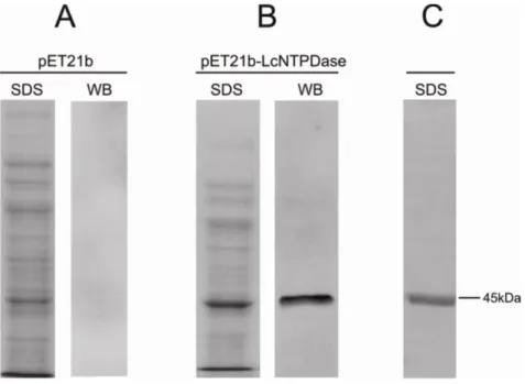
34 affinity chromatography, resulting in a unique band protein with the expected molecular weight of 43.96 kDa as visualized by silver staining (Figure 3C). The expression of rLicNTPDase-2 was confirmed by Western blot (Figure 3B and C).
3.3.3. Biochemical characterization of rLicNTPDase-2
35 Despite the potent rLicNTPDase-2-mediated hydrolysis of GTP, the nucleotides ATP, ADP, UTP and UDP are the most studied due to their important roles in cell signaling processes. We can assume that the hydrolysis of ADP could inhibit the activation of P2Y1, P2Y12 and P2Y13 [34]. These P2 receptors are involved
with the production of the inflammatory mediators TNF- , IL-1 and IL-2 [34-36]. Furthermore, hydrolysis of ADP would provide a substrate for the ecto-5'-nucleotidase, which could hydrolyze AMP to adenosine. A2 receptors are activated by adenosine, leading to anti-inflammatory effects such as inhibition of the production of TNF- , IL-6 and IL-8 [36]. The difference in response to experimental infection with
Leishmania has been attributed to its ability to hydrolyze nucleotides, resulting in decreased production of interferon- and TNF by the lymph nodes and reduced proliferation of spleen cells and germinal centers [10]. Therefore, the mechanism of infection of L. infantum chagasi could involve facilitation by regulation of inflammation through NTPDases and another ectonucleotidase.
36 3.3.4. Enzymatic activity assays with apyrase inhibitors
37 belonging to the same family and having the five ACR conserved regions, there are differences in the sensitivity of the ENTPDases from different parasites to inhibitors.
Our results shown that rLicNTPDase-2 is truly an apyrase, which has five conserved regions and hydrolyzes tri and diphosphate nucleotides. Under the conditions used, all nucleotides tested were hydrolyzed. Because of the importance of uridine and adenine nucleotides in purinergic signaling and the evidence of the ability of this enzyme to hydrolyze them, we suggest that the enzyme could modulate the host's immune system by decreasing the inflammatory response. Additionally, the NTPDase enzymes have been shown to be important in virulence and replication of trypanosomatids [8,11,14,42-44]. In this context, studies of these proteins may help to develop new approaches to therapy. In particular, the study of the crystal structure could contribute to the rational design of new drugs and in better understanding of enzymatic properties of this protein.
3.3.5. Ecto-nucleotidase activity, expression and localization of rLicNTPDases in L. infantum chagasi promastigotes
38 broad ecto-nucleotidase activity on its surface because no permeabilization was made in this assay. Using another L. infantum strain (BH46) and a reaction medium with a different composition, Maia et al. [41] found similar results indicating a broad ecto-nucleotidase activity in L. infantum promastigotes.
The recombinant enzyme activity was compared with the activity of live promastigotes (Figure 6A-inset). The results indicate a similarity in the broad substrate preference pattern, but differences in the rates of hydrolysis, possibly due to the presence of other ectonucleotidases or of unknown parasite surface environment factors. The hydrolysis of AMP occurs only in the parasite and is due the action of other ectonucleotidases because the NTPDases are unable to hydrolyze monophosphate nucleosides.
39 region (Figure 7A-E). Additionally, the gold particles stained the kinetoplast, mitochondria and internal vesicles. No staining was observed in the control assay (Figure 7F). The intracellular localization profile is similar to recent studies of L. braziliensis promastigotes [42] and T. cruzi [28] epimastigotes and reinforces the ubiquitous localization of these proteins and the requirement for further investigations in this area.
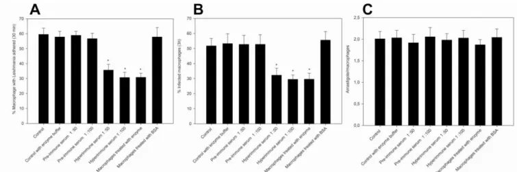
3.3.6. Recombinant rLicNTPDase2 influence L. infantum chagasi adhesion to and infection of macrophages
40 macrophage (Figure 8). However, we cannot exclude other possible explanations as discussed by Mariotini-Moura and co-workers [28]. Anti-rLicNTPDase-2 was also able to significantly reduce the adhesion (48.41% at 1:100 dilution and 40.1% at 1:50 dilution of the hyperimmune serum) and infection (45.4% at 1:100 and 37.7% at 1:50). We can speculate that the antibodies would bind to the parasite rLicNTPDases 1 and/or 2, preventing its interaction with a putative macrophage receptor. However, we cannot ignore other hypothesis such as the presence of unknown molecules that could interfere with the binding of rLicNTPDase to the host cells. Regardless of the mechanism, this area needs to be further investigated.
3.3.7. rLicNTPDases are expressed in naturally infected dogs
In our previous work [21], we demonstrated that the recombinant Lic-NTPDase-2 is a good novel antigen for immunological diagnosis of canine visceral leishmaniasis [21]. That work suggested the presence of active Leishmania in dogs but only via an indirect method because we used the recombinant protein as a target with which to measure the levels of specific antibodies in samples of serum from dogs. Here, we directly investigated the presence of amastigotes in naturally infected dogs.
41 naturally infected dogs and corroborate previous data from our group that demonstrated the potential application of rLicNTPDase-2 in the diagnosis of canine Leishmaniasis [21].
3.4. Conclusions
In this work we expanded the study of ENTPDases from L. infantum chagasi (syn. L. infantum). The bioinformatics studies shown the presence of two ENTPDase paralogs (named here as NTPDase-1 and NTPDase-2) in kinetoplastids, except for
T. cruzi, in which only one member of this family (the known NTPDase-1 described previously) was observed [14]. In the context of the low levels of identity with mammalian ENTPDases and the previous knowledge of the T. cruzi NTPDase-1, we propose here a new nomenclature for the trypanosomatid ENTPDases: TpNTPDase-1 for the trypanosomatid ENTPDases more similar to T. cruzi E-NTPDase-1 (~70 kDa proteins) and TpNTPDase-2 for the lower molecular weight isoform (~40 kDa), which is absent from T. cruzi.
In addition, a new search for TpNTPDases in the molecular protein databanks demonstrated that trypanosomatides, in general, have the two isoforms of TpNTPDases and that T. cruzi remains as a unique trypanosomatide that possesses only the TpNTPDase-1 isoform.
42 nucleotides, is dependent on divalent cations (Mg++ or Ca++) and is partially inhibited by known CD39 family partial inhibitors. In addition, we showed that live L. infantum chagasi promastigotes have ecto-nucleotidase activity, and the immunolocalization shows the expected ENTPDases on the surface of promastigotes. However, our results also indicated a broad intracellular expression of the ENTPDases, opening new fields of investigation into other previously unknown biological roles. This point is interesting because we observed the presence of these enzymes in unexpected localizations such as the kinetoplastids, mitochondria and nuclei.
In addition we investigated the role of rLicNTPDase-2 during the adhesion to and infection of host cells, and our results demonstrated that this protein participates in these processes, most likely acting as a facilitator of infection as previously observed for T. cruzi infection [8,28]. Furthermore, the importance of ENTPDases in natural infection was indicated by direct detection of their expression in tissues from naturally infected dogs. Taken together, these results reinforce the application of rLicNTPDase-2 in the diagnosis of visceral leishmaniasis as previously described [21] and may suggest that the ENTPDases could be used in other biotechnological applications including drug and vaccine development. Both applications are under investigation in our research group.
43 3.5. References
1. Jarvis JN, Lockwood DN (2013) Clinical aspects of visceral leishmaniasis in HIV infection. Curr Opin Infect Dis 26: 1-9.
2. Stauch A, Duerr HP, Dujardin JC, Vanaerschot M, Sundar S, et al. (2012) Treatment of visceral leishmaniasis: model-based analyses on the spread of antimony-resistant L. donovani in Bihar, India. PLoS Negl Trop Dis 6: e1973. 3. Barrett MP, Gilbert IH (2002) Perspectives for new drugs against trypanosomiasis
and leishmaniasis. Curr Top Med Chem 2: 471-482.
4. Marr JJ, Berens RL, Nelson DJ (1978) Purine metabolism in Leishmania donovani and Leishmania braziliensis. Biochim Biophys Acta 544: 360-371.
5. Cohn CS, Gottlieb M (1997) The acquisition of purines by trypanosomatids. Parasitol Today 13: 231-235.
6. Berredo-Pinho M, Peres-Sampaio CE, Chrispim PP, Belmont-Firpo R, Lemos AP, et al. (2001) A Mg-dependent ecto-ATPase in Leishmania amazonensis and its possible role in adenosine acquisition and virulence. Arch Biochem Biophys 391: 16-24.
7. Datta AK, Datta R, Sen B (2008) Antiparasitic chemotherapy: tinkering with the purine salvage pathway. Adv Exp Med Biol 625: 116-132.
8. Santos RF, Possa MA, Bastos MS, Guedes PM, Almeida MR, et al. (2009) Influence of Ecto-Nucleoside Triphosphate Diphosphohydrolase Activity on Trypanosoma cruzi Infectivity and Virulence. PLoS Negl Trop Dis 3: e387. 9. Asai T, Miura S, Sibley LD, Okabayashi H, Takeuchi T (1995) Biochemical and
molecular characterization of nucleoside triphosphate hydrolase isozymes from the parasitic protozoan Toxoplasma gondii. J Biol Chem 270: 11391-11397.
44 11. de Almeida Marques-da-Silva E, de Oliveira JC, Figueiredo AB, de Souza Lima Junior D, Carneiro CM, et al. (2008) Extracellular nucleotide metabolism in Leishmania: influence of adenosine in the establishment of infection. Microbes Infect 10: 850-857.
12. de Souza MC, de Assis EA, Gomes RS, Marques da Silva Ede A, Melo MN, et al. (2010) The influence of ecto-nucleotidases on Leishmania amazonensis infection and immune response in C57B/6 mice. Acta Trop 115: 262-269. 13. Mizumoto N, Kumamoto T, Robson SC, Sevigny J, Matsue H, et al. (2002) CD39
is the dominant Langerhans cell-associated ecto-NTPDase: modulatory roles in inflammation and immune responsiveness. Nat Med 8: 358-365.
14. Fietto JL, DeMarco R, Nascimento IP, Castro IM, Carvalho TM, et al. (2004) Characterization and immunolocalization of an NTP diphosphohydrolase of Trypanosoma cruzi. Biochem Biophys Res Commun 316: 454-460.
15. Robson SC, Sevigny J, Zimmermann H (2006) The E-NTPDase family of ectonucleotidases: Structure function relationships and pathophysiological significance. Purinergic Signal 2: 409-430.
16. Edgar RC (2004) MUSCLE: multiple sequence alignment with high accuracy and high throughput. Nucleic Acids Res 32: 1792-1797.
17. Tamura K, Peterson D, Peterson N, Stecher G, Nei M, et al. (2011) MEGA5: molecular evolutionary genetics analysis using maximum likelihood, evolutionary distance, and maximum parsimony methods. Mol Biol Evol 28: 2731-2739.
18. Abascal F, Zardoya R, Posada D (2005) ProtTest: selection of best-fit models of protein evolution. Bioinformatics 21: 2104-2105.
19. Jones DT, Taylor WR, Thornton JM (1992) The rapid generation of mutation data matrices from protein sequences. Comput Appl Biosci 8: 275-282.
45 21. de Souza RF, Dos Santos YL, de Souza Vasconcellos R, Borges-Pereira L, Caldas IS, et al. (2013) Recombinant Leishmania (Leishmania) infantum Ecto-Nucleoside Triphosphate Diphosphohydrolase NTPDase-2 as a new antigen in canine visceral leishmaniasis diagnosis. Acta Trop 125: 60-66.
22. Sambrook TMEFFJ (2001) Molecular Cloning: a Laboratory Manual. 3 v.: 2231. 23. Bradford MM (1976) A rapid and sensitive method for the quantitation of
microgram quantities of protein utilizing the principle of protein-dye binding. Anal Biochem 72: 248-254.
24. Ekman P, Jager O (1993) Quantification of subnanomolar amounts of phosphate bound to seryl and threonyl residues in phosphoproteins using alkaline hydrolysis and malachite green. Anal Biochem 214: 138-141.
25. Chandler JP (2007) Purification and Characterization of Antibodies. Making and using antibodies: A practical handbook.; Gary C. Howard TJDGI, San Francisco, California, USA; Matthew R. Kaser, editor. New York: CRC Press and Taylor & Francis Group.
26. Russel JSDW (2001) Molecular Cloning: A Laboratory Manual. Cold Spring Harbor Laboratory Press 1, 2 and 3: 2231.
27. Baqui MM, Milder R, Mortara RA, Pudles J (2000) In vivo and in vitro phosphorylation and subcellular localization of trypanosomatid cytoskeletal giant proteins. Cell Motil Cytoskeleton 47: 25-37.
28. Mariotini-Moura C, Bastos MS, de Castro FF, Trindade ML, de Souza Vasconcellos R, et al. (2013) Trypanosoma cruzi nucleoside triphosphate diphosphohydrolase 1 (TcNTPDase-1) biochemical characterization, immunolocalization and possible role in host cell adhesion. Acta Trop 130C: 140-147.
29. Zimmermann H (2001) Ectonucleotidases: some recent developments and a note on nomenclature. Drug Development Research 52: 44-56.
46 31. Handa M, Guidotti G (1996) Purification and cloning of a soluble
ATP-diphosphohydrolase (apyrase) from potato tubers (Solanum tuberosum). Biochem Biophys Res Commun 218: 916-923.
32. Plesner L (1995) Ecto-ATPases: identities and functions. Int Rev Cytol 158: 141-214.
33. Zimmermann H (1999) Two novel families of ectonucleotidases: molecular structures, catalytic properties and a search for function. Trends Pharmacol Sci 20: 231-236.
34. Aslam M, Sedding D, Koshty A, Santoso S, Schulz R, et al. (2013) Nucleoside triphosphates inhibit ADP, collagen, and epinephrine-induced platelet aggregation: role of P2Y(1) and P2Y(1)(2) receptors. Thromb Res 132: 548-557.
35. Burnstock G (2007) Purine and pyrimidine receptors. Cell Mol Life Sci 64: 1471-1483.
36. Abbracchio MP, Burnstock G (1998) Purinergic signalling: pathophysiological roles. Jpn J Pharmacol 78: 113-145.
37. Crack BE, Pollard CE, Beukers MW, Roberts SM, Hunt SF, et al. (1995) Pharmacological and biochemical analysis of FPL 67156, a novel, selective inhibitor of ecto-ATPase. Br J Pharmacol 114: 475-481.
38. Escalada A, Navarro P, Ros E, Aleu J, Solsona C, et al. (2004) Gadolinium inhibition of ecto-nucleoside triphosphate diphosphohydrolase activity in Torpedo electric organ. Neurochem Res 29: 1711-1714.
39. Bisaggio DF, Peres-Sampaio CE, Meyer-Fernandes JR, Souto-Padron T (2003) Ecto-ATPase activity on the surface of Trypanosoma cruzi and its possible role in the parasite-host cell interaction. Parasitol Res 91: 273-282.
47 41. Maia AC, Porcino GN, Detoni Mde L, Emidio NB, Marconato DG, et al. (2013) An antigenic domain within a catalytically active Leishmania infantum nucleoside triphosphate diphosphohydrolase (NTPDase 1) is a target of inhibitory antibodies. Parasitol Int 62: 44-52.
42. Porcino GN, Carvalho-Campos C, Maia AC, Detoni ML, Faria-Pinto P, et al. (2012) Leishmania (Viannia) braziliensis nucleoside triphosphate diphosphohydrolase (NTPDase 1): localization and in vitro inhibition of promastigotes growth by polyclonal antibodies. Exp Parasitol 132: 293-299. 43. Meyer-Fernandes JR, Saad-Nehme J, Peres-Sampaio CE, Belmont-Firpo R,
Bisaggio DF, et al. (2004) A Mg-dependent ecto-ATPase is increased in the infective stages of Trypanosoma cruzi. Parasitol Res 93: 41-50.
48 3.6. Figure legends
50 Figure 2- Alignment of representatives of TpNTPDases-1 and TpNTPDases-2. TpNTPDases from L. infantum (Li), L. braziliensis (Lb), L. major (Lm) and T. cruzi
(Tc) were aligned using the CLC main workbench and manually inspected. The ACRs are shown in dashed line rectangles. The prevalent amino acids are shown in the consensus line and the level of conservation of each position at the primary sequences of proteins is shown in the conservation box.
Figure 3- Analyses of expression and purification of rLicNTPDase-2. (A) Protein extract from E. coli carrying the empty vector pET21b. (B) Protein extract from E. coli
51
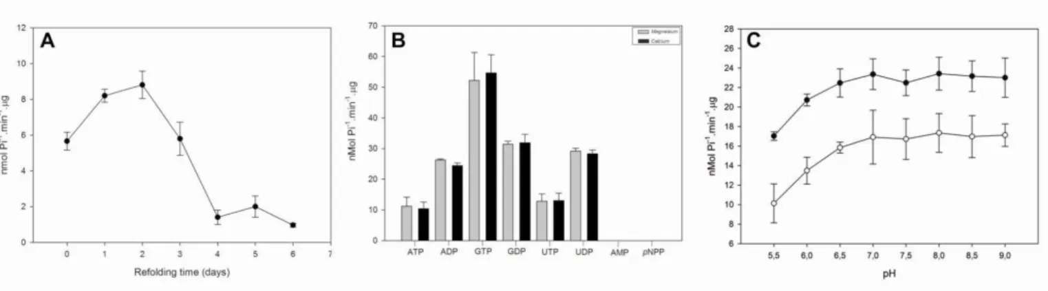
Figure 4 rLic-NTPDase-2: refolding, substrate preference and pH dependence. (A) Refolding assay- the enzymatic activity
was measured just after the refolding (zero point) and up to six days later using ADP as the substrate. (B) The preference for
different substrates was assessed in the presence of the cofactors calcium (grey bars) or magnesium (black bars). (C) ATP (open
circles) and ADP (black circles) were used to evaluate enzymatic activity as a function of pH. The pH-dependence test was
performed in buffer containing 50 mM MES, 50 mM Tris, 50 mM HEPES, 3 mM MgCl2, 116 mm NaCl, 5.4 mM KCl and 2.5 mM
nucleotide. The SDs represent the those from the average of three independent experiments performed in triplicate. The free
52 Figure 5- rLicNTPDase-2 ATPase activity in the presence of known partial Purified rLicNTPDase-2 was assayed in the absence of inhibitors (control column) or in the presence of inhibitors: ARL 67156 (ARL) 300 µM, gadolinium chloride 300 µM, suramin 100 µM,
57
58
Figure 9-Immunohistochemistry using anti-rLicNTPDase-2 in the lymph nodes of naturally infected dogs. (A) Lymph nodes
from 48 Leishmania-positive dogs were evaluated by immunohistochemistry (IHC) using anti-rLicNTPDase-2. The results of the IHC
are compared with ELISA data of the same samples using a Biomanguinhos Kit. (B) An example of IHC result using polyclonal
59 Table 1: rLicNTPDase-2 nucleotidase characterization- Analysis of substrate preference.
Substrate nmol Pi-1.min-1. g
ATP 10.55 ± 2.98
ADP 26.31 ± 0.32
GTP 50.38 ± 9.10
GDP 30.97 ± 1.04
UTP 12.16 ± 2.40
UDP 29.22 ± 0.90
AMP 0.00
pNPP 0.00
* The mean and SD are from three independent experiments.
Table 2: rLicNTPDase-2 nucleotidase: characterization of cofactor preference.
Divalent metal ion nmol Pi-1.min-1. g
Mg2+ 10.55 + 2.98
Ca2+ 10.00 + 2.18
Ni2+ 0.00
Zn2+ 0.00
- 0.00
61
63 CAPÍTULO 2
64 IMPORTANCE OF REFOLDING FOR ENZYMATIC ACTIVITY OF ECTO-NUCLEOSIDE TRIPHOSPHATE DIPHOSPHOHYDROLASE-2 (LicNTPDase-2) FROM Leishmania infantum chagasi
Raphael De Souza Vasconcellos1,2; Christiane Mariotini-Moura1,2; Rafaela Firmino3; Abelardo Silva-Júnior4; Gustavo Costa Bressan3; Márcia Rogéria Almeida3; Juliana Lopes Rangel Fietto, 2,3.
1
Departamento de Biologia Celular e Estrutural, Universidade Federal de Viçosa, 36570-900 Viçosa, MG Brazil.
2
Instituto Nacional de Biotecnologia Estrutural e Química Medicinal em Doenças Infecciosas (INBEQMeDI).
3
Departamento de Bioquímica e Biologia Molecular, Universidade Federal de Viçosa, 36570-900 Viçosa, MG Brazil.
4
Departamento de Veterinária, Universidade Federal de Viçosa, 36570-900, Viçosa, MG Brazil.
65 Abstract
The E-NTPDases (Ectonucleoside Triphosphate Diphosphohydrolases) are important enzymes in the purine ring biosynthesis de novo pathway, fundamental to many parasites the trypanosomatidae family. These enzymes have been related to immune system regulation, platelet aggregation and increased adhesion, infectivity and virulence of many parasites. In this work, the recombinant NTPDase-2 from
66 4.1. Introduction
The E-NTPDase are enzymes from the GDA1/CD39 family, which hydrolyze nucleoside 5'-triphosphates and nucleoside 5'-diphosphates with a broad preference for the type of nucleotide, and with activity dependent on divalent cations. [1]. These family of enzymes play important roles in the regulation of homeostasis and thrombosis [2] and regulating the activity of leukocytes [3, 4]. Several studies have also shown its importance for virulence and survival of some parasites as
Toxoplasma gondii [5], Legionella pneumophila [6], Trypanosoma cruzi [7-9] and
Leishmania [10, 11]
The trypanosomatids, as Leishmania and Trypanosoma, are unable to synthesize the purine ring in the de novo biosynthesis [12], thus depending on the salvage pathway, where the role of NTPDases is described as important [12]. Furthermore, based on the typical function of the apyrase family, it has been proposed that these enzymes can modulate biological responses induced by extracellular nucleotides and its metabolites [13]. Several biological events are modulated by extracellular nucleotides and may be influenced by ectonucleotidases of parasites, such as ADP-dependent platelet aggregation and ATP-dependent inflammatory response [14].
post-67 translational modifications. However, this expression system is generally more time consuming and expensive. Bacteria are still the most suitable expression system due to be ease of cultivation, low cost, rapid growth and good yield expression of recombinant proteins. However, the polypeptides produced by prokaryotic organisms generally suffer incomplete or misfolding, forming insoluble aggregates known as inclusion bodies [15].
When working with inclusion bodies, it is necessary to denature and renature the protein in a controlled environment, so the recombinant protein would be as close as possible to its native form. This step requires hard work and dedication, because a inadequate renaturation can have importante effect on the activity of the enzyme, resulting in data that may not correspond to reality. In this present work we expressed rLicNTPDase-2, a know NTPDase from Leishmania infantum chagasi, a causative agent of visceral leishmaniasis, a neglected tropical disease in new world. This enzymes seems to facilitate mammalian cell infection and is highly expressed in natural infected dogs [11]. Furthermore, we standardized an bioassay to be used in biochemical characterization and search for inhibitors that could be tested as anti-leishmanial drugs against visceral leishmaniasis.
4.2. Material and methods
4.2.1. Cloning and expression of rLicNTPDase-2
The cloning and expression of rLicNTPDase-2 from Leishmania infantum
68 4.2.2. Purification and Renaturation
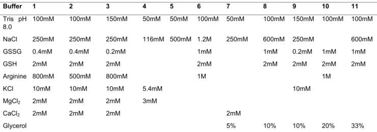
The protein was solubilized from inclusion bodies and purified by affinity chromatography as described by De Souza and cols [16] and and Vasconcellos and cols [11]. To perform renaturation 11 buffers were tested, varying in its composition and / or concentration (table 1).
After purification, the protein was at a concentration of about 1 mg / ml in buffer containing 8M urea, 0.6M NaCl and 0.1M Tris pH 8.0. This sample was then diluted 10-fold in renaturation buffers with vigorous vortexing for about 30 seconds. Then the sample was stored at 4°C for 24h.
4.2.3. Enzymatic activity
The enzyme activity assay was performed by a colorimetric method for the determination of inorganic phosphate (Pi) (malachite green method) [17] as described by Vasconcellos and cols [11].
4.2.4. SDS-PAGE and Western blott analyses
To assess the purity of the protein samples after purification prior to refolding were subjected to electrophoresis with subsequent staining in SDS-PAGE 10%. SDS-PAGE were performed as previously described by Russel et al [18].
Western Blott analysis was performed as previously described by Russel et al
69 and FITC-conjugated secondary antibody (1: 8.000) and was revealed in the equipment Fujifilm phosphorimager 472nm with a blue filter.
4.3. Results and Discussion
After purification of rLicNTPDase-2 by affinity chromatography, an SDS-PAGE was performed to evaluate its purity, showing an intense band with the expected size of 45kDa in the lysate extract (figure 1). This intensity decreases considerably in the soluble fraction of the lysate, suggesting that the amount of protein in the inclusion bodies is greater (figure 1A e B). To remove contaminants from the inclusion bodies, they were washed with a buffer containing 2M urea, sufficient to solubilize protein contaminants and insufficient to solubilize the recombinant protein present in the inclusion bodies [11]. The solubilization step was possible with a buffer containing urea at 8M and prior to the affinity chromatography step, we obtained a visually high purity protein, in a concentration of 0.5 mg/mL (figure 1A). After Ni affinity chromatography we achieved a concentration of 1mg/mL, and the protein was refolded in the buffers described in Table 1. Figure 1 shows samples from differents steps of expression and purification assays.
70 containing arginine were not the best for the refolding of this enzyme. All buffers that contained arginine produced enzymes with activity lower than 4nmol Pi/min/ g (Figure 2). The buffers 1, 2 and 3 had a very similar composition, and even varying the concentration of arginine from 500mM to 800mM did not led to significant improvement in enzyme activity (table 1 and figure 2).
The buffers 1 to 7 showed no significant variation in enzyme activity. The concentration of NaCl in these buffers ranged from 0.25 to 1.2M. The salt concentration can help stabilize nonspecific electrostatic interactions at low concentrations, depending on the ionic strength of the medium. High concentrations of salts may stabilize or destabilize the protein or even denature them [20]. Other salts, such as KCl, MgCl2, CaCl2 were also tested in these buffers and did not
produce significant improvements. The MgCl2, CaCl2. The MgCl2, CaCl2 salts, in
addition to being used as co-solutes in renaturation buffers, act as cofactors of the rLicNTPDase-2 [11] and were tested, because we thought that the presence of the cofactor would aid in the stability of the enzyme during renaturation. But at the tested conditions these cations did not improved the activity of rLicNTPDase-2.