Original article
E-NTPDase (ecto-nucleoside triphosphate diphosphohydrolase) of
Leishmania amazonensis
inhibits macrophage activation
Rodrigo Saar Gomes
a, Luana Cristina Faria de Carvalho
a, Raphael de Souza Vasconcellos
b,
Juliana Lopes Rangel Fietto
b, Lu
í
s Carlos Crocco Afonso
a,*
a
Laboratorio de Imunoparasitologia, Departamento de Ci^encias Biologicas, Instituto de Ci^encias Exatas e Biologicas/NUPEB, Universidade Federal de Ouro Preto, Campus do Morro do Cruzeiro, 35400-000 Ouro Preto, MG, Brazil
bDepartamento de Bioquímica e Biologia Molecular, Laborat
orio de Infectologia Molecular Animal, Instituto de Biotecnologia Aplicadaa Agropecuaria/ BIOAGRO, Universidade Federal de Viçosa, 36570-900 Viçosa, MG, Brazil
Received 21 October 2014; accepted 16 December 2014 Available online 30 December 2014
Abstract
Leishmania amazonensis, the causal agent of diffuse cutaneous leishmaniasis, is known for its ability to modulate the host immune response.
Because a relationship between ectonucleotidase activity and the ability ofLeishmaniato generate injury in C57BL/6 mice has been demon-strated, in this study we evaluated the involvement of ecto-nucleoside triphosphate diphosphohydrolase (E-NTPDase) activity ofL. amazonensis
in the process of infection of J774-macrophages. Our results show that high-activity parasites show increased survival rate in LPS/IFN-g -activated cells, by inhibiting the host-cell NO production. Conversely, inhibition of E-NTPDase activity reduces the parasite survival rates, an effect associated with increased macrophage NO production. E-NTPDase activity generates substrate for the production of extracellular adenosine, which binds to A2Breceptors and reduces IL-12 and TNF-aproduced by activated macrophages, thus inhibiting NO production.
These results indicate that E-NTPDase activity is important for survival ofL. amazonensiswithin macrophages, showing the role of the enzyme in modulating macrophage response and lower NO production, which ultimately favors infection. Our results point to a new mechanism ofL.
amazonensisinfection that may pave the way for the development of new treatments for this neglected disease.
©2015 Institut Pasteur. Published by Elsevier Masson SAS. All rights reserved.
Keywords: Leishmania amazonensis; E-NTPDase; Adenosine; Nitric oxide; Cytokines
1. Introduction
The success of Leishmania infection in macrophages de-pends on the ability of the parasite to adhere to these cells, to be phagocytosed, and to survive the effects of microbicidal mechanisms [1]. After phagocytosis, macrophages are acti-vated to produce nitric oxide and ROS (reactive intermediates of oxygen), which are highly toxic to the parasite[2e4]. NO production by iNOS is stimulated by numerous inflammatory cytokines such as IFN-g, TNF-a, and IL-1, and inhibited by
anti-inflammatory cytokines such as IL-4, TGF-b, and IL-10 [5].
In both animal models and humans infected with Leish-mania major or Leishmania braziliensis, the host immune response is usually able to control the infection. On the other hand, one of the main features of infection by Leishmania amazonensis is its ability to modulate the host immune response and sustain the infection, both in experimental models and in humans [6e8]. Evidence shows that L.
ama-zonensisinhibits macrophage NO production, which favors the survival of the parasite inside the cell[9e11]. The reduction in iNOS expression, and the consequent reduction in NO pro-duction, inL. amazonensisinfection is the result of a number of immune alterations, such as increase in IL-10 and TGF-b
*Corresponding author. Tel.:þ55 31 3559 1701; fax:þ55 31 3559 1680.
E-mail address:[email protected](L.C.C. Afonso).
www.elsevier.com/locate/micinf
http://dx.doi.org/10.1016/j.micinf.2014.12.009
production, reduction in IL-12 JACK/STAT inactivation, activation of phosphotyrosine phosphatases, and inhibition of TNF-aproduction[12].
NTPDases, or nucleoside triphosphate diphosphohy-drolases, are enzymes of the apyrase family with the ability to hydrolyze nucleotide triphosphates to their monophosphate form under stimulation of divalent ions such as calcium (Ca2þ) and magnesium (Mg2þ)[13]. In trypanosomatids, the E-NTPDase is important to the salvation of purine nucleo-tides[14], since these parasites are unable to performde novo
synthesis of these nucleotides [15]. E-NTPDases have been extensively characterized inL. amazonensis[16]as well as in other species of protozoa, such as Toxoplasma gondii [17] and Trypanosoma cruzi [18,19]. Data from our research group have shown the importance of NTPDase activity in establishing Leishmania infection [20e22] and driving the clinical form of the disease[23]. Adenosine, the product of the combined action of E-NTPDase and 50
-ectonucleotidase, is an important molecule in the regulation of macrophage cell function, which is mediated by its binding to specific re-ceptors on the surface of these cells [24]. It has been shown that adenosine can prevent respiratory burst in macrophages [25] and suppress LPS-stimulated cellular NO production [26]. Adenosine, therefore, plays a role in reducing the pro-duction of TNF-aand IL-12, by acting either on A2Aor A2B
receptors[27].
The Leishmania E-NTPDases can be divided into two different groups: E-NTPDase-1, also named guanosine diphosphatase, which is similar to the E-NTPDase-1 of T. cruzi; and E-NTPDase-2, or ATP diphosphohydrolase or nucleoside diphosphatase [28]. Although the role of E-NTPDase of L. amazonensis in developing and maintaining
in vivoinfection is well established, the role of this parasite's enzyme in macrophage infection remains unknown. Here, we observed that the activity of E-NTPDase in L. ama-zonensis plays a critical role in the modulation of the mac-rophage's immune response and increases the parasite's survival.
2. Materials and methods
2.1. Parasites
Promastigotes of Leishmania (Leishmania) amazonensis, PH8 strain (IFLA/BR/67/PH8), and 1IIId clone of the same strain were cultured at 25
C, in Grace's insect medium (SigmaeAldrich, St. Louis, MO, USA) supplemented with 10% heat-inactivated fetal calf serum (FCSeLGC, Cotia, SP, Brazil), 2 mM L-glutamine (SigmaeAldrich), and 100 U/mL
penicillin G potassium (SigmaeAldrich), pH 6.5. The original strain was also maintained in the presence of adenine 5 mM, for a few passages, to reduce ectonucleotidase activity of the parasite, as described previously [16]. Metacyclic promasti-gotes were obtained by gradient centrifugation of parasites at the late log phase of culture (day 5) over Ficoll 400 (Sigma-eAldrich), as previously described[21]. Parasites were kept in culture for no more than 20 passages.
2.2. Ectonucleotidase activity measurement
The activity of ATPase, ADPase and ecto-AMPase was measured by incubation of intact parasites for 1 h at 30
C in reaction buffer containing 116 mM NaCl, 5.4 mM KCl, 5.5 mMD-glucose, 5 mM MgCl2, and 50 mM
Hepes-Tris buffer in the presence of 5 mM of ADP, ATP, or AMP (SigmaeAldrich)[18]. The reaction was initiated with the addition of live metacyclic promastigotes and terminated by addition of 0.2 M HCl [14]. Nonspecific hydrolysis was determined by the addition of parasites after the reaction was stopped. We used a pelleted parasite suspension, and aliquots of the supernatant were used for measuring the liberated inorganic phosphate (Pi) as previously described[29]. Enzyme activities were expressed as nmol of Pi released by 1 108 parasites in 1 h.
2.3. Western blotting
Enriched plasma membrane preparations were obtained by 14,000 g centrifugation of metacyclic promastigote or axenic amastigote extracts as described elsewhere [30] and stored until use at 20
C in the presence of the protease inhibitors: 200mM EGTA, 4 mM PMSF, 40mM TPCK, 40mM TLCK, 4 mM DTT, and 40 mM NEM (SigmaeAldrich). The determination of protein was performed by the Lowry method [31]. For Western blotting analysis, membrane preparations were run on 10% SDSePAGE followed by semi-dry transfer to nitrocellulose membranes. Blotted nitrocellulose mem-branes were incubated with serum from a rabbit immunized with recombinant Leishmania infantum NTPDase (1:2000) followed by peroxidase-goat anti-rabbit IgG conjugate (Zymed Laboratories, San Francisco, CA, USA) (1:10,000), and revealed by reaction with DAB/4-chloro naphtol/meth-anol/H2O2solution.
2.4. Infection of J774 cells
J774 cells were plated at 5 105cells per well (0.5 mL) onto round coverslips in Dulbecco's minimal essential me-dium containing 10% FCS, 2 mM L-glutamine, 100 U/mL
penicillin G potassium, 25 mM N-2-hydroxiethylpiperazine-N9-2-ethanosulfonic acid (HEPES; USBiological, Swamp-scott, MA, USA), and 50 mMb-mercaptoethanol (Pharmacia Biotech AB, Uppsala, Sweden) in 24-well plates. Cells were incubated for 90 min at 37
C, 5% CO2. Non-adherent cells
10 min and then stained with Panotico Rapido kit (Laborclin, Pinhais, PR, Brazil), according to manufacturer's in-structions. The analysis was performed by counting the cells containing adhered or internalized parasites using an optical microscope Olympus BX50 (Olympus, Center Valley, PA, USA). We evaluated the percentage of macrophages con-taining internalized parasites and the number of parasites per 100 cells. At least 200 cells were evaluated.
2.5. Nitric oxide and cytokine quantification
Quantification of NO produced by the cells was performed by the indirect Griess method to detect nitrite [32], and the production of IL-12, IL-10, and TNF-awas evaluated by in-direct ELISA, according to the manufacturer (BD Biosciences).
2.6. Statistical analysis
Statistical analysis was performed by 1way-ANOVA. p <0.05 was considered statistically significant.
3. Results
3.1. Ectonucleotidase activity inL. amazonensisis related to the parasite's survival within macrophages
Given that a relationship has been demonstrated between the level of ectonucleotidase activity and the ability of
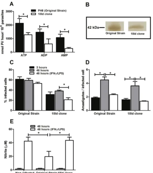
Leishmaniato generate injury in C57BL/6 mice[21,22], here we investigated whether the presence and activity ofL. ama-zonensis'sE-NTPDases also related to the survival of parasites in infected macrophages. To this end, metacyclic Fig. 1.L. amazonensisE-NTPDase activity correlates with survival of parasites within stimulated cells. Parasites were incubated with ATP, ADP, or AMP for 1 h at 30C. (A) Enzymatic activity was evaluated by the measurement of inorganic phosphate released. (B) Analysis of the expression of E-NTPDase by Western
promastigotes of both the original strain ofL. amazonensisand a clone of the same strain (1IIId clone) were incubated with ATP, ADP, or AMP for 1 h and the amount of inorganic phosphate was evaluated. As shown inFig. 1A, the 1IIId clone presents lower ectonucleotidase activity for the three nucleo-tides when compared to metacyclic promastigotes of original strain. In addition, the expression of the protein on the membrane of the parasites is lower in the 1IIId clone than in the original strain (Fig. 1B).
J774 cells were infected with metacyclic promastigotes of the original strain or 1IIId clone of the same strain, for 3 h, washed to remove non-internalized parasites, and stimulated with LPS and IFN-gfor 48 h. Our results show that at three hours of incubation the percentage of macrophages infected by the 1IIId clone was smaller than that of the original strain. More importantly, whereas activation of macrophages infected by the 1IIId clone reduced the percentage of infected cells after 48 h, no effect was observed when infections were initiated by the original strain (Fig. 1C). The number of amastigotes per infected cell was similar in both groups in each incubation condition (Fig. 1D). Given that the percentage of infected macrophages was decreased when 1IIId infected cells were activated, the combined results fromFig. 1C and D indicate that some macrophages were able to complete elim-inate the parasite while in cells infected by the original strain, activation allowed only for a containment of the parasite growth. Interestingly, unlike the 1IIId clone, the original strain reduced NO production by activated cells, which may explain the parasite's survival capacity, even under inflammatory
stimuli (Fig. 1E). This suggests that the level of E-NTPDase activity may influence parasite survival by inhibiting NO production by activated macrophages.
3.2. The modulation of E-NTPDase alters the ability of
L. amazonensisto inhibit NO production by activated J774 cells
Given the role of the E-NTPDase in increasing extracellular AMP, which is hydrolyzed to adenosine by 50
-ectonucleotidase to supply the purine salvage pathway[33], we expected that an increased purine concentration in the culture medium would affect the expression of the E-NTPDase on the parasite sur-face. To modulate the enzyme activity, promastigotes of the original strain were grown in medium supplemented with adenine prior to evaluating enzyme activity. Our data show that maintenance of parasites in the presence of adenine led to a reduction in the activity and expression of E-NTPDase in the original strain (Fig. 2A and B).
To verify if the modulation in the activity of E-NTPDase would also interfere with the ability of the parasite to survive within activated macrophages, the original strain grown in adenine-supplemented medium was used to infect J774 cells. After 3 h, cells were washed to remove non-internalized parasites and cells were incubated for 48 h in the presence of IFN-gand LPS. Our results demonstrate that the original strain grown in the presence of adenine is less able to resist activation by IFN-g and LPS. As shown in Fig. 2C, the percentage of infected cells was reduced after 48 h of Fig. 2. The modulation of E-NTPDase activity alters the ability ofL. amazonensisto inhibit NO production by activated J774 cells. J774-macrophages were incubated for 3 h with metacyclic promastigotes ofL. amazonensis(3 parasites/cell) grown in adenine supplemented medium, washed and incubated for additional 48 h, in presence or not of IFN-g/LPS. (A) Enzymatic activity was evaluated by the measurement of inorganic phosphate released. (B) Analysis of the expression of E-NTPDase by Western blotting on membrane preparations of metacyclic promastigotes. (C) Percentage of infected cells. (D) NO production in 48-h su-pernatants. Bars represent the mean±SD of three independent experiments performed in duplicate. *p<0.05.
activation, when the original strain was grown in the pres-ence of adenine. The decrease in the number of infected cells was associated with an infection rate (amastigotes per infected cell) similar to that observed in infection with the 1IIId strain (Fig. 1D and data not shown) indicating a similar mechanism of parasite control. In fact, the reduced resistance to activation by IFN-g and LPS was accompanied by an increased NO production by cells infected with adenine-treated parasites (Fig. 2D). Our results strongly suggest that the activity of E-NTPDase on the surface of the parasite is involved in the parasite's survival within the macrophage by downmodulating the NO production by activated cells. To our surprise, although the adenine-treated parasites were less phagocytized than the control strain (similar to the 1IIId clone), these parasites seem more capable to disseminate to other cells than the original strain (Fig. 1C). The reason for this behavior remains unclear.
3.3. The ectonucleotidase activity ofL. amazonensisis important for down-modulation of TNF-aand IL-12
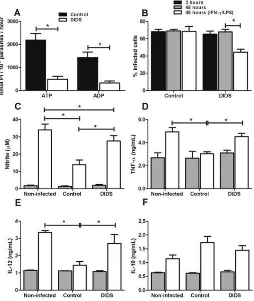
The strong correlation between the activity of E-NTPDase and the ability of L. amazonensis in reducing NO production and favoring the survival of the parasite, as shown above, prompted us to evaluate the effect of the inhibition of this enzyme on the infective capacity of the parasite. Thus, we infected J774 cells with the original strain, which was pre-incubated for 30 min with 100 mM DIDS (4,40
-diisothiocya-natostilbene 2,20
infection. To analyze whether this activity is also important for modulating cytokine production, we used indirect ELISA to investigate the profile of cytokines produced by infected cells. Infection with the original strain reduced the production of IL-12 and TNF-a by stimulated cells. The inhibition of E-NTPDase activity with DIDS reduced L. amazonensis's ca-pacity to down-modulate the release of inflammatory cyto-kines (Fig. 3DeE). No alteration was observed in IL-10 production, and DIDS treatment for 3 h did not alter the production of any of the cytokines or the production of NO by stimulated non-infected cells (data not shown). These data indicate that L. amazonensis ectonocleotidase activity is important for the down-modulation of inflammatory cytokines, which are essential for NO production.
3.4. Adenosine generated by E-NTPDase activity acts on the A2Breceptor
Adenosine plays an immunomodulatory role by binding to specific cell receptors, i.e., A2A and A2B [26]. To confirm
whether E-NTPDase inL. amazonensiswould exert its effects on the inhibition of pro-inflammatory cytokine production by supplying substrate for adenosine production, we used the
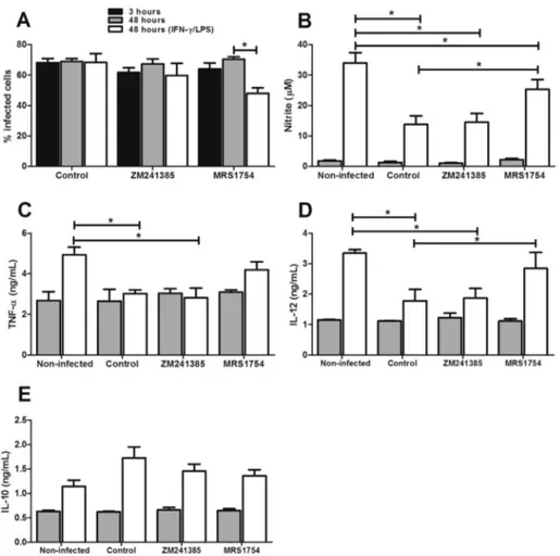
metacyclic promastigotes of the original strain to infect J774 cells that were pre-incubated with specific antagonists of adenosine receptors A2A(ZM241385) or A2B(MRS1754). As
shown in Fig. 4A, blocking A2B receptors reduced parasite
survival within stimulated cells. This result correlates with an increase in NO production (Fig. 4B). No alteration was seen with the A2A antagonist. Interestingly, when we blocked the
A2B receptors, the inhibition of IL-12 and TNF-aproduction
observed in stimulated cells infected with theL. amazonensis
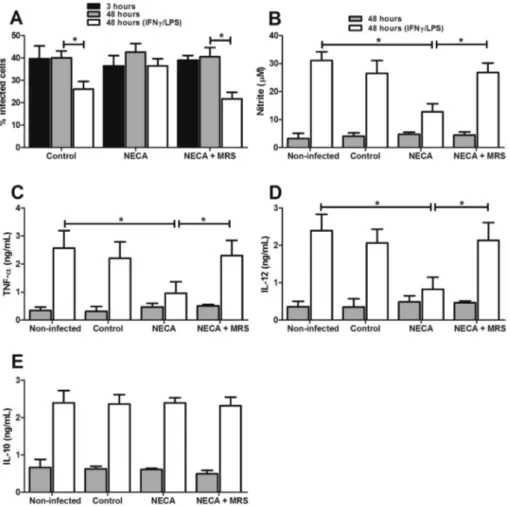
was reverted (Fig. 4CeD). No alteration was observed in IL-10 production (Fig. 4E). Also, treatment of stimulated non-infected cells with either adenosine receptor antagonist for 3 h did not alter the production of any of the cytokines or the production of NO (data not shown). In addition, macrophages were infected with the avirulent clone 1IIId in the presence of 50mM of NECA, an analog of adenosine. As noted earlier, the avirulent clone with low ectonucleotidase activity has a decreased ability to survive in stimulated cells. However, in infections performed in the presence of NECA, the clone is able to survive within stimulated cells (Fig. 5A). This is due to the fact that the production of NO, IL-12 and TNF were reduced by cells stimulated in the presence of NECA (Fig. 5BeD). No change was observed for IL-10 production
Fig. 4. Modulation of infected macrophages is mediated by the activation of the adenosine A2Breceptor. J774-macrophages were pre-incubated with 5mM of
ZM241385 (A2AR antagonist) or MRS1754 (A2BR antagonist) and infected with metacyclic promastigotes ofL. amazonensis(3 parasites/cell) for 3 h, washed and
for additional 48 h, in presence or not of IFN-g/LPS. (A) Percentage of infected cells. (B) NO, (C) TNF-a, (D) IL-12, and (E) IL-10 production in 48-h su-pernatants. Bars represent the mean±SD of three independent experiments performed in duplicate. *p<0.05.
(Fig. 5E). On the other hand, preincubation of these cells with MRS1754 (antagonist of adenosine receptor A2B) reverted the
capacity of avirulent 1IIId clone infected with NECA in reduced inflammatory cytokines and NO production and enhanced survival of this parasite within stimulated cells (Fig. 5). Taken together, these results show that E-NTPDase activity inL. amazonensisis important for generating substrate for adenosine production; furthermore, we show here that adenosine acts on the A2Breceptor, thus reducing production
of IL-12 and TNF-a, which are essential for the release of NO by stimulated cells.
4. Discussion
In this study, we investigated the role ofL. amazonensis E-NTPDase in macrophage infection. We provide evidence that, by decreasing extracellular ATP concentration and allowing for increased adenosine production, this enzyme interferes with the activation of the infected macrophage by IFNg/LPS reducing the production of inflammatory cytokines and NO production. To this aim, we took advantage of a clone of the PH8 strain ofL. amazonensisthat we had previously shown to
have lower infectivity in vivo [22]. We also downmodulated the ectonucleotidase activity of the PH8 strain by culturing the parasites in excess of adenine. Our data show that the presence of E-NTPDase on the surface of the parasite leads to increased in vitro infectivity to J774 cells, which is associated with decreased NO production upon activation by IFNg/LPS (Figs. 1 and 2). In addition to its activity in the hydrolysis of extracellular ATP, E-NTPDase has been shown to facilitate parasite adhesion to the host cell [34,35]. Our data confirm these observations by showing an increased parasite uptake (3 h) by macrophages incubated with the PH8 strain when compared to the 1IIId clone (Fig. 1C) or when the parasite was cultured in the presence of increased adenine concentrations (Fig. 2C).
L. amazonensisE-NTPDase is involved in the hydrolysis of extracellular ATP (which is secreted by macrophages under several circumstances) to AMP, which in turn generates adenosine through the activity of a 50
-nucleotidase [16,21]. While extracellular ATP has been shown to increase activated macrophage NO production [36,37], adenosine has been shown to down-modulate macrophage NO production by acting on two surface receptors (A2A and A2B) [38e41].
Inhibition of the parasite's ectonucleotidase activity resulted in reduced parasite survival in activated J774 cells that was associated with increased NO and inflammatory cytokine production (TNF-aand IL-12) (Fig. 3).
It has been shown that TNF-ais essential for the activation of iNOS in macrophages and with subsequent production of NO [3,42]. Furthermore, low IL-12 production should reduce NK and T cell generation of IFN-y required for macrophage activation [43]. Thus, we demonstrate that ectonucleotidase activity inL. amazonensisis also linked to a reduction in the macrophage inflammatory profile, when stimulated with IFN-g and LPS. This shows that E-NTPDase expression on the surface of the parasite plays a key role in macrophage mod-ulation and infection.
The fact that E-NTPDase provides AMP for adenosine production [16] suggests that modulation of the macrophage depends on adenosine production. In fact, blocking A2B
re-ceptors increased NO and inflammatory cytokine production resulting in reduced survival of the parasite in activated cells (Fig. 4). Moreover, addition of NECA to low activity parasites resulted in recovery of the ability of these cells to sustain macrophage activation (Fig. 5). This demonstrates that binding of adenosine to the A2Breceptors is critical for the modulation
of the infected macrophage. Interestingly, this effect was not observed for A2Areceptors. Therefore, adenosine acts on A2B
receptors and, by increasing cAMP inside cells, reduces the production of inflammatory cytokines and increases the pro-duction of regulatory cytokines [44,45]. This study shows an important association between adenosine production and E-NTPDase activity inL. amazonensis and that this association modulates infected macrophages.
It has been extensively shown that L. amazonensis down-modulates several macrophage functions [9,46,47], including NO production, which is one of the main leishmanicidal mechanisms of the infected cell [4,48]. In addition, several studies demonstrate that the initial steps of the interaction between Leishmania and the host cells are important in determining the outcome of the infection [7]. Our research group has demonstrated that E-NTPDase of L. amazonensis
interferes with the outcome of infection in mice[21,22]. The present results extend these findings by showing that E-NTPDase activity is another mechanism by which this para-site interferes with the host immune response by down-modulating macrophage production of important cytokines such as IL-12 and TNF-a, as well as NO production. Furthermore, our data did not demonstrate an involvement of IL-10 in the control of the activation of the macrophage, a finding that is in agreement with previous findings in the literature that indicate that, in the case of the infection by L. amazonensis, IL-10 does not seem to control the response to the parasite [49].
The present work support the evidences for a correlation between activity/expression of E-NTPDase and the capacity of the parasite to induce injuryin vivo, since the production of adenosine in the early stages of the infection may alter the ability of macrophages to be activated later, preventing the action of inflammatory stimulus on the host cell. This new
mechanism represents an important target for therapeutic measures and paves the way for the development of new treatments for this neglected disease.
Conflict of interest
The authors declare no conflict of interest.
Acknowledgments
The authors wish to thank Leandro H. dos Santos, Mar-corelio Divino de Souza, Amanda Braga de Figueiredo and Hellem Damazo for technical and scientific support in the laboratory. Luís C. C. Afonso is a research fellow from CNPq. This work received financial support from Coordenaç~ao de Aperfeiçoamento de Pessoal de Nível Superior (scholarship to RSG), Conselho Nacional de Desenvolvimento Científico e Tecnologico (304556/2013-0), Fundaç~ao de Amparo a Pes-quisa do Estado de Minas Gerais (CBB-APQ-01144-11), Rede de Pesquisas em Doenças Infecciosas Humanas e Animais no Estado de Minas Gerais (Rede 20/12), and Rede Mineira de Bioterismo (Rede 31/11).
References
[1]Alexander J, Russell DG. The interaction of Leishmania species with macrophages. Adv Parasitol 1992;31:175e254.
[2]Liew FY, Millott S, Parkinson C, Palmer RM, Moncada S. Macrophage killing of Leishmania parasite in vivo is mediated by nitric oxide from L-arginine. J Immunol 1990;144:4794e7.
[3]Bogdan C. Nitric oxide and the immune response. Nat Immunol 2001;2:907e16.
[4]Green SJ, Nacy CA, Meltzer MS. Cytokine-induced synthesis of nitrogen oxides in macrophages: a protective host response to Leishmania and other intracellular pathogens. J Leukoc Biol 1991;50:93e103. [5]Jorens PG, Matthys KE, Bult H. Modulation of nitric oxide synthase
activity in macrophages. Mediators Inflamm 1995;4:75e89.
[6]Afonso LC, Scott P. Immune responses associated with susceptibility of C57BL/10 mice to Leishmania amazonensis. Infect Immun 1993;61:2952e9.
[7]Ji J, Sun J, Soong L. Impaired expression of inflammatory cytokines and chemokines at early stages of infection withLeishmania amazonensis. Infect Immun 2003;71:4278e88.
[8]Pereira BA, Alves CR. Immunological characteristics of experimental murine infection withLeishmania (Leishmania) amazonensis. Vet Para-sitol 2008;158:239e55.
[9]Balestieri FM, Queiroz AR, Scavone C, Costa VM, Barral-Netto M, Abrahamsohn Ide A.Leishmania (L.) amazonensis-induced inhibition of nitric oxide synthesis in host macrophages. Microbes Infect 2002;4:23e9.
[10]Almeida TF, Palma LC, Mendez LC, Noronha-Dutra AA, Veras PS. Leishmania amazonensisfails to induce the release of reactive oxygen intermediates by CBA macrophages. Parasite Immunol 2012;34:492e8. [11]Calegari-Silva TC, Pereira RM, De-Melo LD, Saraiva EM, Soares DC, Bellio M, et al. NF-kappaB-mediated repression of iNOS expression in Leishmania amazonensis macrophage infection. Immunol Lett 2009;127:19e26.
[12]Olivier M, Gregory DJ, Forget G. Subversion mechanisms by which Leishmania parasites can escape the host immune response: a signaling point of view. Clin Microbiol Rev 2005;18:293e305.
[13]Zimmermann H. Extracellular metabolism of ATP and other nucleotides. Naunyn Schmiedebergs Arch Pharmacol 2000;362:299e309.
[14] Fietto JL, DeMarco R, Nascimento IP, Castro IM, Carvalho TM, de Souza W, et al. Characterization and immunolocalization of an NTP diphosphohydrolase of Trypanosoma cruzi. Biochem Biophys Res Commun 2004;316:454e60.
[15] Marr JJ, Berens RL, Nelson DJ. Purine metabolism in Leishmania donovani and Leishmania braziliensis. Biochim Biophys Acta 1978;544:360e71.
[16] Berredo-Pinho M, Peres-Sampaio CE, Chrispim PP, Belmont-Firpo R, Lemos AP, Martiny A, et al. A Mg-dependent ecto-ATPase in Leish-mania amazonensisand its possible role in adenosine acquisition and virulence. Arch Biochem Biophys 2001;391:16e24.
[17] Asai T, Miura S, Sibley LD, Okabayashi H, Takeuchi T. Biochemical and molecular characterization of nucleoside triphosphate hydrolase iso-zymes from the parasitic protozoanToxoplasma gondii. J Biol Chem 1995;270:11391e7.
[18] Bisaggio DF, Peres-Sampaio CE, Meyer-Fernandes JR, Souto-Padron T. Ecto-ATPase activity on the surface of Trypanosoma cruzi and its possible role in the parasite-host cell interaction. Parasitol Res 2003;91:273e82.
[19] Meyer-Fernandes JR, Saad-Nehme J, Peres-Sampaio CE, Belmont-Firpo R, Bisaggio DF, Do Couto LC, et al. A Mg-dependent ecto-ATPase is increased in the infective stages ofTrypanosoma cruzi. Parasitol Res 2004;93:41e50.
[20] Maioli TU, Takane E, Arantes RM, Fietto JL, Afonso LC. Immune response induced by New World Leishmania species in C57BL/6 mice. Parasitol Res 2004;94:207e12.
[21] de Almeida Marques-da-Silva E, de Oliveira JC, Figueiredo AB, de Souza Lima Junior D, Carneiro CM, Rangel Fietto JL, et al. Extracellular nucleotide metabolism in Leishmania: influence of adenosine in the establishment of infection. Microbes Infect 2008;10:850e7.
[22] de Souza MC, de Assis EA, Gomes RS, Marques da Silva Ede A, Melo MN, Fietto JL, et al. The influence of ecto-nucleotidases on Leishmania amazonensis infection and immune response in C57B/6 mice. Acta Trop 2010;115:262e9.
[23] Leite PM, Gomes RS, Figueiredo AB, Serafim TD, Tafuri WL, de Souza CC, et al. Ecto-nucleotidase activities of promastigotes from Leishmania (Viannia) braziliensis relates to parasite infectivity and dis-ease clinical outcome. PLoS Negl Trop Dis 2012;6:e1850.
[24] Ralevic V, Burnstock G. Receptors for purines and pyrimidines. Phar-macol Rev 1998;50:413e92.
[25] Si QS, Nakamura Y, Kataoka K. Adenosine inhibits superoxide pro-duction in rat peritoneal macrophages via elevation of cAMP level. Immunopharmacology 1997;36:1e7.
[26] Hasko G, Szabo C, Nemeth ZH, Kvetan V, Pastores SM, Vizi ES. Adenosine receptor agonists differentially regulate IL-10, TNF-alpha, and nitric oxide production in RAW 264.7 macrophages and in endo-toxemic mice. J Immunol 1996;157:4634e40.
[27] Hasko G, Kuhel DG, Chen JF, Schwarzschild MA, Deitch EA, Mabley JG, et al. Adenosine inhibits IL-12 and TNF-[alpha] production via adenosine A2a receptor-dependent and independent mechanisms. FASEB J 2000;14:2065e74.
[28] de Souza RF, Dos Santos YL, de Souza Vasconcellos R, Borges-Pereira L, Caldas IS, de Almeida MR, et al. Recombinant Leishmania (Leishmania) infantum ecto-nucleoside triphosphate diphosphohydrolase NTPDase-2 as a new antigen in canine visceral leishmaniasis diagnosis. Acta Trop 2012;125:60e6.
[29] Ekman P, Jager O. Quantification of subnanomolar amounts of phosphate bound to seryl and threonyl residues in phosphoproteins using alkaline hydrolysis and malachite green. Anal Biochem 1993;214:138e41. [30] Zingales B, Carniol C, Abramhamsohn PA, Colli W. Purification of an
adenylyl cyclase-containing plasma membrane fraction from Trypano-soma cruzi. Biochim Biophys Acta 1979;550:233e44.
[31] Lowry OH, Rosebrough NJ, Farr AL, Randall RJ. Protein measurement with the Folin phenol reagent. J Biol Chem 1951;193:265e75. [32] Green LC, Ruiz de LK, Wagner DA, Rand W, Istfan N, Young VR,
et al. Nitrate biosynthesis in man. Proc Natl Acad Sci U S A 1981;78:7764e8.
[33] Robson SC, Sevigny J, Zimmermann H. The E-NTPDase family of ectonucleotidases: structure function relationships and pathophysiolog-ical significance. Purinergic Signal 2006;2:409e30.
[34] Pinheiro CM, Martins-Duarte ES, Ferraro RB, Fonseca De Souza AL, Gomes MT, Lopes AH, et al. Leishmania amazonensis: biological and biochemical characterization of ecto-nucleoside triphosphate diphos-phohydrolase activities. Exp Parasitol 2006;114:16e25.
[35] Santos RF, Possa MA, Bastos MS, Guedes PM, Almeida MR, DeMarco R, et al. Influence of ecto-nucleoside triphosphate diphospho-hydrolase activity onTrypanosoma cruziinfectivity and virulence. PLoS Negl Trop Dis 2009;3:e387.
[36] la SA, Ferrari D, Di VF, Idzko M, Norgauer J, Girolomoni G. Alerting and tuning the immune response by extracellular nucleotides. J Leukoc Biol 2003;73:339e43.
[37] Langston HP, Ke Y, Gewirtz AT, Dombrowski KE, Kapp JA. Secretion of IL-2 and IFN-gamma, but not IL-4, by antigen-specific T cells requires extracellular ATP. J Immunol 2003;170:2962e70.
[38] Raskovalova T, Huang X, Sitkovsky M, Zacharia LC, Jackson EK, Gorelik E. Gs protein-coupled adenosine receptor signaling and lytic function of activated NK cells. J Immunol 2005;175:4383e91. [39] Panther E, Corinti S, Idzko M, Herouy Y, Napp M, la SA, et al.
Aden-osine affects expression of membrane molecules, cytokine and chemo-kine release, and the T-cell stimulatory capacity of human dendritic cells. Blood 2003;101:3985e90.
[40] Hasko G, Cronstein B. Regulation of inflammation by adenosine. Front Immunol 2013;4:85.
[41] Hasko G, Pacher P. Regulation of macrophage function by adenosine. Arterioscler Thromb Vasc Biol 2012;32:865e9.
[42] Fonseca SG, Romao PR, Figueiredo F, Morais RH, Lima HC, Ferreira SH, et al. TNF-alpha mediates the induction of nitric oxide synthase in macrophages but not in neutrophils in experimental cuta-neous leishmaniasis. Eur J Immunol 2003;33:2297e306.
[43] Wan YY, Flavell RA. How diverse eCD4 effector T cells and their functions. J Mol Cell Biol 2009;1:20e36.
[44] Hasko G, Csoka B, Nemeth ZH, Vizi ES, Pacher P. A(2B) adenosine receptors in immunity and inflammation. Trends Immunol 2009;30:263e70.
[45] Kreckler LM, Wan TC, Ge ZD, Auchampach JA. Adenosine inhibits tumor necrosis factor-alpha release from mouse peritoneal macrophages via A2A and A2B but not the A3 adenosine receptor. J Pharmacol Exp Ther 2006;317:172e80.
[46] Gregory DJ, Olivier M. Subversion of host cell signalling by the protozoan parasite Leishmania. Parasitology 2005;130(Suppl.):S27e35.
[47] Camacho M, Forero ME, Fajardo C, Nino A, Morales P, Campos H. Leishmania amazonensis infection may affect the ability of the host macrophage to be activated by altering their outward potassium currents. Exp Parasitol 2008;120:50e6.
[48] Mauel J, Corradin SB, Buchmuller Rouiller Y. Nitrogen and oxygen metabolites and the killing of Leishmania by activated murine macro-phages. Res Immunol 1991;142:577e80.