Submitted27 February 2016 Accepted 31 May 2016 Published23 June 2016 Corresponding author Martin R. Langer,
martin.langer@uni-bonn.de
Academic editor James Reimer
Additional Information and Declarations can be found on page 15
DOI10.7717/peerj.2157
Copyright
2016 Förderer and Langer
Distributed under
Creative Commons CC-BY 4.0 OPEN ACCESS
Five new species and one new genus of
recent miliolid foraminifera from Raja
Ampat (West Papua, Indonesia)
Meena Förderer and Martin R. Langer
Steinmann Institut, Paleontology, University of Bonn, Bonn, NRW, Germany
ABSTRACT
Raja Ampat is an archipelago of about 1,500 small islands located northwest off the Bird’s Head Peninsula of Indonesia’s West Papua province. It is part of the Coral Triangle, a region recognized as the ‘‘epicenter’’ of tropical marine biodiversity. In the course of a large-scale survey on shallow benthic foraminifera we have discovered one new genus and five new species of recent miliolid benthic foraminifera from the highly diverse reefal and nearshore environments. The new fischerinid genus
Dentoplanispirinellais characterized by its planispiral coiling and by the presence of a simple tooth, that differentiate it from Planispirinella Wiesner. It is represented in our sample material by the new species Dentoplanispirinella occulta. The other four species described herein are Miliolinella moia, Miliolinella undina, Triloculina kawea and Siphonaperta hallocki.All new species are comparatively rare and occur sporadically in the sample material. Detailed morphological descriptions, scanning electron microscopy pictures of complete and dissected specimens as well as micro-computed tomography images are provided.
SubjectsBiodiversity, Paleontology, Taxonomy
Keywords Foraminifera, Coral Triangle, Raja Ampat, Indonesia, Tropical reefs, Benthic, Protists
INTRODUCTION
The Raja Ampat Archipelago (West Papua, Indonesia) off the northwestern coast of New Guineas Bird’s Head Peninsula (Fig. 1B) is one of the most species rich marine environments (Erdmann & Pet,2002;McKenna, Allen & Suryadi,2002), situated in the Indo-Pacific’s ‘‘epicenter’’ of biodiversity, commonly referred to as the Coral Triangle (Hoeksema,2007). The Coral Triangle encompasses a large part of the tropical marine waters of Indonesia, the Philippines, Malaysia, the Solomon Islands, Papua New Guinea and Timor-Leste (Fig. 1A). It includes ecoregions that are each home to at least 500 species of hermatypic corals and also show extraordinary diversity among coral associated species (Veron,1995; Veron et al.,2009;Roberts et al., 2002; Bellwood & Hughes, 2001; Tittensor et al.,2010). The region is recognized as a ‘‘species factory’’ and functions as the most significant net exporter of biodiversity for adjacent reef regions (Briggs & Bowen,2013;Ekman,1953).
Figure 1 Maps of the sampling area.(A) Area of the Coral Triangle (shaded) in the Central Indo-Pacific; (B) location of Raja Ampat northwest of the Bird’s Head Peninsula (West Papua, Indonesia); (C) location of sample stations where the species described herein occur (for details seeTable 1).
(Cushman,1921;Graham & Militante,1959), in the Papuan Lagoon near Port Moresby, Papua New Guinea (Haig,1988a;Haig,1988b;Haig,1993), in the Timor Sea and Sahul Shelf (Loeblich & Tappan,1994), in Madang, eastern Papua New Guinea (Langer,1992; Langer & Lipps,2003) and more recently in the Ningaloo Reef area at Australia’s northwest coast (Parker,2009), at Chuuk Island of the Caroline reefs (Makled & Langer,2011) and around New Caledonia (Debenay,2012). Recent environmental and biogeographic studies on larger benthic foraminifera in the tropical waters of the central Indo-Pacific were conducted by Langer & Hottinger(2000),Renema(2003),Renema & Hohenegger(2005), Renema(2006),Renema(2010),Renema & Troelstra(2001),Hohenegger(2004),Hohenegger (2011),Weinmann et al.(2013) andPrazeres, Uthicke & Pandolfi(2016).
To date, however, large-scale systematic studies on benthic foraminifera from Raja Ampat are lacking. The archipelago consists of the four main islands Waigeo, Batana, Salawati, and Misool, and hundreds of small satellite islets, which are largely uninhabited. Due to its remote location and difficult access conditions the coral reefs of the region remained relatively unexplored and pristine. However, increasing exposure to exploitation have required the establishment of several marine protected areas (Agostini et al.,2012). The first and to date only report on benthic foraminifera from Raja Ampat is that ofHofker (1927) andHofker (1930), who examined the material taken by the Siboga Expedition (1899–1900) that included five samples from Raja Ampat. He documented nine species of benthic foraminifera including eight rotalid taxa and the miliolid symbiont bearing species
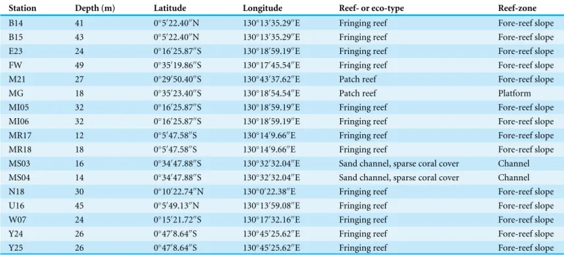
Table 1 Collection sites.Sample site information for collection locations from Raja Ampat (Indonesia) including environmental information on reefal habitat type.
Station Depth (m) Latitude Longitude Reef- or eco-type Reef-zone
B14 41 0◦5′22.40′′N 130◦13′35.29′′E Fringing reef Fore-reef slope B15 43 0◦5′22.40′′N 130◦13′35.29′′E Fringing reef Fore-reef slope E23 24 0◦16′25.87′′S 130◦18′59.19′′E Fringing reef Fore-reef slope FW 49 0◦35′19.86′′S 130◦17′45.54′′E Fringing reef Fore-reef slope M21 27 0◦29′50.40′′S 130◦43′37.62′′E Patch reef Fore-reef slope
MG 18 0◦35′23.40′′S 130◦18′54.54′′E Patch reef Platform
MI05 32 0◦16′25.87′′S 130◦18′59.19′′E Fringing reef Fore-reef slope MI06 32 0◦16′25.87′′S 130◦18′59.19′′E Fringing reef Fore-reef slope MR17 12 0◦5′47.58′′S 130◦14′9.66′′E Fringing reef Fore-reef slope MR18 18 0◦5′47.58′′S 130◦14′9.66′′E Fringing reef Fore-reef slope MS03 16 0◦34′47.88′′S 130◦32′32.04′′E Sand channel, sparse coral cover Channel MS04 14 0◦34′47.88′′S 130◦32′32.04′′E Sand channel, sparse coral cover Channel N18 30 0◦10′22.74′′N 130◦0′22.38′′E Fringing reef Fore-reef slope U16 45 0◦5′49.13′′N 130◦13′59.08′′E Fringing reef Fore-reef slope W07 24 0◦15′21.72′′S 130◦17′32.16′′E Fringing reef Fore-reef slope Y24 26 0◦47′8.64′′S 130◦45′25.62′′E Fringing reef Fore-reef slope Y25 26 0◦47′8.64′′S 130◦45′25.62′′E Fringing reef Fore-reef slope
MATERIAL AND METHODS
This study was conducted with 30 sediment samples from the Raja Ampat Archipelago (New Guinea, Indonesia) from around the islands of Waigeo, Batana, Kawe, Fam and adjacent small islets in an area that covers about 2,500 km2(Fig. 1C). The archipelago is located in the central Indo-Pacific warm pool with an average annual sea surface temperature of 29◦C
(Mangubhai et al.,2012). Raja Ampat is further situated in the passage way of the Indonesian Throughflow, a major ocean current that leads water masses from the western Pacific to the eastern Indian Ocean. Previous studies have shown that the reef fauna of Raja Ampat is strongly current dependent (Devantier, Turak & Allen,2009;Turak & Souhoka,2003).
The samples were collected by snorkeling and SCUBA diving in September 2011 by M Langer. Sediment surface samples from the top 2 cm were collected from the fore-reef slope of fringing reefs, with two samples from a patch reef, and two samples from a sandy channel with sparse coral cover (Table 1). The sediment was predominantly carbonaceous (∼90%) and included fine-grained sediments as well as coarse reef rubble. All samples were washed through a 63µm sieve and dried at 50◦C in an oven overnight. Foraminifera
Repository of the Material: the holotypes and topotypic paratypes of the new species are deposited in the micropaleontology collection of the Steinmann Institute of Paleontology at the University of Bonn, Germany (MaLaPNG 2011–10, MaLaPNG 2011–11, MaLaPNG 2011–12, MaLaPNG 2011–13, MaLaPNG 2011–14).
The electronic version of this article in Portable Document Format (PDF) will represent a published work according to the International Commission on Zoological Nomenclature (ICZN), and hence the new names contained in the electronic version are effectively published under that Code from the electronic edition alone. This published work and the nomenclatural acts it contains have been registered in ZooBank, the online registration system for the ICZN. The ZooBank LSIDs (Life Science Identifiers) can be resolved and the associated information viewed through any standard web browser by appending the LSID to the prefixhttp://zoobank.org/. The LSID for this publication is: urn:lsid:zoobank.org: pub:FB001C3C-AEA9-45D5-9224-EDD084378897. The online version of this work is archived and available from the following digital repositories: PeerJ, PubMed Central and CLOCKSS.
RESULTS
Smaller miliolid benthic foraminifera are typical dwellers in surface sediments of shallow water reefal and lagoonal habitats. By studying the highly diverse assemblages of benthic foraminifera from Raja Ampat, taken from different locations around the islands (Fig. 1C), we recorded a total of 455 species among them 249 miliolid species, of which five are described here as new. Four species belong to the widely distributed miliolid genera of
MiliolinellaWiesner,Triloculinad’Orbigny andSiphonapertaVella. As the morphological properties of the fifth species differentiate it from any previously known genera, we designate and describe it as the new genusDentoplanispirinella.
SYSTEMATIC DESCRIPTIONS
Subclass MiliolanaSaidova,1981
Order MiliolidaDelage & Hérouard,1896 Suborder MiliolinaDelage & Hérouard,1896 Superfamily CornuspiraceaSchultze,1854
Family Fischerinidae Millett, 1898
Subfamily Fischerininae Millett, 1898
GenusDentoplanispirinella Förderer and Langer gen. nov. urn:lsid:zoobank.org:act:98A1DD41-C0AE-4401-830B-0D189E70661A
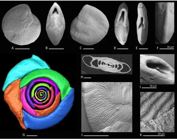
Figure 2 Holotype, paratype, CT scans and details ofDentoplanispirinellagen. nov.occultasp. nov.
(A) Side view and (B) apertural view of a more juvenile specimen with a nearly triangular aperture and weakly developed tooth (paratype); (C) side view and (D) apertural view (holotype); (E) apertural view of a specimen with a well-developed tooth and elongated aperture; (F) detail of a well-developed periph-eral keel; (G) CT scan reconstruction of the chamber cavities revealing the presence of 2.5 – 3.5 chambers per whorl in an adult specimen (note that penultimate chamber is broken); (H) CT scan showing planispi-rally arranged chambers; (I) detail of an aperture with a very well-developed tooth; (J) detail of the striate surface ornamentation; (K) detail of the construction of the outer wall layer showing randomly arranged calcite needles in the lower part (test surface removed) and longitudinally arranged calcite needles on the outer test surface. Scale bar is 100µm (unless indicated).
overlap the umbilical region in each whorl. Sutures oblique, thin and irregular. Aperture arch-shaped, triangular in juvenile specimens, high and subtriangular in adult specimens, tapering apically, on the base connected with the peripheral margin of the proceeding chamber and provided with a very small and thin tooth. In juvenile specimens the tooth appears just like a little knob or slightly raised spine.
Type species.Dentoplanispirinella occultasp. nov.
Dentoplanispirinella occultaFörderer and Langersp. nov.
Figures 2A–2K
urn:lsid:zoobank.org:act:7E132939-5284-484D-9B50-BC79A0B52D0A
Etymology.From the Latin ‘‘occultare’’ meaning for ‘‘hiding.’’
Material.28 specimens from nine samples (MR18, MI05, MI06, MS03, MS04, MG, M21,
U16, Y24;Fig. 1C;Table 1), recent.
Holotype. The specimen illustrated here asFigs. 2C and2D(sample MS03; MaLaPNG
2011–10).
Paratype. The specimen illustrated here asFigs. 2A and2B(sample MS03; MaLaPNG 2011–10).
Type locality. The holotype and the paratype are from sample station MS03 (16m), a sand channel between Arborek Island and Pulau Mansuar; Raja Ampat, New Guinea (Indonesia).
Diagnosis. A species ofDentoplanispirinellagen. nov. with a discoidal to biconvex test shape, a slightly keeled periphery, a radial oriented, finely striate surface ornamentation and an arch-shaped, triangular aperture, provided with a small tooth.
Dimensions.Observed range of test dimensions: diameter 285–704µm (lateral view), test
width 100–193µm (apertural view).
Occurence.Dentoplanispirinella occultais widely distributed in the Raja Ampat area in fine to coarse coral rubble samples from depths of 14 to 45 m.
Remarks.Dentoplanispirinella occultasp. nov. differs fromPlanispirinella involutaCollins (1958, p. 374, pl. 4,Figs. 2Aand2B) by its more lenticular biconvex shape in horizontal section, the subtriangular shape of the aperture, the presence of a small tooth, and the striate surface ornamentation.
Superfamily MiliolaceaEhrenberg,1839
Family HauerinidaeSchwager,1876
Subfamily HauerininaeSchwager,1876
GenusMiliolinella(Wiesner,1931)
Miliolinella moiaFörderer and Langersp. nov.
Figures 3A–3L
urn:lsid:zoobank.org:act:D8184E0C-2805-40D7-BCCB-492D74216168
Etymology.The new species is named after the indigeneous Moi people from Malaumkarta, a Papuan tribe from the north coast near Sorong.
Material. 11 specimens from six samples (B14, B15, E23, MR17, N18, U16;Fig. 1C; Table 1), recent.
Holotype.The specimen illustrated here asFigs. 3A–3C(sample B14; MaLaPNG 2011–11).
Paratypes.The specimens illustrated here asFigs. 3D–3F(sample B14),Figs. 3G–3Iand Figs. 3J–3J(sample ER23; MaLaPNG 2011–11).
Type locality.The holotype and the paratype are from sample station B14 (41 m), Bag Island, east of Pulau Uranie; Raja Ampat, New Guinea (Indonesia).
Diagnosis.A slightly enlongated, medium-sized species ofMiliolinellaWiesner,1931with a compressed, angular and slightly slanted outline, a smooth and shiny wall, and a high subcircular opening.
Dimensions. Observed range of test dimensions: test height 409–554 µm, test width
278–396µm (lateral view), 166–250µm (apertural view).
Occurence.This species is widely distributed in the Raja Ampat area in fine to coarse coral rubble samples and occurs at depths between 12 and 45 m.
Remarks.Miliolinella moiasp. nov. differs fromMiliolinella pilasensisMcCulloch,1977(p. 566, pl. 238, Fig. 16 andLoeblich & Tappan,1994, p. 57, pl. 99, Figs. 1–9) in its angular and more compressed outline, and the large subcircular opening. Millet (1898) depicted a species ofMiliolina valvularis(Reuss) from the Malay Archipelago (p. 11, Figs. 5A–5C) that shows a high degree of similarity toMiliolinella moia, but his specimen has a more rounded periphery. The original description ofTriloculina valvularisbyReuss(1851, p. 85, pl. 7, Fig. 56) shows a specimen with a broadly rounded periphery and inflated chambers without angles.Miliolinellasp. 2 figured inParker,2009from Ningaloo Reef, Australia (p. 128, Figs. 92A–92I, 93A–93J, 94A–94K) differs fromMiliolinella moiaby the low apertural opening and the broadly rounded and more inflated chambers.
Miliolinella undinaFörderer and Langersp. nov.
Figure 4A–4I
urn:lsid:zoobank.org:act:D11E1426-9DCC-41B8-A992-27D974A92520
1988aMiliolinellasp. B—Haig, Papuan Lagoon, Port Moresby, p. 224, pl. 2, Figs. 23 and 24.
1992Miliolinellasp.—Hatta & Ujiié, Ryukyu Islands, p. 72, pl. 10, Fig. 6. ?2012Miliolinellacf.M. semicostata(Wiesner)—Debenay, New Caledonia, p. 110, 275.
Etymology.After the undulate ornamentation of the test. From the Latin ‘‘unda’’ meaning wave and mythological ‘‘Undine,’’ a term established by the Renaissance alchemist Paracelcus for water spirits.
Material.Three specimens from three samples (MR18, N18, U16;Fig. 1C;Table 1), recent.
Holotype. The specimen illustrated here asFigs. 4A–4C(sample MR18; MaLaPNG
2011–12).
Paratypes.The specimens illustrated here asFigs. 4D–4F(sample N18) andFigs. 4G–4I (sample U16; MaLaPNG 2011–12).
Type locality.The holotype is from sample station MR18 (18 m), east of Kawe Island. The paratypes are from sample stations N18 (30 m), south-west coast of Pulau Wayag, and U16 (45 m), between Pulau Uranie and Bag Island; Raja Ampat, New Guinea (Indonesia).
Diagnosis.A small quinqueloculine species ofMiliolinellaWiesner with inflated chambers, a rounded outline and an undulate to reticulate surface ornamentation.
large parts of the test. Outer-wall layer constructed of needle-shaped crystals that are primarily aligned in longitudinal direction. Aperture basal, a large semicircularMiliolinella -type opening, provided with a thickened and everted peristomal rim and a broad, slightly excavated basal flap.
Dimensions. Observed range of test dimensions: test height 146–162 µm, test width
114–224µm (lateral view), 81–119µm (apertural view).
Occurence.Miliolinella undinais present with one specimen in each of three highly diverse, miliolid-rich, fine coral rubble samples from depths of 18 to 45 m.
Remarks.Specimens ofMiliolinella undina sp. nov. have been previously documented by Haig,1988aas Miliolinellasp. B from the Papuan Lagoon, Port Moresby and by Hatta & Ujiié,1992asMiliolinella sp.from the Ryukyus. Hatta & Ujiié mentioned the species to occur rarely in their assemblages. The new species has also been recorded in samples from northern Palawan (M Förderer, 2016, unpublished data). Miliolinellacf.
M. semicostata (Wiesner) depicted by Debenay from New Caledonia (2012, p. 110, 275) may also belong toMiliolinella undina, but shows a less undulated test ornamentation. Test shape, apertural and ornamental features are more similar to our holotype (Figs. 4A–4C) than toMiliolinella semicostata(Wiesner,1923) from the Mediterranean Sea (see Cimerman & Langer,1991, p. 42, pl. 38, Figs. 10–15).Miliolinella semicostatahas less inflated chambers and the ornamentation is not reticulate but longitudinally striate and restricted to the angles.Miliolinella undinaalso resemblesMiliolinellasp. 4 depicted by Parker from the Ningaloo Reef in Western Australia (2009, p. 136, Figs. 97A–97H), but his specimen has a less undulated and more striate alignment of costae. The new species also resembles
Miliolinella flintiana(Cushman,1932) (p. 55, pl. 12: 4A–4C) in size, test shape, chamber arrangement and apertural features. However it differs in its surface ornamentation, that is distinctly longitudinal costate inMiliolinella flintianaand undulate and more irregular inMiliolinella undina.Miliolinella flintianaalso occurs in our assemblages.
GenusTriloculina D’Orbigny,1826
Triloculina kaweaFörderer and Langersp. nov.
Figs. 5A–5H
urn:lsid:zoobank.org:act:6F5B38CE-88B3-4FBE-9329-8483756158E1
2009 Triloculina? sp. 2—Parker, Ningaloo Reef, p. 372, Figs. 271F–271J.
Etymology.This species is named in honor of the indigeneous people of West Papua after the Kawe tribe, that owns and protects a highly diverse marine protected area of Raja Ampat.
Material.12 specimens from seven samples (B15, FW, M05, MS04, N18, U16, Y25;Fig. 1C;Table 1), recent.
Holotype.The specimen illustrated here asFigs. 5A–5C(sample FW; MaLaPNG 2011–13).
Figure 5 Holotype, paratype, cross section and detail ofTriloculina kaweasp. nov.(A–C) Holotype: (A) lateral view of more involute side; (B) apertural view; (C) lateral view of more evolute side; (D) cross section of a specimen; (E–G) Paratype: (E) lateral view of more evolute side; (F) apertural view; (G) lateral view of more involute side; (H) detail of the irregular test surface. Scale bar is 100µm (unless indicated).
Type locality.The holotype and the paratype are from sample station FW (49 m), south-east Penemu, Fam Islands; Raja Ampat, New Guinea (Indonesia).
Diagnosis. A medium-sized species ofTriloculinad’Orbigny with a slightly elevated
‘‘Lachlanella’’-type aperture, rounded periphery, blunt angles and a roughly textured wall.
Dimensions. Observed range of test dimensions: test height 377–439 µm, test width
200–245µm (lateral view), 162–195µm (apertural view).
Occurence.This species is widely distributed in our sampling area in fine to coarse coral rubble samples from depths of 14 to 49 m.
Remarks.The speciesTriloculina? sp. 2 reported byParker,2009from Western Australia differs fromTriloculina kaweasp. nov. in its less triangular shape and less elongated outline. We consider Parker’s specimen a juvenile individual ofTriloculina kawea. The aperture of Parker’s specimen is not intact but resembles very well the apertural features ofTriloculina kawea. The outer wall layer appears identical (Fig. 5H). Parker mentioned the species to be possibly cryptoquinqueloculine.Figures 5Band5Fand the horizontal section (5D) show the triloculine chamber arrangement. Triloculinasp. 1, reported by Debenay 2012 from New Caledonia (p. 139, 278) is very similar in shape and surface ornamentation to
Triloculina kawea, but has significantly more acute angles and a Y-shaped tooth.Triloculina kaweafurther differs fromTriloculina linneianad’Orbigny depicted byBaccaert,1987from the Great Barrier Reef (p. 128, pl. 57, Figs. 3 and 4) in the less striate ornamentation and more acute angles.
Subfamily SiphonapertinaeSaidova,1975
GenusSiphonapertaVella,1957
Siphonaperta hallockiFörderer and Langersp. nov.
Figs. 6A–6F
urn:lsid:zoobank.org:act:DD4F0DB3-1355-4BB1-841A-FFE32E0F6455
?1988aQuinqueloculinasp. C—Haig, Papuan Lagoon, Port Moresby, p. 234, pl. 9, Figs. 7–10.
?2009Quinqueloculinasp. 13—Parker, Ningaloo Reef, p. 311, Figs. 224A–224J, 225A–225G.
Etymology. In honor of Pamela Hallock Muller for her extensive work on tropical foraminifera.
Material.Four specimens from three samples (MS03, N18, W07;Fig. 1C;Table 1), recent.
Holotype. The specimen illustrated here asFigs. 6A–6C(sample MS03; MaLaPNG 2011–14).
Paratype.The specimen illustrated here asFigs. 6D–6F(sample N18; MaLaPNG 2011–14).
Type locality.The holotype is from sample station MS03 (18 m), a sand channel between Arborek Island and Pulau Mansuar. The paratype is from sample station N18 (30 m), south-west coast of Pulau Wayag; Raja Ampat, New Guinea (Indonesia).
Diagnosis.A medium-sized species ofSiphonapertaVella with a finely agglutinated wall, carinate shoulders, a short neck and a circular aperture with a small bifid tooth.
Figure 6 Holotype and paratype ofSiphonaperta hallockisp. nov.(A–C) Holotype: (A) lateral view of more evolute side; (B) apertural view; (C) lateral view of more involute side; (D–F) paratype: (D) lateral view of more evolute side; (E) apertural view; (F) lateral view of more involute side. Scale bar is 100µm (unless indicated).
(in well preserved specimens). Aboral end rounded and produced; oral end becoming more slender and leading into a short produced neck. Aperture terminal, a wide circular opening with a short T-shaped, bifid tooth, that reaches more than one third of the apertural diameter. Apertural opening surrounded by a slightly thickened and everted peristomal rim.
Dimensions. Observed range of test dimensions: test height 240–442 µm, test width
132–233µm (lateral view), 87–119µm (apertural view).
Remarks.Very similar specimens were previously documented asQuinqueloculinasp. C from the Papuan Lagoon (Haig,1988a) andQuinqueloculinasp. 13 from Ningaloo Reef (Parker,2009). Test shape, wall texture and apertural features appear to be identical to our specimens from Raja Ampat.Quinqueloculinasp. 4 documented byParker,2009from the Ningaloo Reef appears very similar toSiphonaperta hallocki, but differs in its more elongated shape, more rounded and inflated chambers and the cryptoquinqueloculine coiling. In addition, Parker describes the wall as roughly textured with some agglutinated grains.Quinqueloculina tropicalisCushmanfrom Samoa (1924, p. 63, pl. 23, Figs. 9 and 10) differs from our new species by its more compressed shape and more elongated broadly rounded chambers without any angles or costae.Quinqueloculina polygona D’Orbigny (1839, p. 198,pl. 12, Figs. 21–23) differs fromSiphonaperta hallockiin its smooth and shiny surface, the pronounced carinae and the less inflated chambers.Langer et al.,2013depicted a specimen of Quinqueloculina polygonad’Orbigny from Bazaruto (Langer et al.,2013, p. 163, Fig. 5: 14) that resembles our new species in size, shape and apertural features. However, it is unlikely that this species from Bazaruto belongs toSiphonaperta hallocki, as its outer wall layer is not agglutinated.
ACKNOWLEDGEMENTS
The authors gratefully acknowledge Dr. Stephanie Pietsch for assistance with the collection of the samples, Georg Oleschinski for help with the SEM and Peter Göddertz and Kai Jäger for support with CT scan imaging. We thank Justin H. Parker, Tomas Cedhagen and an anonymous reviewer for constructive and helpful comments on the manuscript.
ADDITIONAL INFORMATION AND DECLARATIONS
Funding
Collection and study of the material was supported by grants from the German Science Foundation (La-884/10-1/13- 1) and the University of Bonn. The funders had no role in study design, data collection and analysis, decision to publish, or preparation of the manuscript.
Grant Disclosures
The following grant information was disclosed by the authors: German Science Foundation: La-884/10-1/13-1.
University of Bonn.
Competing Interests
The authors declare there are no competing interests.
Author Contributions
• Martin R. Langer conceived and designed the experiments, performed the experiments, analyzed the data, contributed reagents/materials/analysis tools, wrote the paper, reviewed drafts of the paper.
Data Availability
The following information was supplied regarding data availability:
1) Micropaleontological Collection Steinmann Institute of Paleontology at the University of Bonn, Germany
2) MaLaPNG 2011-10, MaLaPNG 2011-11, MaLaPNG 2011-12, MaLaPNG 2011-13, MaLaPNG 2011-14.
New Species Registration
The following information was supplied regarding the registration of a newly described species:
Dentoplanispirinellagen. nov. urn:lsid:zoobank.org:act:98A1DD41-C0AE-4401-830B-0D189E70661A
Dentoplanispirinella occultasp. nov.
urn:lsid:zoobank.org:act:7E132939-5284-484D-9B50-BC79A0B52D0A
Miliolinella moiasp. nov.
urn:lsid:zoobank.org:act:D8184E0C-2805-40D7-BCCB-492D74216168
Miliolinella undinasp. nov.
urn:lsid:zoobank.org:act:D11E1426-9DCC-41B8-A992-27D974A92520
Triloculina kaweasp. nov.
urn:lsid:zoobank.org:act:6F5B38CE-88B3-4FBE-9329-8483756158E1
Siphonaperta hallockisp. nov.
urn:lsid:zoobank.org:act:DD4F0DB3-1355-4BB1-841A-FFE32E0F6455
The LSID for this publication is: urn:lsid:zoobank.org:pub:FB001C3C-AEA9-45D5-9224-EDD084378897.
REFERENCES
Agostini VN, Grantham HS, Wilson J, Mangubhai S, Rotinsulu C, Hidayat N, Muljadi A, Muhajir, Mongdong M, Darmawan A, Rumetna L, Erdmann MV, Possingham HP. 2012.Achieving fisheries and conservation objectives within marine protected areas: zoning the Raja Ampat network. Indo-Pacific Division, Denpasar: The Nature Conservancy Report No 2/12.Available athttps:// www.conservationgateway.org/ Documents/ Agostini%20etal12_Raja%20Ampat%20Zoning%20REPORT.pdf (accessed 24 February 2016).
Baccaert J. 1987.Distribution patterns an taxonomy of benthic foraminifera in the Lizard Island reef complex, northern great barrier reef, australia. PhD thesis, Université de Liège.
Brady HB. 1884.Report on the foraminifera dredged by H.M.S. Challenger during the years 1873–1876.Reports of the Scientific Results of the Voyage of H. M. S. Challenger, Zoology9:1–814.
Briggs JC, Bowen BW. 2013.Marine shelf habitat: biogeography and evolution.Journal of Biogeography40:1023–1035.DOI 10.1111/jbi.12082.
Cimerman F, Langer MR. 1991.Mediterranean Foraminifera. Ljubljana: Slovenska akademija znanosti in umetnosti. Academia Scientiarum et Artium Slovencia Classis IV: Historia Naturalis.
Collins AC. 1958. Foraminifera. In:Great Barrier Reef Expedition 1928–1929. British Museum of Natural History Scientific reports, vol. 6. London: British Museum (Natural History), 335–437.
Cushman JA. 1921.Foraminifera of the Philippine and adjacent seas.Bulletin of the United States National Museum100(4):1–608.
Cushman JA. 1924. Samoan foraminifera. In:Department of marine biology of the Carnegie Institution of Washington. Vol. XXI. Washington, D.C.: Carnegie Institute of Washington, 1–85.
Cushman JA. 1932.The foraminifera of the tropical Pacific collections of the ‘‘Alba-tross,’’ 1899–1900. Part 1. Astrorhizidae to Trochamminidae.Bulletin of the United States National Museum161:1–88.
Debenay JP. 2012.A Guide to 1,000 Foraminifera from Southwestern Pacific, New Caledonia. Paris: Editions IRD Marseille/Publications Scientifiques du Muséum.
Delage Y, Hérouard E. 1896.Traité de Zoologie Concrète, La Cellule et les Protozoaires. Vol. 1. Paris: Schleicher Frères.
Devantier L, Turak E, Allen G. 2009.Reef-scapes, reef habitats and coral communities of Raja Ampat, Birds’s Head Seascape, Papua, Indonesia. Report to The Nature Conservancy, Bali, Indonesia.
D’Orbigny A. 1826.Tableau méthodique de la classe des Céphalopodes.Annales des Sciences Naturelles7:245–314.
D’Orbigny A. 1839. Foraminifères. In: Ramon de la Sagra, ed.Histoire physique, politique et naturelle de l’île de Cuba. Paris: Arthus Bertrand, 1–224.
Ehrenberg CG. 1839. Über die Bildung der Kreidefelsen und des Kreidemergels durch unsichtbare Organismen.Physikalische Abhandlungen der Königlichen Akademie der Wissenschaften zu Berlin, 1838 [1840: separate 1839]. Berlin: Königlichen Akademie der Wissenschaften, 59–147.
Ekman S. 1953.Zoogeography of the sea. London: Sidgwick and Jackson.
Erdmann MV, Pet JS. 2002.A rapid marine survey of the northern Raja Ampat islands (Eastern Indonesia). Report from Henry Foundation/The Nature Conservancy/N-RM/EPIQ, 36 pp, The Nature Conservancy.Available athttp:// www.rajaampat.org/ downloads/ Raja4%20trip%20report.pdf (accessed 24 February 2016).
Graham JJ, Militante PJ. 1959.Recent foraminifera from the Puerto Galera Area, northern Mindoro, Philippines.Stanford University Publications, Geological Sciences
Haig DW. 1988a.Miliolid foraminifera from the inner neritic sand and mud facies of the Papuan Lagoon, New Guinea.Journal of Foraminiferal Research18:203–236 DOI 10.2113/gsjfr.18.3.203.
Haig DW. 1988b.Distribution of miliolid foraminifera in marine sediments around Motupore Island, Papua New Guinea.Science in New Guinea14:54–94.
Haig DW. 1993.Buliminid foraminifera from inner neritic and mud facies of the Papuan Lagoon.Journal of Foraminiferal Research23:162–179DOI 10.2113/gsjfr.23.3.162.
Hatta A, Ujiié H. 1992.Benthic foraminifera from coral seas between Ishigaki and Iriomote Islands, Southern Ryukyu Island Arc, Northwestern Pacific. part 1.
Systematic descriptions of textulariina and Miliolina.Bulletin of the College of Science, University of Ryukyus53:49–119.
Hoeksema BW. 2007. Delineation of the Indo-Malayan centre of maximum marine
biodiversity: the Coral Triangle. In: Renema W, ed.Biogeography, Time, and Place: Distributions, Barriers, and Islands. Dordrecht: Springer, 117–178.
Hofker J. 1927. The foraminifera of the Siboga Expedition. Part 1. Families Tinoporidae, Rotaliidae, Nummulitidae, Amphisteginidae.Siboga Expeditie, Monographie IVa. Leiden: E. J. Brill, 1–78.
Hofker J. 1930. The foraminifera of the Siboga Expedition. Part 2. Families Astorhizidae, Rhizamminidae, Reophacidae, Anomalinidae, Peneroplidae.Siboga Expeditie, Monographie IVa. Leiden: E. J. Brill, 79–170.
Hohenegger J. 2004.Depth coenoclines and environmental considerations of Western Pacific larger foraminifera.Journal of Foraminiferal Research34:9–33 DOI 10.2113/0340009.
Hohenegger J. 2011.Large Foraminifera—Greenhouse constructions and gardeners in the oceanic microcosm. Kagoshima: Kagoshima University Museum.
Langer MR. 1992.New recent foraminiferal genera and species from the lagoon at Madang, Papua New Guinea.Journal of Micropaleontology11:85–93
DOI 10.1144/jm.11.1.85.
Langer MR, Hottinger L. 2000.Biogeography of selected ‘‘larger’’ foraminifera. Biology of Foraminifera.Micropaleontology46 Suppl.1:105–126.
Langer MR, Lipps JH. 2003.Foraminiferal distribution and diversity, Madang Reef and Lagoon, Papua New Guinea.Coral Reefs22:143–154
DOI 10.1007/s00338-003-0298-1.
Langer MR, Thissen JM, Makled WA, Weinmann AE. 2013.The foraminifera from the
Bazaruto Archipelago (Mozambique).Neues Jahrbuch für Geologie und Paläontologie. Abhandlungen Band267/2:155–170DOI 10.1127/0077-7749/2013/0302.
Loeblich AR, Tappan H. 1987.Foraminiferal genera and their classification. New York: Springer Verlag.
Loeblich AR, Tappan H. 1994.Foraminifera of the Sahul Shelf and Timor Sea.Cushman Foundation for Foraminiferal Research31:1–661.
Mangubhai S, Erdmann MV, Wilson JR, Huffard CL, Ballamu F, Hidayat NI, Hitipeuw C, Lazuardi ME, Muhajir, Pada D, Purba G, Rotinsulu C, Rumetna L, Sumolang K, Wen W. 2012.Papuan Bird’s Head Seascape: emerging threats and challenges in the global center of marine biodiversity.Marine Pollution Bulletin64:2279–2295 DOI 10.1016/j.marpolbul.2012.07.024.
McCulloch I. 1977.Qualitative observations on recent foraminiferal tests with emphasis on the eastern Pacific: parts I, II, III. Los Angeles: University of Southern California.
McKenna S, Allen G, Suryadi S. 2002. A marine rapid assessment of the Raja Ampat Islands, Papua Province, Indonesia. In:RAP Bulletin of Biological Assessment, Vol. 22. Washington, D.C.: Conservation International.Available athttp:// www. conservation.org/ publications/ Documents/ RAP_Reports/ RAP22_Raja_Ampat_ Indonesia_Apr-2002.pdf (accessed 24 February 2016).
Millett FW. 1898–1904.Report on the recent Foraminifera of the Malay Archipelago contained in anchor-mud collected by Mr. A. Durrand, F. R. M. S. Parts I-XVII.
Journal of Royal Microscopical Society1898:258–269 (Part I), 499–513 (Part II), 607– 614 (Part III); 1899:249–255 (Part IV), 357–365 (Part V), 557–564 (Part VI); 1900:6– 13 (Part VII), 273–281 (Part VIII), 539–549 (Part IX); 1901:1–11 (Part X), 485–497 (Part XI), 619–628 (Part XII); 1902:509–528 (Part XIII); 1903:253–275 (Part XIV), 685–704 (Part XV); 1904, 489–506 (Part XVI), 597–609 (Part XVII).
Parker J. 2009.Taxonomy of foraminifera from Ningaloo Reef, Western Australia.
Memoirs of the Association of Australasian Paleontologists36:1–810.
Prazeres M, Uthicke S, Pandolfi JM. 2016.Influence of local habitat on the physiological responses of large benthic foraminifera to temperature and nutrient stress.Scientific Reports6: 21936DOI 10.1038/srep21936.
Renema W. 2003.Larger foraminifera on reefs around Bali (Indonesia).Zoologische Verhandelingen345:337–366.
Renema W. 2006.Large benthic foraminifera from the deep photic zone of a mixed siliciclastic-carbonate shelf off East Kalimantan, Indonesia.Marine Micropaleontology
58:73–82DOI 10.1016/j.marmicro.2005.10.004.
Renema W. 2010.Is increased calcarinid (foraminifera) abundance indicating a larger role for macro-algae in Indonesian Plio-Pleistocene coral reefs?Coral Reefs
29:165–173DOI 10.1007/s00338-009-0568-7.
Renema W, Hohenegger J. 2005.On the identity ofCalcarina spengleri(Gmelin, 1791).
Journal of Foraminiferal Research35:15–21DOI 10.2113/35.1.15.
Renema W, Troelstra SR. 2001.Larger foraminifera distribution on a mesotrophic carbonate shelf in SWSulawesi (Indonesia).Palaeogeography, Palaeoclimatology, Palaeoecology175:125–147 DOI 10.1016/S0031-0182(01)00389-3.
Reuss AE. 1851.Ueber die fossilen Foraminiferen und Entomostraceen der Septharien-thone der Umgegend von Berlin.Zeitschrift der Deutschen Geologischen Gesellschaft
Roberts CM, McClean CJ, Veron J, Hawkins JP, Allen GR, McAllister DE, Mittermeier CG, Schueler FW, Spalding M, Wells F, Vynne C, Werner TB. 2002.Marine biodi-versity hotspots and conservation priorities for tropical reefs.Science295:1280–1284 DOI 10.1126/science.1067728.
Saidova KM. 1975.Bentosnye Foraminifery Tikhogo Okeana [Benthonic foraminifera of the Pacific Ocean], Vol. 3. Moscow: Institut Okeanologii P. P. Shirshova, Akademiya Nauk SSSR.
Saidova KM. 1981.O sovremennom sostoyanii sistemy nadvidovykh taksonov Kayno-zoyskikh bentosnykh foraminifer [On an up-to-date system of supraspecific taxonomy of Cenozoic benthonic foraminifera]. Moscow: Institut Okeanologii P. P. Shirshova, Akademiya Nauk SSSR.
Schultze MS. 1854.Über den Organismus der Polythalamien (Foraminiferen), nebst Bemerkungen über die Rhizopoden im Allgemeinen. Leipzig: Wilhelm Engelmann.
Schwager C. 1876.Saggio di una classificazione dei foraminiferi avuto riguardo alle loro famiglie naturali.Bolletino R. Comitato Geologico d’Italia7:475–485.
Tittensor DP, Mora C, Jetz W, Lotze HK, Ricard D, Vanden Berge E, Worm B. 2010.Global patterns and predictors of marine biodiversity across taxa.Nature
466:1098–1011DOI 10.1038/nature09329.
Turak E, Souhoka J. 2003. Coral diversity and the status of coral reefs in the Raja Ampat Islands. In: Donnelly R, Neville D, Mous P, eds.Report on a rapid eco-logical assessment of the Raja Ampat Islands, Papua, Eastern Indonesia, held Octo-ber 30–NovemOcto-ber 22, 2002. Available athttp:// www.rajaampat.org/ downloads/ RajaAmpatREAFinalDraft%20Screen.pdf (accessed 24 February 2016).
Vella P. 1957.Studies in New Zealand foraminifera.Paleontological Bulletin, Wellington
28:1–64.
Veron JEN. 1995.Corals in space and time: the biogeography and evolution of the Sclerac-tinia. Ithaca: Cornell University Press.
Veron JEN, DeVantier LM, Turak E, Green AL, Kininmonth S, Stafford-Smith M, Peterson N. 2009.Delineating the Coral Triangle.Galaxea, Journal of Coral Reef Studies11:91–100.
Weinmann AE, Rödder D, Lötters S, Langer MR. 2013.Heading for new shores:
Projecting marine distribution ranges of selected larger foraminifera.PLoS ONE
8(4):e62182DOI 10.1371/journal.pone.0062182.
Wiesner H. 1923.Die Milioliden der östlichen Adria. Prag-Bubenec.
Wiesner H. 1931.Die foraminiferen der deutschen Südpolar Expedition 1901–1903.