doi:10.1017/S0967199407004261 Printed in the United Kingdom
Structural and ultrastructural characteristics of the yolk syncytial
layer in
Prochilodus lineatus
(Valenciennes, 1836) (Teleostei;
Prochilodontidae)
A. Ninhaus-Silveira
1, F. Foresti
2, A. de Azevedo
2, C.A. Agostinho
3and R. Ver´ıssimo-Silveira
2 Universidade Estadual Paulista (UNESP), S˜ao Paulo, BrasilDate submitted: 13.04.06. Date accepted: 12.07.06
Summary
The yolk syncytial layer (YSL) has been regarded as one of the main obstacles for a successful cryopreservation of fish embryos. The purpose of this study was to identify and characterize the YSL in
Prochilodus lineatus, a fish species found in southeastern Brazil and considered a very important fishery
resource. Embryos were obtained through artificial breeding by hormonal induction. After fertilization, the eggs were incubated in vertical incubators with a controlled temperature (28◦C). Embryos were
collected in several periods of development up to hatching and then fixed with 2% glutaraldehyde and 4% paraformaldehyde in 0.1 M sodium phosphate buffer (pH 7.3). Morphological analyses were carried out under either light, transmission or scanning electron microscopy. The formation of the YSL in
P. lineatusembryos starts at the end of the cleavage stage (morula), mainly at the margin of the blastoderm,
and develops along the embryo finally covering the entire yolk mass (late gastrula) and producing a distinct intermediate zone between the yolk and the endodermal cells. The YSL was characterized by the presence of microvilli on the contact region with the yolk endoderm. A cytoplasmic mass, full of mitochondria, vacuoles, ribosomes, endomembrane nets and euchromatic nuclei, indicated a high metabolic activity. This layer is shown as an interface between the yolk and the embryo cells that, besides sustaining and separating the yolk, acts as a structure that makes it available for the embryo. The structural analyses identified no possible barriers to cryoprotectant penetration.
Keywords: Cryopreservation, Embryo, Morphology, Periblast, Teleost
Introduction
The periblast or yolk syncytial layer (YSL) is a continued cytoplasmic layer located between the blastoderm disc and the yolk, and resulting from the incomplete division of the blastomeres. Initially, it is a ring-shaped layer around the yolk that it spreads
All correspondence to: A. Ninhaus-Silveira, Departamento de Biologia e Zootecnia - Universidade Estadual Paulista/Ilha Solteira, Av. Brasil, 56 – Centro Postal Box 31, CEP: 15385–000 Ilha Solteira, S˜ao Paulo, Brasil. Tel: +55 02118 37431285. Fax: +55 02118 37431186. e-mail: [email protected]
1Universidade Estadual Paulista (UNESP) – Depto. de
Biologia e Zootecnia–Ilha Solteira–SP.
2Universidade Estadual Paulista (UNESP) – Depto. de
Morfologia–Botucatu–SP.
3Universidade Estadual Paulista (UNESP) – Depto. de
Produc¸˜ao e Explorac¸˜ao Animal–Botucatu–SP.
eventually under the entire blastoderm (Kimmelet al., 1995). This layer is greatly important for the embryonic development of teleost eggs. Due to its location, all the nutrients from the yolk pass through this layer to reach the blastoderm (Trinkaus, 1951, 1993; Devillers, 1961). In addition, the YSL works as a force for the epiboly movement of the blastoderm (Trinkaus, 1984a, b, 1993).
Recent research on the epiboly and somite stages, suggests that the major problem for cryopreservation of zebrafish embryos is the difficulty in gaining sufficient cryoprotectant penetration, particularly in the yolk compartment (Rawson et al., 2000). Hagedorn et al.
yolk, in order to identify its structural, functional and physiological features (Rawsonet al., 2000).
The knowledge of YSL characteristics has great importance for the development of cryopreservation techniques for fish embryos. Inasmuch as there is no detailed study about this subject in Neotropical teleosts, the present study has aimed at anylysing the structure and ultrastructure of YSL, using as a model
Prochilodus lineatus, a Neotropical fish that belongs to
theProchilodontidaefamily, with a wide distribution in
southeastern Brazil, and found in the entire Paran´a-Paraguay and Para´ıba river basins (Corrˆea e Castro, 1990).
Materials and methods
Embryos were obtained through artificial fertilization of mature adult individuals ofProchilodus lineatusfrom the broodstock kept at Aquaculture Section, in the Animal Production Department of the Faculdade de Medicina Veterin´aria e Zootecnia–UNESP, Botucatu. The material was further processed and analyzed at the Biology and Fish Genetics Laboratory at the Morphology Department of the Biosciences Institute– UNESP, Botucatu, Brazil.
Embryos were collected at different developmental stages up to hatching and fixed with 2% glutaraldehyde and 4% paraformaldehyde in 0.1 M sodium phosphate buffer (pH 7.3). To carry out the light microscopic (LM) analyses, representative embryo samples from each embryonal stage were dehydrated in a series of increasing alcohol concentrations. They were sequentially and individually put into glycol-methacrylate for further microtomy, in order to obtain transverse and sagittal serial sections of 3 to 5µm. Subsequently, the sections were stained with
Harris eosin hematoxylin and toluidine blue, analyzed and then photographed under a photomicroscope (Axiophot, Zeiss).
To perform the transmission electron microscopy (TEM), the samples were post-fixed in 1% osmium tetroxide for 2 h, contrasted with an aqueous solution of 0.5% uranyl acetate, dehydrated in acetone and placed in epoxy resin. For contrast, ultra-fine sections were counter-stained with uranyl acetate (Watson, 1958), washed in 50% alcohol and then stained with lead citrate (Reynolds, 1963). The material was analyzed and photographed using transmission electron microscopy (Phillips, CM100).
In the scanning electron microscopy (SEM) process, pre-fixed embryos were previously cryofractured, after placing the samples in a 30% sucrose solution until they settled, washing in 1% phosphate buffer, cryofractured in liquid nitrogen and submerging in
a 2.5% glutaraldehyde solution. After 24 h, these embryos were transferred to a 13 mm cover slide coated with 1% poly-L-lysine, post-fixed in 0.5% osmium tetroxide, dehydrated in alcohol, dried in Balzer’s critical point dryer CPD-20, coated with 10 nm of gold in Balzer’s MED-010 sputtering unit, and then observed and photographed under a scanning electron microscope (Phillips SEM 515).
Results
YSL formation in P. lineatus started at the end of the cleavage stage (morula) and at the beginning of blastula stage, on the margin of blastoderm, giving rise to a surrounding ridge (Fig. 1a, c). It was characterized as a cytoplasmic layer bearing several nuclei with no separating cytoplasmic membranes and possibly showed fractionated yolk globules within its cytoplasm (Fig. 1c,d,f,g). The first nuclei were derived from peripheral blastomeres of incomplete cytokinesis that, when in contact with the cytoplasmic layer surrounding the yolk (YCL), released their contents into this layer (Fig. 1f).
Initially, the peripheral YSL presented a thicker aspect (Fig. 1a). However, as long as the nuclei underwent divisions and migrated to the YCL portion located below the blastoderm, its peripheral portion became thinner (Fig. 1b). At the end of blastula stage, a distinctive multinucleated intermediate layer was observed, arranged between the yolk vacuoles and blastoderm cells (Fig. 1c).
The nuclei under LM were presented as basophilic and uncondensed structures, showing a high metabolic activity (Fig. 1a,d,f). During epiboly, the YSL spread along the embryo until it encompassed the yolk mass entirely, forming the yolk plug (late gastrula) (Fig. 1e). The YSL was present as a thin layer involving the yolk completely at the segmentation stage (Fig. 1g,h) and, at the larval stage, it reached the primitive intestine (Fig. 1i).
The YSL analysis under TEM at several embryonal development phases revealed that this layer contained many ribosomes, a large net of endomembranes, several mitochondria, vacuoles and some multivacuole bodies (Fig. 2e,f). Individualized Golgi complexes were not found. The nuclei were elongated and euchromatic, presenting uneven contours and a high amount of pores, which indicated high metabolic activity (Fig. 2c–
Figure 1 Photomicrographies of P. lineatus embryo cuts: (a, b) transversal sections of embryo at the initial and late stage, respectively (toluidine blue); (c) lateral transversal section showing the yolk syncytial layer (YSL) covering the entire surface under the blastoderm (toluidine blue); (d) gastrula stage (Toluidine blue; 50% epiboly), pointing out the euchromatic nuclei of the yolk syncytial layer (YSL); (e) yolk plug made up by YSL (90% epiboly) (HE); (f) blastomere releasing its content into YSL (toluidine blue); (g) yolk globules within the YSL (HE); (h) view of YSL enveloping the yolk completely (segmentation stage) (HE); (i) longitudinal cut of the embryo at the larval stage showing the YSL enveloping the primitive intestine (HE). b, blastomere; bl, blastoderm; n, nucleus; no, notochord; s, somite; ycl, yolk cytoplasmic layer; yg, yolk globules; ysl, yolk syncytial layer. Open blastomere to the ycl; pi, primitive intestine. Bars: (a,b) 50µm; (c) 50µm; (d) 50µm; (e) 25µm; (f) 6.25µm; (g) 25µm; (h) 100µm; (i) 333.33µm.
Figure 2 Ultrastructure (TEM) of the yolk syncytial layer: (a) view of arrangement between the endoderm, the yolk syncytial layer (YSL) and the yolk; (b) detail of microvilli present between the YSL and the endoderm; (c,d) show that the nuclei of the YSL are cylindrical and uneven; (e,f) point out the large quantity of mitochondrias and extensive net of endomembranes found in the cytoplasm of the YSL as well as fibres, multivacuole bodies and yolk vacuoles. ed, endomembranes; en, endoderm; f, fibres; m, mitochondria; mv, microvilli; n, nucleus; vb, vacuole body; yg, yolk globules; ysl, yolk syncytial layer. Bars: (a) 2.3µm; (b) 0.59µm; (c,d) 3.1µm; (e) 0.75µm; (f) 0.59µm.
mostly from the YSL, were detected in the PF (Fig. 2a,
b). In addition, yolk globules were observed throughout the cytoplasm and their size decreased towards PF (Fig. 2c,d).
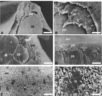
Figure 3 Eletronmicrographies (SEM) of cryofractured P. lineatus embryos (4–6 somite phase): (a, b) show the overlapping of ectoderm layer, endoderm, yolk syncytial layer and the yolk; (c, d) show the cytoplasm of the YSL and its arrangement with the yolk globules; (e, f) show the microvilli of the YSL and the stains on its surface. ec, ectoderm; en, endoderm; mv, microvilli; n, nucleus; st, stains; yg, yolk globules; ysl, yolk syncytial layer. Bars: (a) 30.77µm; (b) 13.89µm; (c) 2.35µm; (d) 3.97µm; (e) 4.5µm; (f) 0.87µm.
Discussion
Observations made on the start of YSL formation in
P. lineatus were supported by previous analyses in
Fundulus heteroclitus and Brachydanio rerio (Lentz &
Trinkaus, 1967; Kimmelet al., 1985; Trinkaus, 1993) and confirmed that YSL nucleation is observed at the end of the cleavage (morula) stage, through the releasing of the nucleus and the cytoplasmic content of some peripheral blastomeres within the yolk cytoplasmic layer (YCL). The YSL undergoes the epiboly process regardless of the blastoderm (Trinkaus, 1993; Devillers, 1961) and it acts as a primary force for blastoderm epiboly (Betchaku & Trinkaus, 1986; Trinkaus, 1993). The peripheral YSL undergoes contraction, resulting in several events such as facilitating the migration of the nuclei to its inner part and completes its formation with thinning towards the vegetal pole along the blastoderm, to which it is firmly adhered, leading to the epiboly movement (Trinkaus, 1993). Analyses in
P. lineatuscorroborate the observations of the authors
mentioned above, pointing out that the YSL appears clearer from nucleation onwards, as was also reported by Lentz & Trinkaus (1967) for F. heteroclitus, which suggests that besides the volume increase, there is a
change in membrane characteristics, making it more visible.
Regarding the importance of YSL in the yolk incorporation by P. lineatus embryos, the presence of microvilli in its upper part and the membrane projections between the yolk globules, as well as the lack of yolk globules close to the blastoderm, suggest that the vitelline material is degraded by hydrolytic enzymes, as reported by Lentz & Trinkaus (1967), and then transferred to the blastoderm (Walzer & Sch ¨onenberger, 1979). Nevertheless, as pinocytic or phagocytic vacuoles were not observed close to the microvilli, we could infer that the nutritive material passes through the plasmatic membrane as small molecules, as reported by Lentz & Trinkaus (1967).
TEM analysis showed some different external structure characteristics in the YSL of the plasma membrane in P. lineatus in relation to those found by Rawson et al. (2000) in B. rerio. For instance, the membrane in P. lineatus is wrinkled and covered by microvilli and, at ultrastructural level (TEM, SEM), presented neither pores nor similar structures. On the other hand, our observations are in agreement with those reported by Rawsonet al.(2000) on the granular aspect of the cytoplasm of the YSL.
Inversely to the impermeability of the YSL to cryoprotectants proposed by Hagedorn et al. (1998), the present analysis of YSL ultrastructure showed no morphological evidence that it could act as a barrier to cryoprotectant substances, as was reported by Rawson
et al. (2000). However, there is evidence that the YSL,
besides connecting, sustaining and separating the yolk and the embryo cells, makes the yolk content available to the embryo. Furthermore, this finding also suggests that the YSL has some control over the entrance and exit of substances to the vitelline vacuole.
Acknowledgements
This work was supported by Aquaculture Division of the Faculdade de Medicina Veterin´aria e Zootecnia/ UNESP/Botucatu/S˜ao Paulo/Brazil, which provided the fish and the facilities used in this study, and also by CAPES (Coordenac¸˜ao de Aperfeic¸oamento de Pessoal de N´ıvel Superior) and CNPq (Conselho Nacional de Pesquisa).
References
Devillers, C. (1961). Structural and dynamics aspects of the development of the teleostean egg.Adv. Morphol.1, 379– 428.
Fowler, H.W. (1954). Os peixes de ´agua doce do Brasil. Arq. Zool.2, 1–400.
Hagedorn, M., Hsu, E.W., Pilatus, U., Wildt, D.E., Rall, W.F. & Blackband, S.J. (1996). Magnetic resonance microscopy and spectroscopy reveal kinetics of cryoprotectant permeation in a multicompartimental biological system. Proc. Natl. Acad. Sci. USA93, 7454–9.
Hagedorn, M., Kleinhans, E.W., Wildt, D.E. & Rall, W.E. (1997). Chill sensitivity and cryoprotectant permeability of dechorionated zebrafish embryos, Brachydanio rerio. Cryobiology34, 251–63.
Hagedorn, M., Kleinhans, E.W., Artemov, D. & Pilatus, U. (1998). Characterization of a major permeability barrier in the zebrafish embryo.Biol. Reprod.59, 1240–50.
Kimmel, C.B. & Law, R.D. (1985). Cell lineage of zebrafish blastomeres. I. Cleavage pattern and cytoplasmic bridges between cells.Dev. Biol.108, 78–85.
Kimmel, C.B., Ballard, W.W., Kimmel, S.R. & Ullmann, B. (1995). Stages of embryonic development of zebrafish. Dev. Dyn.203, 253–10.
Lentz, T.L. & Trinkaus, J.P. (1967). A fine structural study of cytodifferentiation during cleavage, blastula and gastrula stages ofFundulus heteroclitus. J. Cell. Biol.32, 121–38. Rawson, D.M., Zhang, T., Kalicharan, D. & Jongebloed,
W. L. (2000). Field emission scanning electron microscopy and transmission electron microscopy studies of the chorion, plasma membrane and syncytial layers of the
gastrula stage embryo of the zebra fish.Brachydanio rerio: a consideration of the aspect structural and functional relationships with respect to cryoprotectant penetration. Aquac. Res.31, 325–36.
Reynolds, E.S. (1963). The use of lead citrate at light pH an electron-opaque stain for electron microscopy.J. Cell. Biol. 17, 208–15.
Trinkaus, J.P. (1951). A study of mechanism of epiboly in the egg ofFundulus heteroclitus. J. Exp. Zool.118, 269–320. Trinkaus, J.P. (1984a).Cells into Organs: the Forces that Shape the
Embryo, 2nd edn, Prentice-Hall, Englewood Cliffs, NJ. Trinkaus, J.P. (1984b). Mechanism of Fundulus epiboly – a
current review.Am. Zool.24, 673–88.
Trinkaus, J.P. (1993). The yolk syncytial layer of Fundulus heteroclitus: origin and history and its significance for early embryogenesis.J. Exp. Zool.265, 258–84.
Walzer, C. & Sch ¨onenberger, N. (1979). Ultrastructure and cytochemistry study of the yolk syncytial layer in the alevin of trout (Salmo fariotrutta1.) after hatching.Cell Tissue Res. 196, 59–73.
Watson, M.L. (1958). Staining of tissue sections for electron microscopy with heavy metals.J. Biophys. Biochem. Cytol.4, 5–8.
Zhang, T.T. & Rawson, D.M. (1996). Permeability of the vilelline membrane of zebrafish (Brachydanio rerio) embryos to methanol and propane-1,2-diol.Cryo. Lett.17, 273–80. Zhang, T.T. & Rawson, D.M. (1998). Permeability of the