179 DOI: 10.1590/1982-0224-20130127
The effect of water pH on the incubation and larviculture
of curimbatá
Prochilodus lineatus
(Valenciennes, 1837)
(Characiformes: Prochilodontidae)
David Augusto Reynalte-Tataje
1, Bernardo Baldisserotto
2and Evoy Zaniboni-Filho
3Different pH levels of water in the incubation and larviculture of ‘curimbatá’ (Prochilodus lineatus) were tested to evaluate the effect of this variable on the survival and development of eggs and larvae. During incubation, pH of 5.0, 6.0, 7.0, 8.0, and 8.5 was tested. To observe the effects of pH on larvae with different hatchery histories, the following experimental design was utilised (pH incubation - pH larviculture): L 6-6; L 6-7; L 7-6; L 7-7; L 7-8; L 8-7; L 8-8; L 8.5-7, and L 8.5-8.5. At pH 5.0 and four hours after incubation, an almost total mortality of incubated eggs was seen. Eggs incubated at pH 6.0 to 8.5 had similar fertilization and survival rates at the end of incubation, although eggs and larvae incubated at pH 6.0 had a smaller diameter and total length than larvae incubated at neutral and alkaline pH. During larviculture, it was observed the highest post-larvae survival rates with incubation at pH values of 7.0 (93 ± 7%) and the largest post-larvae lengths from the L 8.5-7 treatment (9.29 ± 0.8 mm). It can be concluded that hatchery and larviculture pH directly affects the initial developmental stages of P. lineatus.
Diferentes níveis de pH da água foram testados na incubação e na larvicultura do curimbatá (Prochilodus lineatus) para avaliar seu efeito na sobrevivência e desenvolvimento de ovos e larvas. Durante a incubação, pH de 5,0; 6,0; 7,0; 8,0 e 8,5 foram testados. Para observar o efeito do pH nas larvas com diferentes históricos de incubação, foi utilizado o seguinte desenho experimental (pH incubação – pH larvicultura): L 6-6; L 6-7; L 7-6; L 7-7; L 7-8; L 8-7; L 8-8; L 8,5-7 e L 8,5-8,5. Quatro horas depois de iniciada a incubação, foi observada a mortalidade de todos os ovos incubados em pH 5,0. Ovos incubados em pH 6,0 a 8,5 tiveram taxas de fertilização e eclosão semelhantes, embora, ovos e larvas incubados em pH 6,0 apresentaram diâmetro e comprimento total inferiores às larvas incubadas em pH’s neutro e alcalinos. Durante a larvicultura foi observada uma maior taxa de sobrevivência das pós-larvas que tinham sido incubadas em pH 7,0 (93 ± 7%) e pós-larvas com maior comprimento no tratamento L 8,5-7 (9,29 ± 0,8 mm). Conclui-se que o pH da incubação e da larvicultura afeta diretamente a sobrevivência e o crescimento dos estágios iniciais de P. lineatus.
Keywords: Fish culture, Fish larval physiology, Fish eggs physiology, Hatchery, Water quality.
1Universidade Federal Fronteira Sul, Laboratório de Zoologia, Ciências Biológicas, Campus Cerro Largo, Bloco A, 97900-000 Cerro
Largo-RS, Brasil. david.tataje@uffs.edu.br
2Universidade Federal de Santa Maria, Centro de Ciências da Saúde, Departamento de Fisiologia e Farmacologia, 97105-900 Santa
Maria-RS, Brasil. bbaldisserotto@gmail.com
3Programa de Pós-Graduação em Aquicultura, Universidade Federal de Santa Catarina, Centro de Ciências Agrárias, Departamento de
Aquicultura. Rodovia Admar Gonzaga, 1346, 88034-001 Florianópolis-SC, Brasil. evoy@lapad.ufsc.br
Introduction
The ‘curimbatá’, Prochilodus lineatus, is the most
important commercial fish species in the rivers of the La
Plata basin based on biomass, and it constitutes, together
with other species of the same genus, the largest fishing
resource in South American Rivers (Smolders et al., 2000). However, in spite of the remarkable abundance of ‘curimbatá’ in neotropical basins in recent years, its natural stocks are
starting to decline due to human population growth, which is associated with pollution, ciliary deforestation and the construction of river dams (Zaniboni-Filho & Schulz, 2003; Agostinho et al., 2007). Consequently, the production of ‘curimbatá’, as well as other migratory species, in captivity is gaining importance.
Information on the incubation of eggs and on the
larviculture of South American migratory fish is scarce.
cultivated species is lacking, most authors agree that the
influence of abiotic factors in the initial growing phases
on cultivated species is significant (Ferreira et al., 2001;
Handeland et al., 2008; Zaniboni-Filho et al., 2009) and crucial to the survival of eggs and larvae (Lin et al., 2006). One of the critical factors that limit the rearing of aquatic organisms is pH. According to Kleerekoper (1990), the acidic, alkaline or neutral nature of the medium in which these organisms live is important because the proportion of H+ and OH- ions in solution extensively regulates several physiological processes that are critical to animals and plants. The same author also states that most water organisms show a remarkable preference for a particular pH in the environment. Therefore, the pH values of water have a decisive role in the composition of a biotic community.
Studies carried out in neotropical environments have shown that a preference for a pH value between 6 and 8 exists in many species (Lopes et al., 2001; Townsend & Baldisserotto, 2001; Baumgartner et al., 2008), although this preference can vary substantially between species and
between the different stages of fish’s life cycle (Ferreira et
al., 2001; Parra & Baldisserotto, 2007). Thus, for the rearing of a native species, it is necessary to know the appropriate parameter ranges for the culture water. The present study aims to answer two questions: (1) the ideal pH for the development of eggs and larvae; and (2) if the incubation
pH influences larval development during larviculture. To answer these questions, the present study verified
the effect of pH during incubation and larviculture on the performance of ‘curimbatá’ eggs and larvae.
Material and Methods
The experiment was carried out in the Laboratory of Biology and Rearing of Fresh Water Species, in the Federal University of Santa Catarina’s Aquaculture Department.
To evaluate how the pH of water during incubation and larviculture affects the development of ‘curimbatá’ (P. lineatus), the study was divided into two stages.
Stage I. Incubation. The first stage was measured from
egg fertilization to the beginning of the first feeding of the
‘curimbatá’ larvae. In this stage, the following pH values were tested: 5.0 ± 0.1; 6.0 ± 0.1; 7.0 ± 0.1; 8.0 ± 0.2 and 8.6 ± 0.4. The pH treatments for the water during Stage I will hereafter be referred to as I 5; I 6; I 7; I 8, and I 8.5, respectively. These pH values were chosen based on the range of pH values generally found in the La Plata basin, natural environment of ‘curimbatá’.
All eggs were obtained from the same breeders, which were induced to reproduce by the hypophysation method with previous doses (Zaniboni-Filho & Barbosa, 1996). After the extrusion and dry mixture of gametes, samples of 3.0 ± 0.1 g (approximately 3.400 eggs) were transferred
to fifteen 500-mL beakers. Then water with different pH values specific to each treatment were added to allow
fertilization and hydration. The eggs were then washed and transferred to the hatching units.
Incubators of 20-L black fibreglass funnel-type were
used. To make the control of the pH easier, hatching was carried out in a closed system. The oxygenation and agitation
of the eggs was accomplished by artificial aeration. For
each treatment, were used three replicate incubators. The pH of the water in the incubators was adjusted prior to the introduction of the eggs.
Water pH was monitored every four hours with a pHmeter YSI-60. The water pH in the different treatments was regulated with sulphuric acid, H2SO4 (5%), and sodium hydroxide, NaOH (10%) (Kugel & Peterson, 1989; Lopes et al., 2001). Treatments were continuously aerated, but water was not changed during the experiment. Water temperature and dissolved oxygen concentration were monitored every day at 08:00 and 17:00 h with an YSI-55 oximeter. Alkalinity, hardness, and ammonia and nitrite concentrations were measured every day using colorimetric methods. The Na+
levels in the hatching were measured with a B262 flame
spectrophotometer (precision 1 mg L-1) at the beginning and at the end of hatching.
To follow egg’s development of the ‘curimbatá’ in the different treatments, samples containing 8-10 eggs were removed from each hatchery at 1, 3, 9, and 15 h after fertilization. The diameter of the hydrated egg, perivitelline space and the size of the yolk sac were measured, according to recommendations of Ahlstrom et al. (1976). The fertilization rate was checked according to Godinho
(2007). The hatched time was recorded when the first larvae
hatched.
To characterize the larvae in each treatment, biometric measurements were conducted at the moment of hatching and at the beginning of external feeding of the larvae (the end of incubation) to 20 larvae from each hatchery. The
sampled larvae were fixed in 4% buffered formol for the
subsequent measurement of the total length, total weight and volume of the yolk sac, according to Pavlov & Moksness
(1995). The final survival rate for this stage was estimated
73 hours after fertilization by counting all the larvae.
Stage II. Larviculture. Larval growth was evaluated from
the beginning of the first feeding to the 16th day of rearing.
In this stage, nine treatments were tested (Fig. 1).
Once the survival rate for the incubation stage was recorded, the larvae were installed in continuously-aerated 200-L black plastic tanks in a closed system and with a total of 220 larvae tank-1.
was supplied. To evaluate the growth of the post-larvae, seven individuals from each tank were randomly captured every four days and measured.
The water pH values, the oxygen level, and the temperature in the different treatments were regulated with the same criteria and methodology used for incubation. The Na+ levels in the incubator water were measured with
a B262 flame spectrophotometer (precision 1 mg L-1) at the
beginning and at the end of larviculture. The photoperiod was 16 h of light: 8 h of darkness.
Fig. 1. Experimental design elaborated for the incubation and larviculture stages of Prochilodus lineatus. 1 In the
treatments tested in the larviculture, the first number
represents the incubation pH history and the second the pH in which the larviculture took place. The larviculture treatments will be described in this fashion throughout the text. * Treatment not accomplished due to an almost total mortality in the incubation stage.
Data analysis. To evaluate differences in eggs (total diameter and perivitelline space), larvae (total length, volume of the yolk sac and total weight) and biomass, Analysis of Variance (one-way ANOVA) was applied, with treatment as an independent factor. To evaluate the fertilization and hatching rates at the incubation stage and the survival rate in the larviculture stage, the percent data were arc-sin transformed and one-way ANOVA was used, with treatment as independent factor. The data meet the assumptions for ANOVA (normality and homocedasticity).
When ANOVA results were significant, Tukey’s test
was applied a posteriori to evaluate differences between
treatments. The significance level was α=0.05. All analyses
were carried out in the software Statistica 7.0.
Results
Stage I. Incubation. By carrying out the experiment in a closed environment, abrupt temperature variations were avoided (24.9 ± 0.4°C). Oxygen levels were always
higher than 7.5 mg L-1, and total ammonia and nitrite values were never higher than 0.4 mg L-1 and 0.01 mg L-1, respectively, over the course of the entire incubation stage. The alkalinity during this stage averaged 20 mg CaCO3 L-1 and hardness averaged 35 mg CaCO
3 L
-1. The water pH values remained at the desired values. The adjustment of tank water to alkaline pH with NaOH increased Na+ levels from 0.112 to 0.174 mEq L-1 in treatment I 8 and 0.478 mEq
L-1 in treatment I 8.5 (F=4.128; P=0.004); in the others
treatments, the Na+ levels were similar (F=0.912; P=0.338) to the initial hatching level.
One hour after the beginning of the experiment, it was also noticed that the eggs from treatment I 5 had withered, were presenting white stains in the chorion and were clumped together. At this pH, most of the eggs
floated or sank to the bottom of the incubator. Two hours
after fertilization, almost all of the eggs were whitish in colour.
After 9 hours, it was noticed that in spite of a small tendency towards increased fertilization in treatment I 8.5, there were no differences among treatments I 6, I 7,
I 8 and I 8.5 (F=5.612; P=0.111). Treatment I 5 had the
lowest fertilization rate (Table 1). After 72 hours, by the end of the incubation stage, none of the eggs had hatched in treatment I 5, but the survival rates were similar in the
other treatments (F=0.912; P=0.308) (Table 1).
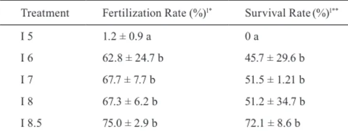
Table 1. Average values ± standard deviation of the fertilization and survival rates during the incubation of ‘curimbatá’ Prochilodus lineatus. 1Different letters in the columns indicate statistical difference in the averages by the Tukey test (P < 0.05). *Fertilization Rate was measured that according to Godinho (2007). **Survival Rate was measured by counting all larvae.
Treatment Fertilization Rate (%)1* Survival Rate(%)1**
I 5 1.2 ± 0.9 a 0 a
I 6 62.8 ± 24.7 b 45.7 ± 29.6 b
I 7 67.7 ± 7.7 b 51.5 ± 1.21 b
I 8 67.3 ± 6.2 b 51.2 ± 34.7 b
I 8.5 75.0 ± 2.9 b 72.1 ± 8.6 b
During incubation, it was noticed that the eggs incubated in treatment I 6 had average of diameter of 3.29 ± 0.05 mm,
which was significantly smaller (F=5.891; P=0.001) than
that of eggs incubated with pH values equal to or higher than 7 (3.87 ± 0.06 mm). The difference was due to the
existence of significantly smaller perivitelline space for
treatment I 6 (0.96 ± 0.09 mm) compared to treatments I 7 (1.49 ± 0.04 mm), I 8 (1.53 ± 0.07 mm) and I 8.5 (1.50
± 0.08 mm) (F=6.274; P=0.001) (Table 2). These different
egg diameters were verified during most of the embryonic stage, except for the first two hours, in which treatment
The average values for the length of P. lineatus larvae at hatching varied according to the water pH, presenting similar values when the water pH ranged from 7 to 8.5. However, total length was shorter for larvae incubated in
treatment I 6 (F=5.498; P=0.001; Table 2).
The average volume for the yolk sac was 0.55 ± 0.07
mm3 and was not significantly different (F=1.529; P=0.214)
among treatments. In the present study, hatching took place 19.5 hours after fertilization, but a delay of one hour was observed in the hatching of larvae incubated in I 6.
In the final yolk sac absorption stage, larvae cultivated
in treatment I 6 were smaller (5.66 ± 0.09 mm). There was no statistical difference in the length of larvae kept at pH values between 7 and 8.5 (5.83 ± 0.03 mm; F=0.628;
P=0.587). However, no difference was recorded in the
volume of the yolk sac of larvae kept in all treatments at the end of the incubation (0.04 ± 0.01 mm; Table 2).
Stage II. The pH values during larviculture matched the values registered in the experimental protocol, except for the most alkaline pH, which presented notorious variations during the day (Table 3).
Similar to what was observed during the incubation stage, the temperature remained between 24°C and 25°C
during the 16 days of larviculture. The dissolved oxygen values were always higher than 7.5 mg L-1, and total ammonia and nitrite were not higher than 0.3 mg L-1 and 0.01 mg L-1,respectively. The alkalinity was 20 mg CaCO
3 L-1 and the hardness was 30 mg CaCO
3 L
-1. The adjustment of tank water to alkaline pH with NaOH increased Na+ levels from 0.98 to 0.315 mEq L-1 in treatments L 7-8 and L 8-8 and 0.821 mEq L-1 in treatment L 8.5-8.5 (F=6.032;
P=0.001). For the others treatments, the Na+ levels were
similar (F=1.304; P=0.217) to the initial larviculture
level.
Higher survival rates were observed in larvae incubated
at pH 7 (Table 3). The final length measurements showed
that larvae cultivated in treatment L 8.5-7 had the highest
length values (F=3.041; P=0.029) while treatments L 6-6
and L 8-8 had the lowest values (Table 3; Fig. 2). The two treatments incubated at pH 6.0 (L6-6 and L6-7) and the L8-8
treatment had the lowest final biomass values (F=4.339; P=0.018), and the highest value was observed in treatment L 8.5-7 (F=4.538; P=0.014). The final weight of the larvae
did not show any statistical differences between treatments
(F=1.583; P=0.091). The results showed that, in general, the
larvae of the treatments carried through larviculture at pH 7 showed the best performance (Table 3).
Table 2. Average values ± standard deviation of the Diameter of the egg, Perivitelline Space (P.S), Length of the larvae (initial
and final), Volume of the Yolk Sac (initial and final) during the incubation of Prochilodus lineatus at the different pH values
tested. 1Different letters in the same column indicate statistical differences in the averages by the Tukey test (P < 0.05).
pH
Diameter (mm) 1 Length (mm) 1 Volume of the yolk sac (mm3) 1
Egg P.S Initial Final Initial Final
I 5 3.22±0.09a 0.92±0.07a - - -
-I 6 3.29±0.05a 0.96±0.09a 4.33±0.03a 5.66±0.09a 0.54±0.02 0.04±0.01
I 7 3.87±0.06b 1.49±0.04b 4.48±0.03b 5.84±0.07b 0.56±0.04 0.04±0.01
I 8 3.90±0.10b 1.53±0.07b 4.53±0.03b 5.85±0.08b 0.54±0.07 0.04±0.01
I 8.5 3.88±0.04b 1.50±0.08b 4.50±0.05b 5.81±0.10b 0.57±0.03 0.04±0.01
Table 3. Average values ± standard deviation of water pH, weight, length, biomass, and survival at the end of 16 days of larviculture for Prochilodus lineatus in the different treatments. 1Different letters in the same column indicate statistical differences in the averages for the Tukey test (P < 0.05).
Treatment pH Final weight (mg) 1 Final length (mm) 1 Survival (%)1 Final biomass (mg)1
L 6-6 6.0 ± 0.2 4.4 ± 1.9 8.19 ± 0.5a 56 ± 5a 541.2 ± 132.9a
L 6-7 7.0 ± 0.1 3.7 ± 1.4 8.56 ± 1.1ab 69 ± 4b 562.0± 124.5a
L 7-6 6.0 ± 0.2 4.5 ± 1.0 8.75 ± 0.6ab 98 ± 3c 972.0± 118.9bc
L 7-7 7.0 ± 0.1 4.2 ± 0.9 8.45 ± 0.8ab 90 ± 9c 831.6± 87.4b
L 7-8 8.0± 0.1 4.0 ± 0.7 8.25 ± 0.8ab 92 ± 12c 808.0± 171.6ab
L 8-7 7.0 ± 0.1 4.6 ± 2.1 8.50 ± 1.1ab 64 ± 9ab 648.6± 316.4ab
L 8-8 8.0 ± 0.1 3.9 ± 1.3 8.16 ± 0.6a 68 ± 10ab 585.0± 138.2a
L 8.5-7 7.0 ± 0.1 5.7 ± 0.4 9.29 ± 0.8b 82 ± 10bc 1026.0± 142.2c
Fig. 2. Growth curves in total length of the Prochilodus lineatus larvae after 16 days of larviculture. The results are grouped in the different pH values used in the incubation.
Discussion
The successful incubation and larviculture of fish
requires careful management of water quality parameters, since these are not only critical for the survival of eggs and larvae, but also for obtaining high quality fry.
Several studies have identified that incubations carried
out in acidic waters, with pH lower than 5.5, may cause high mortality during this phase. Korwin-Kossakonski (1988) noticed in laboratory experiments that Cyprinus carpio larvae died after nine days in water with pH lower than 5.0. Atlantic salmon (Salmo salar) eggs, when exposed to water with pH between 4.0 and 5.5 in the eye pigmentation stage, had lowered hatching rates (Peterson et al., 1980). Swarts et al. (1978) have also registered lethal results for brook trout (Salvelinus fontinalis) eggs incubated at pH 4.75. The harmful effects of an acidic pH may be easily observed in the egg membranes through the emergence
of bubbles that stick to them. This makes the eggs float
on the surface of the incubator (Zaniboni-Filho, 2000). Westernhagen (1988) noticed that damage to the osmotic activity of perivitelline colloids reduced water uptake of eggs cultivated at low pH values. Together with the lower water uptake, the ability of resisting to deformations by mechanical actions decreases (Eddy & Talbot, 1983). Eddy & Talbot (1983) found an inhibition of water uptake by eggs of Atlantic salmon and rainbow trout (Oncorhynchus mykiss) that had recently been fertilized in water with a pH equal to or lower than 5.5.
In the present study, it was noticed that, one hour after fertilization, the different pH levels tested allowed for the fertilization of P. lineatus eggs. This could indicate that the
pH range that was evaluated does not significantly affect
the activity of spermatozoids and oocytes. Fertilization was also observed in eggs incubated at pH 5.0. These eggs, although withered and stuck to each other, presented
the first embryonic stages. Four hours after fertilization,
the great majority of the eggs incubated at this pH were decomposing.
In the current study, it was observed a smaller diameter of eggs incubated at pH 6.0 and observed a lower hydration of the perivitelline space compared to eggs incubated in waters with neutral to basic pH values.
The perivitelline fluid tends to allow cations such as Na+,
K+, Ca2+, Mg2+, and H+ to penetrate under normal conditions of external and internal pH, where these conditions are optimal in freshwater with neutral pH, and most detrimental in acidic media (Alderdice, 1988). Moreover, the development of embryos in acidic waters could result in a decrease of ion accumulation and, according to Westernhagen (1988), delayed overall development.
Based on the results obtained in this study, the fertilization of ‘curimbatá’ eggs may be possible in a pH ranging from 5 to 8.5. However, an embryonic development at pH 5.0 is harmful to the embryos and causes mortality.
Exposure to low pH prolongs the period between fertilization and hatching (Kelley, 1946; Westernhagen, 1988), and one of the effects of a pH lower than 5.5 is the inactivation of the enzyme chorianase, which promotes hatching. The pH of the incubation medium has a strong
influence on the activity of hatching enzymes, which
present highest activity over rainbow trout eggs at pH 8.5 (Hagenmaier, 1974), whereas this value stays between 7.5
and 8 for the Pacific salmon (Oncorhynchus keta) (Bell et
al., 1969). A low pH delayed the development of Pacific
herring (Clupea pallasii) embryos (Kelley, 1946), and a high pH (pH 10) slightly accelerated the development of zebra
fish, Danio rerio (Johansson et al., 1973). In the current
study, ‘curimbatá’ larvae incubated at pH 6.0 hatched one hour later than larvae in alkaline and neutral treatments. In addition, eggs incubated at pH 6.0 resulted in smaller larvae than those obtained from the remaining treatments.
The effect of pH on the size of recently hatched larvae has already been investigated. According to Johansson & Kihlstrom (1975), Northern pike (Esox lucius) larvae incubated at pH 4.2 were smaller than those cultivated at neutral pH, however they had larger yolk sacs, indicating a decreased use of this reserve. The delay in the absorption of the yolk sac at pH 5.0 was also observed in brook trout (Menendez, 1976). In rainbow trout larvae, a low pH affects metabolism by decreasing the heart rate, decreasing the time of bone formation, reducing size, lowering pigmentation and increasing the mortality rate (Nelson, 1982).
The use of a yolk sac reserve is necessary for the survival and production of healthy larvae. Larvae unable to use these reserves could die, particularly between the yolk
sac absorption period and first feeding, which is a critical stage for most fish species (May, 1974). In this work, no significant difference was observed in the survival rate of
‘curimbatá’ eggs incubated between pH 6 to 8.5.
Neutral and slightly alkaline pH values have been recommended as appropriate for the rearing of commercial freshwater species (Boyd, 1982; Michaels, 1988). Most
studies to date have been developed with fish from rivers
many environments present extreme pH conditions. In the Amazon area, there are marginal ponds in the rio Negro basin that are highly acidic, with pH values below 3. In
contrast, ponds subject to isolation in flooded plains in the
Pantanal (wetlands) in Mato-Grosso have been found to have pH values of 11 (Esteves, 1988). In spite of the existence of
extreme values, there are fish populations adapted to these
conditions (Zaniboni-Filho, 2000).
Lopes et al. (2001) found a significantly higher survival
rate, length and weight of silver catfish (Rhamdia quelen)
larvae that were maintained at a pH between 8.0-8.5 at the end of 21 days of rearing, compared to those kept at pH values of 5.0, 6.0 and 7.0. The growth and survival of larvae
is significantly reduced in flagfish Jordanella floridae (Craig
& Baksi, 1977) and in brook trout (Kwain & Rose, 1985) at
pH values between 4.5 and 5.5, but flagfish larvae presented
similar growth when cultivated in pH 6.0 with pH 6.8 (Craig & Baksi, 1977). According to Zaniboni-Filho et al. (2009), ‘curimbatá’ larvae presented high mortality when raised at pH values higher than 9.7.
In this study, the highest post-larvae survival values of ‘curimbatá’ were recorded after incubation at pH 7 and the longest post-larvae lengths were observed for the L8.5-7 treatment. The worst values for survival and length were registered for the L6-6 treatment.
The low survival and length values obtained at the end of larviculture in the treatments with a pH incubation of 6.0
may be a reflection of the negative effects of incubation at
this pH, which might have directly affected the development of embryos and larvae. The difference in the larval survival between the treatments with slightly acidic (L6-6, 56%), and neutral (L7-6, 98%) incubation pH seems to clearly indicate that the difference was due to the pH in which the incubation took place, with detrimental effects on incubation evident even at a slightly acidic pH. The high survival rate obtained in treatment L7-6 may indicate that P. lineatus larvae may adapt to pH 6.0 during larviculture and to neutral or alkaline
pH values, which are acknowledged as ideal fish rearing
media (Boyd, 1982; Michaels, 1988), as long as they have a neutral or alkaline pH incubation history.
Treatments L6-6 and L6-7 present statistical differences in survival rate. The considerable difference among the survival averages of L6-7 (69%) and L6-6 (56%) could indicate a light recovery of the post-larvae incubated in slightly acidic pH when they were transferred to a neutral pH during larviculture and may be explained by the capacity of larvae to capture ions that are important to form their body
structures in the first few days of life, since the assimilation
of these ions is maximized at neutral pH (Alderdice, 1988).
In the present study, significant differences were found
in the length of the larvae obtained from the different treatment groups at the end of the larviculture stage and observed certain trends: (a) The larvae with incubation pH values of 8.5 were largest at the end of the larviculture stage; (b) Comparing the growth of the larvae coming from the same incubation pH, there was a slight tendency to obtain
larger post-larvae specimens when the larviculture pH was lower, where this fact was observed for most treatments, except those where the eggs were incubated at pH 6.0.
It was observed a tendency for higher fertilization and survival rates for the I 8.5 incubation group, as well as an increased growth of the larvae during the rearing stage after that incubation, where this may have been caused by the interference of a higher waterborne Na+ concentration in this treatment, especially during the incubation stage. The amount of NaOH that was introduced in the incubators of treatment I 8.5 was necessary for the maintenance of pH values within the range established for this treatment
and was significantly higher than the amount added in
the remaining treatments, thus increasing the availability of Na+ in the most alkaline treatment. The effect of this
ion might have acquired significance due to the low
alkalinity of the water. According to Rudy & Potts (1969), salmonid embryos capture and accumulate Na+ in their
perivitelline fluid until the eye formation stage, which is an
advantage, especially when rearing takes place in waters of low alkalinity. According to Brow & Lynam (1981), the incubation of brown trout (Salmo trutta) eggs in water with a pH of 4.5 was most successful in water containing 10 mg L-1 of calcium, apart from the concentration of Na+ (0, 1 or 10 mg L-1). After incubation, they observed that the survival of fry was higher in water containing Na+ at 1 or 10 mg L-1 (Brow & Lynam, 1981).
Each species has an ideal growth range within each physiochemical parameter, where this is related to its natural history. According to Leis & Trnski(1989), larvae and adults are often totally different ecologically, and may even be considered echo-species, presenting peculiarities for habitat type, feeding, and behaviour.
The results obtained in this study agree with the environmental conditions of growth and reproduction of the ‘curimbatá’. According to Agostinho et al. (1993),
South American migratory species, among which it is find
the ‘curimbatá’, usually spawn in open waters of the main channel or tributary where the pH is usually neutral or close to neutral. The annual pH average is 7.4 ± 0.3 for the Paraná River, 7.0 ± 0.3 for the Ivinhema River (Thomaz et al., 1997), and 7.1 ± 0.2 for the Uruguay River (Larenze & Arrigoni, 1993). Once spawning has taken place, eggs
are transported by the stream towards flooded areas and
marginal ponds, where the larvae usually arrive shortly before their digestive system is totally formed (Domingues & Hayashi, 1998).
Flooded locations are seasonally submitted to harsh
modifications in their physical, chemical and hydrological
characteristics, which change the animal and vegetal communities. These changes are incorporated into the life
cycles of several species. In flood periods, when the volume
introduced in the process. This factor significantly favours
the proliferation of bacteria, phytoplankton and zooplankton
in the entire flooded environment, and it also influences pH
reduction, through the accumulation of organic matter, as well as the wide variations of this variable due to the high concentration of phytoplankton. In adjacent ponds of the Paraná River, the natural environment of ‘curimbatá’, the pH presents an annual average of 6.6 ± 0.5, varying from
5.1 to 9.1. In the temporary ponds, where it can also find
the initial stages of this species, the annual pH average is of 6.2 ± 0.5, with variations that oscillate between 4.9 and 6.8 (Thomaz et al., 1997). The data found in the present study agrees with what takes place in nature, where the incubation of eggs and larvae occur in a neutral pH and the larviculture is accomplished in environments where the pH values are lower, in spite of large variations in time.
It can be concluded that water pH directly affects the initial stages of ‘curimbatá’ growth and survival during incubation and larviculture. The best incubation pH for the eggs is neutral and alkaline, whereas acidic and neutral pH values are recommended for larviculture. It can be also
conclude that the incubation pH strongly influences the
future performance of ‘curimbatá’ larvae in larviculture.
Acknowledgments
The authors would like to thank the coworkers from
LAPAD (CCA/UFSC) for their help in field sampling and
sample sorting. This work is part of the project “Monitoring and management of the ichthyofauna at Itá hydroelectric power station” (Monitoramento e Manejo da Ictiofauna da UHE Itá), supported by TRACTEBEL ENERGIA. Bernardo Baldisserotto and Evoy Zaniboni-Filho are recipients
of National Council for Scientific and Technological
Development (CNPq) research fellowship grants.
References
Agostinho, A. A., A. E. A. de M. Vazzoler, L. C. Gomes & E. K. Okada. 1993. Estratificação espacial y comportamiento de
Prochilodus scrofa em distintas fases del ciclo de vida, em la planície de inundación del alto rio Paraná y embalse de Itaipu, Paraná, Brasil. Revue D’Hydrobiologie Tropicale, 26: 79-90. Agostinho, A. A., L. C. Gomes & F. M. Pelicice. 2007. Ecologia e
Manejo de Recursos Pesqueiros em Reservatórios do Brasil. Maringá, EDUEM.
Ahlstrom, E. H., J. L. Butler & B. Y. Sumida. 1976. Pelagic stromateoid fishes (Pisces, Perciformes) of the eastern Pacific: kinds, distributions, and early life histories and observations on five of these from the Northwest Atlantic. Bulletin of Marine Science, 26: 285-402.
Alderdice, D. F. 1988. Osmotic and ionic regulation in teleost eggs and larvae. Pp. 407–466. In: Hoar, W. S. & D. J. Randall (Eds.). Fish Physiology. Boston, Academic Press. v. 11A. Baumgartner, G., K. Nakatani, L. C. Gomes, A. Bialetzki, P.
V. Sanches & M. C. Makrakis. 2008. Fish larvae from the upper Paraná River: Do abiotic factors affect larval density? Neotropical Ichthyology, 6: 551-558.
Bell, G. R., G. E. Hoskins & J. W. Bagshaw. 1969. On the structure and enzymatic degradation of the external membrane of the salmon egg. Canadian Journal of Zoology, 47: 146-148.
Boyd, C. E. 1982. Water Quality Management for Pond Fish Culture. Amsterdam, Elsevier.
Brow, D. J. A. & S. Lynam. 1981. The effect of sodium and calcium concentrations on the hatching of eggs and the survival of yolk sac fry of brown trout, Salmo trutta L. at low pH. Journal of Fish Biology,19: 205-211.
Craig, G. R. & , W. F. Baksi. 1977. The effects of depressed pH on
flagfish reproduction, growth and survival. Water Research,
11: 621-626.
Domingues, W. M. & C. Hayashi. 1998. Estudo experimental sobre anéis diários em escamas nas fases iniciais do
desenvolvimento do curimbatá, Prochilodus lineatus
(Valenciennes, 1836) (Characiformes, Prochilodontidae).
Revista Brasileira de Biologia, 58: 609-617.
Eddy, F. B. & , C. Talbot. 1983. Formation of the perivitelline
fluid in Atlantic salmon eggs (Salmo salar) in fresh water and
in solutions of metal ions. Comparative Biochemistry and Physiology, Part C, 75: 1-4.
Esteves, F. 1988. Fundamentos de Limnologia. Rio de Janeiro, Ed. Interciência-FINEP.
Ferreira, A. A., A. P. O. Nuñer & J. R. Esquivel. 2001. Influencia do pH sobre ovos e larvas de jundiá, Rhamdia quelen
(Osteichthyes, Siluriformes). Acta Scientiarum, 23: 477-481. Godinho, H. P. 2007. Estratégias reprodutivas de peixes
aplicadas à aqüicultura: bases para o desenvolvimento de tecnologia de produção. Revista Brasileira de Reprodução Animal, 31: 351-360.
Hagenmaier, H. E. 1974. The hatching process in fish embryos IV. The enzymological properties of a highly purified enzyme (chorionase from the hatching fluid of the rainbow trout.
Salmo gairdneri Rich.). Comparative Biochemistry and Physiology, 49: 313-324.
Handeland, S. O., A. K. Imsland & S. Stefansson. 2008. The effect of temperature and fish size on growth, feed intake, food conversion efficiency and stomach evacuation rate of Atlantic salmon post-smolts. Aquaculture, 283: 36-42. Johansson, N., J. E. Kihlstrom & A. Wahlberg. 1973. Low pH
values shown to affect developing fish eggs (Brachydanio
rerio Ham-Buch.). Ambio, 2: 42-43.
Johansson, N. & J. E. Kihlstrom. 1975. Pikes (Esox lucius L.) shown to be affected by low pH values during first weeks after hatching. Environmental Research, 9: 12-17.
Kelley, A. M. 1946. Effect of abnormal carbondioxide tension on development of herring eggs. Journal of the Fisheries
Research Board of Canada,6: 435-440.
Kleerekoper, H. 1990. Introdução ao Estudo da Limnologia. Porto Alegre, Ed. da UFRGS.
Korwin-Kossakonski, M. 1988. Larval development of carp,
Cyprinus carpio L. in acid water. Journal of Fish Biology, 32: 17-26.
Kugel, B. & R. H. Peterson. 1989. Perivitelline fluid pH of rainbow trout (Oncorhynchus mykiss) eggs in relation to ambient pH. Canadian Journal of Fisheries and Aquatic Sciences, 46: 2070-2073.
Larenze, G. & S. Arrigoni. 1993. II Seminario de Calidad de Aguas y Control de la Contaminación del Rio Uruguay. Colon, Publicación de la Comisión Administrativa del Rio Uruguay. Leis, J. M. & T. Trnski. 1989. The Larvae of Indo-Pacific
Shorefishes. Honolulu, University of Hawaii Press.
Lin, Q., L. Junyi, Y. Gao, L. Shen, J. Cai & J. Luo, 2006. The effect of temperature on gonad, embryonic development and survival rate of juvenile seahorses, Hyppocampus kuda
Bleeker. Aquaculture, 254: 701-713.
Lopes, J. M., L. V. M. Silva & B. Baldisserotto. 2001. Survival and growth of silver catfish larvae exposed to different water pH.Aquaculture International, 9: 73-80.
May, R. C. 1974. Larval mortality in marine fishes and the critical period concept. Pp. 3-19. In: Blaxter, J. H. S. (Ed.). The Early Life History of Fish. Berlin and New York, Springer-Verlag. Menendez, R. 1976. Chorionic effects of reduced pH on brook
trout. Journal of the Fisheries Research Board of Canada, 33: 118-123.
Michaels, V. K. 1988. Carp Farming. England, Fishing News Brooks.
Nelson, J. A. 1982. Physiological observations on developing rainbow trout, Salmo gairdneri (Richardson), exposed to low pH and varied calcium ion. Journal of Fish Biology, 20: 359-372.
Parra, J. E. G. & B. Baldisserotto. 2007. Effect of water pH and hardness on survival and growth of freshwater teleosts. Pp.135-150. In: Baldisserotto, B., J. M. Mancera & B. G Kapoor (Eds.). Fish Osmorregulation. Enfield, Science Publishers.
Pavlov, D. A. & E. Moksness. 1995. Development of wolfish eggs at different temperature regimes. Aquaculture International, 3: 315-335.
Peterson, R. H., P. G. Daye & J. L. Metcalfe. 1980. Inhibition of Atlantic salmon (Salmo salar) hatching at low pH. Canadian Journal of Fisheries and Aquatic Sciences,37: 770-774. Rudy, P. P. & W. T. W. Potts. 1969. Sodium balance in the eggs
of Atlantic salmon, Salmo salar. Journal of Experimental Biology, 50: 239-246.
Smolders, A. J. P., G. Van Der Velde, J. G. M. Roelfofs & M. A. Guerrero. 2000. El Niño caused collapse of the Sábalo fishery (Prochilodus lineatus, Pisces: Prochilodontidae) in South American river. Naturwissenschaften, 87: 30-32.
Swarts, F. A., W. A. Dunson & J. E. Wright. 1978. Genetic and environmental factors involved in increased resistance of brook trout to sulfuric acid solutions and mine acid polluted waters. Transactions of the American Fisheries Society, 107: 651-677.
Thomaz, S. M., M. C. Roberto & L. M. Bini. 1997. Caracterização limnológica dos ambientes aquáticos e influência dos níveis fluviométricos. Pp.73-102. In: Vazzoler, A. E. A. de M., A. A. Agostinho & N. H. Segatti (Eds.). A Planície de Inundação do Alto Rio Paraná. Maringá, EDUEM.
Townsend, C. R. & B. Baldisserotto. 2001. Survival of silver catfish fingerlings exposed to acute changes of water pH and hardness. Aquaculture International, 9: 413-419.
Westernhagen, H. V. 1988. Effects of pollutants on fish eggs and larvae. Pp. 407-466. In: Hoar, W. S. & D. J Randall (Eds.). Fish Physiology. Boston, Academic Press. v. 11A.
Zaniboni-Filho, E. & N. D. Barbosa, de C. 1996. Priming hormone administration to induce spawning of Brazilian migratory fish. Revista Brasileira de Biologia, 56: 655-659.
Zaniboni-Filho, E. 2000. Larvicultura de Peixes de Água Doce. Informe Agropecuario, 21: 69-77.
Zaniboni-Filho, E. & U. H. Schulz. 2003. Migratory Fishes of the Uruguay River. Pp.157-194. In: Carolsfeld, J., B. Harvey, C. Ross & A. Baer (Eds.). Migratory Fishes of South America. Victoria, World Bank.
Zaniboni-Filho, E., A. P. O. Nuñer, D. A. Reynalte-Tataje & R.
L. Serafini. 2009. Water pH and Prochilodus lineatus larvae
survival. Fish Physiology and Biochemistry, 35: 151-155.