doi:10.1017/S096719940600373X Printed in the United Kingdom
Structural and ultrastructural analysis of embryonic
development of
Prochilodus lineatus
(Valenciennes, 1836)
(Characiforme; Prochilodontidae)
Alexandre Ninhaus-Silveira
1, Fausto Foresti
2and Alexandre de Azevedo
2Universidade Estadual Paulista (UNESP), Ilha Solteira and Botucatu, S˜ao Paulo, Brazil
Date submitted: 08.11.05. Date accepted: 22.12.05
Summary
This survey was performed to characterize the embryogenesis of Prochilodus lineatus. Seven stages of embryo development were identified – zygote, cleavage, blastula, gastrula, segmentation, larval and hatching – after a period of incubation of 22 h (24◦C) or 14 h (28◦C). The following cleavage pattern
was identified: the first plane was vertical (2 blastomeres); the second was vertical and perpendicular to the first (4 blastomeres); the third was vertical and parallel to the first (4×2); the fourth cleavage was vertical and parallel to the second (4×4); the fifth was vertical and parallel to the first (4×8); and the sixth cleavage was horizontal (64 blastomeres). At the blastula stage (3.0–4.0 h (24◦C); 1.66–2.0 h
(28◦C)) irregular spaces were detected and periblast structuring was initiated. At the gastrula stage (4.0–
8.0 h (24◦C); 3.0–6.0 h (28◦C)) the epiboly, convergence and cell movements, as well as the formation
of embryonic layers, had begun. The segmentation stage (10.0–15.0 h (24◦C); 7.0–10.0 h (28◦C)) was
characterized by a rudimentary formation of organs and systems (somites, optic vesicle and intestinal delimitation). The embryo at the larval stage (16.0–21.0 h (24◦C); 11.0–13.0 h (28◦C)) showed a free tail,
more than 25 somites, an optic vesicle and a ready-to-hatch larval shape. The blastomeres at cleavage stage had disorganized nuclei indicating high mitotic activity. At gastrula, the blastomeres and the periblast had euchromatic nuclei and a large number of mitochondria and vesicles. The yolk was organized into globose sacs, which were dispersed into small pieces prior to absorption.
Keywords: Characiforme, Embryo, Embryogenesis,Prochilodus lineatus, Teleost
Introduction
The ‘curimbata’,Prochilodus lineatus, a member of the family Prochilodontidae, is an iliophagous fish species with wide distribution over southeastern Brazil that undergoes reproductive migration and total spawning (Fowler, 1951). According to Corrˆea & Castro (1990), this species is recorded along the entire Paran´a– Paraguay and Paraiba river basins. It represents an
All correspondence to: A. Ninhaus-Silveira, Departamento de Biologia e Zootecnia, Universidade Estadual Paulista/Ilha Solteira, Av. Brasil, 56, Centro, Postal Box 31, CEP: 15385-000, Ilha Solteira, S˜ao Paulo, Brazil. Tel/Fax: +55 02118 3743-1285/3743-1186. e-mail: [email protected]
1Universidade Estadual Paulista (UNESP), Depto. de Biologia
e Zootecnia, Ilha Solteira, S˜ao Paulo, Brazil.
2Universidade Estadual Paulista (UNESP), Depto. de
Morfologia, Botucatu, S˜ao Paulo, Brazil.
economically and ecologically important native species of medium to large size.
Knowledge of the embryonic development of diffe-rent fish species is useful to the management of fishery resources, as well as to surveys related to fish culture, since it provides additional information about species’ life cycles. In addition, embryological analysis is helpful in studies about evolutionary relationships, heredity, developmental mechanisms and environ-mental influences over structural features of distinct organisms (Lagler, 1959).
The embryonic development of fishes is a complex phenomenon, useful for ontogeny studies, experi-mental modelling, and evaluation of environexperi-mental quality and effects of toxic substances on aquatic fauna (Floreset al., 2002), as well as for experiments onex situ species preservation.
Table 1Embryonic development ofProchilodus lineatusat 24◦C
Time (h) Stage Description
0–0.75 Zygote Cytoplasm streams towards the animal pole to form the blastodisc 1.0 Cleavage 50% with 2 cells; 36% with 4 cells; 12% with 8 cells; 2% with 16 cells 1.25 Cleavage 42% with 4 cells; 52% with 5–7 cells; 6% with 8 cells
1.5 Cleavage 2% with 4 cells; 6% with 6 cells; 46% with 8 cells; 10% with 12–14 cells; 15% with 10 cells; 24% with 16 cells
1.75 Cleavage 75% with 16 cells; 25% with 16+ cells
2.0 Cleavage 5% with 16 cells; 95% with 32 cells
2.25 Cleavage 5% with 32 cells; 75% with 64 cells
3.0 Blastula 100% morula
4.0 Blastula; Gastrula 75% morula; 25% gastrula
5.0 Gastrula 100% gastrula (25% epiboly)
6.0 Gastrula 75% gastrula (25% epiboly); 25% gastrula (50% epiboly)
7.0 Gastrula 10% gastrula (25% epiboly); 50% gastrula (50% epiboly); 40% gastrula (75% epiboly)
8.0 Gastrula 20% gastrula (50% epiboly); 80% gastrula (75% epiboly)
9.0 Gastrula 100% epiboly, neural tube
10.0 Segmentation 100% neurula
11.0 Segmentation 4 somites
12.0 Segmentation 100%, otic vesicle, 9 somites and tail attached
13.0 Segmentation 100%, Kupffer and optic vesicles, 13 somites and tail attached 14.0 Segmentation 100%, Kupffer and optic vesicles, 19 somites and tail attached 15.0 Segmentation 100%, optic and otic vesicles, 22 somites and tail attached 16.0 Larval 100%, optic and otic vesicles, 25+ somites and free tail 17.0 Larval 100%, optic and otic vesicles, 30+ somites and free tail
18.0–21.0 Larval 100%, embryo growth
22.0 Hatching 100% hatching
Table 2Embryonic development ofProchilodus lineatusat 28◦C
Time (h) Stage Description
0–0.34 Zygote Cytoplasm streams towards the animal pole to form the blastodisc
0.50 Cleavage 100% with 2 cells
0.66 Cleavage 10% with 2 cells; 90% with 4 cells
0.84 Cleavage 100% with 8 cells
1.00 Cleavage 15% with 8 cells; 85% with 16 cells
1.16 Cleavage 10% with 16 cells; 90% with 32 cells
1.34 Cleavage 100% with 64 cells
1.50 Blastula 64+ cells (morula)
1.66 Blastula 100% morula
1.84 Blastula 100% morula
2.00 Blastula 100% morula
3.00 Gastrula 100% gastrula (25% epiboly)
4.00 Gastrula 100% gastrula (50% epiboly)
5.00 Gastrula 100% gastrula (75% epiboly); early neural tube
6.00 Gastrula 10% gastrula (100% epiboly); 90% gastrula (90% epiboly) 7.00 Segmentation 35% with 4 somites; 65% neurula
8.00 Segmentation 13 somites; optic and Kupffer vesicles and tail attached 9.00 Segmentation 19 somites, optic and Kupffer vesicles and tail attached
10.00 Segmentation 24 somites, optic and otic vesicles, Kupffer vesicle missing and free tail 11.00 Larval 28 somites, optic and otic vesicles and free tail
12.00 Larval 30+ somites; embryo growth
13.00 Larval 100%, embryo growth
Figure 1Phases of the embryonic development ofProchilodus lineatus.(A), (A’) Post-fertilization without chorion; (B), (B’) 2-cell embryo; (C), (C’) 4-cell embryo; (D), (D’) 8-cell embryo; (E), (E’) 16-cell embryo; (F), (F’) 32-cell embryo; (G), (G’) 64-cell embryo; (H) morula; (I) gastrula (25% epiboly); (J) gastrula (50% epiboly); (K) gastrula (75% epiboly); (L), (L’) gastrula (90%). Scale bars represent 113.6µm.
Brazil et al. (2002), which studied egg morphology modifications just after fertilization, and that of Castellaniet al. (1994), who carried out observations of the embryonic development ofP. lineatusunder the light microscope.
Therefore, given on the ecological and economic value of Prochilodus lineatus, the present work was performed to analyse the morphological events during the embryonic development of this species at the structural and ultrastructural levels.
Materials and methods
Adult individuals of Prochilodus lineatus from the broodstock at the Aquaculture Division of the Faculdade de Medicina Veterin´aria e Zootecnia, UNESP, Botucatu, S˜ao Paulo, Brazil were used to obtain embryos.
Figure 2(A), (A’) Neurula; (B) in the presence of about 13 somites, optic vesicle, attached tail; (C) embryo bearing nearly 19 somites, optic vesicle, Kupffer’s vesicle and attached tail; (D) 24 somites, presence of optic and otic vesicles, absence of Kupffer’s vesicle and free tail. s, somite; arrow, neural keel; vk, Kupffer’s vesicle; op, optic vesicle. Scale bars represent: (A), (A’) 55µm; (B) 57.1µm; (C) 70.4µm; (D) 76.3µm.
able to reproduce naturally and hormonal induction is necessary. Breeders were stimulated with carp pituitary extract by inoculating 3 mature females (3 years old) with 0.5 mg and 5 mg/kg body weight, respectively, at an interval of 10 h, and 6 mature males (2 years old) with 1 mg/kg body weight at the time of the second female inoculation. The extrusion of eggs and sperm was performed about 6 h after the last induction.
The dry method was employed for the fertilization process, in which the eggs are mixed with the sperm avoiding contact with water. After that, water was added in order to activate spermatozoa and allow egg hydration. The excess semen was rinsed off and the eggs were incubated in 200 l vertical incubators. To verify a putative effect of water temperature on embryo development, the eggs were divided into two groups incubated at different temperatures (24◦C and 28◦C).
The incubators were connected to a closed heated water system coupled with a thermostat.
Embryonic development
To evaluate the possible temporal morphological vari-ation of P. lineatusembryos, about 200 embryos were collected at different development stages, defining the moment of fertilization as time zero. The first samples were collected within intervals of 15 and 10 min for eggs incubated at 24 and 28◦C, respectively, until 2 h of
em-bryonic development, while subsequent samples were taken at intervals of 1 h until the point of hatching. The embryo samples were fixed in a solution of 2% glutaral-dehyde, 2% paraformaldehyde diluted in sodium phos-phate buffer 0.1 M, pH 7.3, for 24 h prior to analyses.
Stuctural and ultrastructural analysis
Twenty representative individuals from each develop-ment stage were carefully selected and embedded in glycol methacrylate. These samples were submitted to microtomy to obtain serial transverse and longitudinal cuts of from 3 to 5µm. After that, they were stained
with Harries haematoxylin–eosin or toluidine blue and analysed and photographed using a Zeiss Axiophot photomicroscope.
The embryos were postfixed in 1% osmium tetroxide for 2 h, counterstained with an aqueous solution of 0.5% uranyl acetate, dehydrated with acetone and embedded in epoxy resin for analysis by transmission electron microscopy (TEM). The ultrafine sections were caught on a copper net, counterstained with uranyl acetate (Watson, 1958), washed in 50% alcohol and re-counterstained in lead citrate (Reynolds, 1963). The material was analysed and electromicrographed using a Philips CM100 transmission electron microscope.
For analyses by scanning electron microscopy (SEM), the embryos, prefixed in 2.5% glutaraldehyde, were transferred to a 13 mm coverslip, embedded with 1% poly-L-lysine, postfixed in 0.5% osmium tetroxide, dehydrated with ethanol and dried in a critical-point dryer (Balzers CPD-20). The samples were covered with a 10 mm gold pellicle in a Balzers Metalizer MED-010 and observed and electromicrographed using a Philips 515 scanning electron microscope.
Results
Embryogenesis
The duration of embryonic development inProchilodus lineatus, from fertilization until hatching, has been shown to be dependent on the water temperature. At 24◦C, the incubation period was 22 h, and at 28◦C, it
was 14 h. The following stages were identified in the embryonic development ofP. lineatusafter fertilization: zygote, cleavage, blastula, gastrula, segmentation, larval and hatching (Tables 1, 2; Figs. 1, 2, 3).
A higher heterogeneity in embryo development was observed at 24◦C, i.e. embryos at different stages of
embryogenesis were detected at the same time, espe-cially at the beginning of cleavage (1–1.5 h) (Fig. 4A). Furthermore, embryos displaying 5, 7, 10, 12 or 15 blastomeres were also found (Table 1; Fig. 4A, B). At 28◦C, embryo development was homogeneous, despite
slight variations during the first cleavage phases (0.66– 1.66 h) (Table 2; Fig. 4C,D).
1: Zygote stage (0–0.75 h (24◦C); 0–0.34 h (28◦C))
After fertilization, hydration of the eggs could be observed by the increase in the perivitelline space,
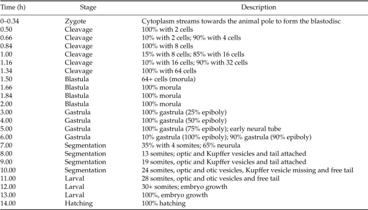
Figure 3 (A) 30+ somites, growing larva; (B) pre-hatchery embryo; (C) hatched embryo. Scale bars represent: (A) 63.3µm; (B) 75.6µm; (C) 69.4µm.
pronuclear fusion, and cytoplasm reorganization with the establishment of vegetal and animal poles (Fig. 5A– C). The animal pole was composed of active cytoplasm and a nucleus, allowingin vivoand light microscopic identification, since it is slightly transparent. On the other hand, the vegetal pole was denser at in vivo observation and weakly stained in total preparations (Fig. 5C), being composed of yolk vesicles (Fig. 6A– C). Moreover, a thin layer of cytoplasm involving the whole yolk was observed, comprising several central alveoli reminiscent of the cortical reaction during fertilization (Fig. 6A).
2: Cleavage stage (1.0–2.25 h (24◦C); 0.50–1.34 h (28◦C))
0% 10% 20% 30% 40% 50% 60% 70% 80% 90% 100%
0 0,16 0,34 0,5 0,66 0,84 1,0 1,16 1,34
Time (Hours)
Embryos/Embryonary Stage at
28 ° C 0% 10% 20% 30% 40% 50% 60% 70% 80% 90% 100%
3 4 5 6 7 8 9 10
Time (Hours)
Embryos/Embryonary Stage at
24
°
C
s/cl 2bl 4bl 5bl 8bl 10bl 12-14bl 16bl 32bl 64bl
Morula Gastrula 25% epi 50% epi 75% epi 90% epi 100% epi
0% 10% 20% 30% 40% 50% 60% 70% 80% 90% 100%
0 0,25 0,5 0,75 1 1,25 1,5 1,75 2 2,25
Time (Hours)
Embryos/Embryonary Stage at
24 ° C 0% 10% 20% 30% 40% 50% 60% 70% 80% 90% 100%
1,5 1,66 1,84 2 3 4 5 6
Time (Hours)
Embryos/Embryonary Stage at
28 ° C A D C B
Figure 4Analysis of the embryonic development ofProchilodus lineatusunder two temperature conditions (24◦C and 28◦C). (A), (B) Segmentation period. (C), (D) Morphogenesis period. s/s, not segmented; bl, blastomeres; epi, epiboly.
Figure 5(A) Fertilized and non-hydrated egg (×129); (B), (C) hydrated egg, showing well-defined animal and vegetal poles (×55). arrowhead, chorion;∗, perivitelline space; arrow, animal pole; v, yolk. Scale bars represent: (A) 77.5µm; (B), (C) 181.8µm.
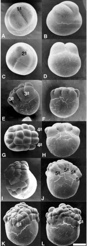
perpendicular to the first, giving rise to 4 blastomeres; the third was vertical and parallel to the first, giving rise to 8 blastomeres displaying a 4×2 arrangement; the fourth was vertical and parallel to the second, originating 16 blastomeres in a 4×4 formation; the fifth plane was vertical and parallel to the first cleavage,
originating 32 blastomeres in a 4×8 formation; and the sixth cleavage plane was horizontal, giving rise to two cell layers comprising 64 blastomeres (Fig. 8A–H).
Figure 6Analysis under a light microscope of embryos ofProchilodus lineatus,stained with basic toluidine blue. (A) 0.25 h after fertilization, showing the yolk cytoplasmic layer; (B) cleavage phase (1.5 h), revealing the penetration of yolk globules into blastomeres; (C) detail of the formation of the yolk syncytial layer in an embryo at the blastula stage; (D) 4.25 h of development (gastrula stage), characterized by the presence of blastomeres with euchromatic nuclei, yolk syncytial layer and high mitotic activity (MO); (E) embryo section at 50% of epiboly, stained with basic toluidine blue; (F) embryo section at 90% of epiboly, stained with H&E. gv, yolk globules; ycl, yolk cytoplasmic layer; b, blastomere; bl, blastoderm; ysl, yolk syncytial layer; n, nucleus; gv, yolk globules. Scale bars represent: (A) 11.5µm; (B) 17.5µm; (C) 16.7µm; (D) 3.6µm; (E) 80µm; (F) 5.7µm.
after the fourth cleavage, blastomeres of distinct sizes can be observed (Fig. 8A–H).
No distinctive layer was observed between the blastoderm and yolk. It was verified that yolk globules penetrate into blastomeres in a fragmented way, probably to facilitate their absorption by cells (Fig. 7C). Analyses under the light microscope showed that, during the cleavage stage, individualized nuclei were
absent (Fig. 6C) while TEM showed blastomeres with a large number of mitochondria, euchromatic nuclei and free ribosomes (Fig. 7B,C), indicating a high cell metabolism, typical of high mitotic activity.
3: Blastula stage (3.0–4.0 h (24◦C); 1.66–2.0 h (28◦C))
Figure 7Analysis under an electron microscope of embryos ofProchilodus lineatus. (A) Embryo blastomeres at cleavage stage, showing euchromatic nucleus and a large number of yolk vesicles in the cytoplasm (TEM); (B) Detail of yolk globules (SEM); (C) Embryos ofP. lineatusat the gastrula stage showing the periblast with euchromatic nucleus, cytoplasm with several vesicles, mitochondria and some yolk granules (TEM); (D) ultrastructure (TEM) showing irregular nuclei of the yolk syncytial layer and subjacent yolk globules. n, euchromatic nucleus; gv, yolk globules; b, blastomeres; ysl, yolk syncytial layer. Scale bars represent: (A) 6.1µm; (B) 20.4µm; (C), 3.8µm; (D) 5.7µm.
underwent divisions, but the cleavage planes were undetermined. As the number of cell increased, the blastoderm changed into a half-moon shape.
The main characteristics of this stage are the irregular spaces among blastomeres (blastocoele) and the beginning of the formation of a periblast or yolk syncytial layer (Fig. 7D,E). At the end of this stage, the first epiboly movements could be identified.
4: Gastrula stage (4.0–8.0 h (24◦C); 3.0–6.0 h (28◦C))
The gastrula stage was characterized by epiboly movement and the occurrence of morphogenetic movements of convergence and cell migration that give rise to the first layers and to the head–tail and latero-lateral embryonic axes.
The epiboly movement started after 4 h at 24◦C and
after 3 h at 28◦C. It was observed as a fringe, formed by
the yolk syncytial layer, across the blastoderm border, from its formation to the closure of the blastopore (Fig. 6C, E, F). The morphogenetic movements of convergence and cell migration began at the border of blastoderm, at about 50% of epiboly (Fig. 6E),
originating the germ ring and the embryonic shield and culminating with the formation of the two embryonic layers, the epiblast and hypoblast.
The epiboly movement goes on alongside the closure of the yolk plug by the yolk syncytial layer, which is delimited by the blastopore (Fig. 6F), following the total recovery of the blastoderm plug.
Ultrastructural analyses demonstrated that the blastomere cytoplasm contains a large number of mitochondria and vesicles, several filled with yolk material. These yolk granules, prior to absorption by blastoderm cells, are fragmented at the periblast region. The blastomeres and periblast nuclei were euchromatic (uncondensed), indicating high metabolic activity (Fig. 7C,D).
5: Segmentation and organogenesis stage (10.0–15.0 h (24◦C); 7.0–10.0 h (28◦C))
Figure 8 Observation of the first six cleavage planes in embryos ofProchilodus lineatusunder a scanning electronic microscope. Scale bars represent 263.1µm.
the initial delimitation of the intestines, were observed, leading to the subsequent growth and elongation of the embryo, particularly along the head–tail axis.
After 10 h of development at 24◦C and 7 h at 28◦C,
it was possible to identify the neural keel, the neural plate, the mesendoderm notochord (Fig. 9A) and Kupffer’s vesicle at the tail region (Fig. 9B), as well as segmented somites in some embryos (28◦C) (Fig. 2A).
In this phase, the formation of neural tube is initiated (Fig. 10E) and as long as its components show differential growth, it is possible to identify the prosencephalon, mesencephalon and rhombenceph-alon regions (Figs. 2B, 9A).
After 12 h of development at 24◦C and 8 h at 28◦C,
the embryos already contained an optic vesicle and several somites (Fig. 9D, F). After 15 h at 28◦C and
10 h at 24◦C, the otic vesicle was present, as well as
a complete neural tube (Fig. 9C, D), a rudimentary digestive system and free tail; in addition Kupffer’s vesicle was absent (Fig. 2D).
6: Larval stage (16.0–21.0 h (24◦C); 11.0–13.0 h (28◦C))
A free tail, the presence of more than 25 pairs of somites and a ready-to-hatch larval shape, characterized the embryos at the larval stage. The embryos showed a well-developed optic calyx, crystalline lens and optic vesicle (Fig. 10A, D). The notochord extended from the cephalic region to the tail (Fig. 10E). The somites showed the beginning of the myogenesis process for the formation of muscles (Fig. 10B,C) and the posterior primitive intestine was well defined (Fig. 10E). Another feature of this stage is the occurrence of spasmodic movements, which increased as embryonic development proceeded.
7: Hatching (22.0–23.0 h (24◦C); 13.0–14.0 h (28◦C))
In the hatching phase the larvae demonstrated vigorous swimming movements, important for chorion rupture. Full larval hatching was observed after nearly 23 and 14 h of development at 24 and 28◦C, respectively
(Tables 1, 2).
Discussion
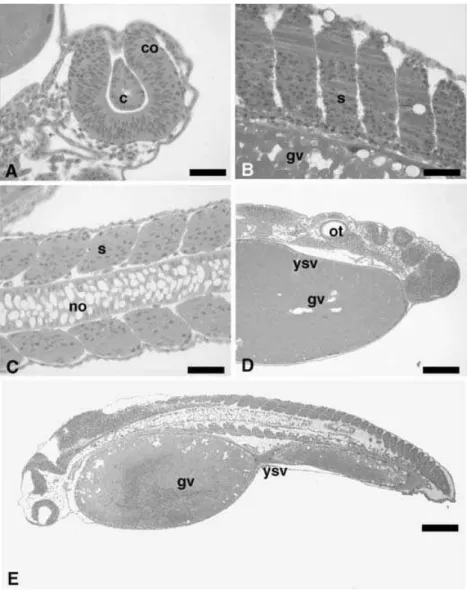
Figure 9Details under a light microscope of embryos ofProchilodus lineatusat the segmentation stage. (A) Section showing the notochord, the neural keel, the mesendoderm and the neural plate (H&E); (B) detail of the structure of Kupffer’s vesicle; (C) longitudinal section of the optic vesicle (H&E); (D) longitudinal section of somites (toluidine blue); (E) transverse section, detailing the notochord, somites and the neural tube (H&E); (F) longitudinal section detailing the presence of the optic vesicle (H&E). gv, yolk globules; ysl, yolk syncytial layer; no, notochord; ot, optic vesicle; tn, neural tube; s, somite; me, mesendoderm; pn, neural plate; sn, neural keel; vk, Kupffer’s vesicle. Scale bars represent: (A) 7.5µm; (B) 7.7µm; (C) 9.1µm; (D) 14.6µm;
(E) 22.5µm; (F) 23.4µm.
Fat droplets inside the yolk sac were absent in P. lineatus, similar to what has been observed in other Characiformes, such as Brycon orbignyanus (Ganeco, 2003) andBrycon insignis(Andrade-Talmelliet al., 2001). The morphological events identified during the embryogenesis of P. lineatus, as well as the short duration of embryonic development, were similar to those reported in other teleosts (Ganeco, 2003; Flores et al., 2002; Andrade-Talmelliet al., 2001; Cardosoet al., 1995; Kimmelet al., 1995; Ribeiroet al., 1995).
The pattern of egg segmentation in vertebrates depends on the amount and distribution of yolk and its proportion in relation to the cytoplasm that composes the blastodisc (Gilbert, 1991). The cleavage of P. lineatusfollows a meroblastic or partial pattern, as commonly observed in most teleosts (Lagleret al., 1977, Leme dos Santos & Azoubel, 1996). The
arrangement of blastomeres (4×2; 4×4; 4×8) at the early cleavage stages is similar to that reported in Catostomus commersoni (Long & Ballard, 1976), Danio rerio (Kimmel et al., 1995) and Oreochromis niloticus (Morrisonet al., 2001), but it differs from those observed inRhamdia sapo(Matkoviket al., 1985),Alosa sapidissima (Shardo, 1995) andBrycon orbignyanus(Ganeco, 2003).
During the cleavage of fish embryos the number of cells increases while their size decreases, a feature previously reported by Castellane et al. (1994) in P. lineatusand also observed in the present study.
Figure 10Longitudinal sections of embryos ofP. lineatusat the larval stage, stained with H&E. (A) Detail of the optic calyx and the crystalline lens; (B) somites at myogenesis; (C) details of somites and notochord; (D) details of the optic vesicle and the regions comprising the prosencephalon, mesencephalon and rhombencephalon; (E) general details of the embryo, revealing a primitive gut. gv, yolk globules; ysl, yolk syncytial layer; no, notochord; co, optic calyx; c, crystalline lens; s, somite; ot, optic vesicle; mes, mesencephalon; rom, rhombencephalon; pro, prosencephalon; ip, rudimentary intestine. Scale bars represent: (A) 18.2µm; (B) 39.1µm; (C) 18.1µm; (D) 51.5µm; (E) 130.2µm.
under optimal conditions, there is asynchrony of embryonic development. Hisaoka & Firlit (1960) reported that, in zebrafish, the mitotic divisions are synchronous until 32 cells but become asynchronous at 64 cells. Arezonet al.(2002) reported that embryos ofCynolebias melanotaeniadevelop faster at 25◦C, but
severe abnormalities occur, in contrast to what is detected at lower temperatures (20 and 16◦C).
In the present experiment with P. lineatus, despite the utilization of young breeders (2 years old) and a constant water temperature, incubation at 24◦C led
to a higher asynchrony of embryonic development and a variation in blastomere division, in some cases, following a non-geometric progression after the third and fourth planes. At 28◦C this variability was reduced
and nearly restricted to the cleavage stage. In addition, cleavage progression was geometric, at least until the first 64 cells. These data corroborate the observations of previous reports and stress the important role of incubation temperature in the embryonic development of fish species.
Hisaoka & Firlit (1960) suggested that the proto-plasmic movement between the yolk and blastomeres ceases after the 64-cell stage. This fact could be related to the beginning of periblast formation, which occurs at the beginning of the blastula stage and morula stage inP. lineatus.
(Kimmel & Law, 1985; Trinkaus, 1984; Kimmel et al., 1995; Ganeco, 2003) in other teleost species. How-ever, in some species, such as Oreochromis niloticus (Morrison, 2001), no characteristic cavity was identi-fied, whereas others, such asHippocampus reidi(Silveira, 2001) and trout (Lagler, 1977), a typical blastocoele cavity was seen between the blastoderm and the periblast.
The gastrula stage begins with the first epiboly movements (Leme dos Santos & Azoubel, 1996) and is completed by closure of the blastopore by the blastoderm and the formation of a tail button (Kimmel et al., 1995). These observations are in accordance with what was detected in P. lineatus and also reported by Ganeco (2003) inBrycon orbignyanus. On the other hand, in Oreochromis niloticus, Morrison et al. (2001) reported that, due to the size of yolk, the embryo is not able to extend over the entire vegetal pole and, thus, rudimentary organogenesis (somite segmentation) starts before the epiboly movement is finished.
In Prochilodus lineatus, the formation of the em-bryonic shield is visible when the blastoderm involves 50% of the yolk sphere, similar to previous observations in other species (Kimmel, 1995; Firlit & Hisaoka, 1960; Ganeco, 2003).
According to Hisaoka & Firlit (1960), Kupffer’s vesicle represents a remnant structure of the archenteron that is located over the periblast and below the notochord. In Oncorhynchus keta(Mahon & Hoar, 1956 cited in Hisaoka & Firlit, 1960) Kupffer’s vesicle is described as an oblique and elongated cavity, with walls of columnar epithelium, which is separated from the periblast by a layer of endoderm cells. The histology of this structure in P. lineatus was similar to that reported inO. keta.
Following Kimmel (1995), the neurula phase was included in the segmentation stage. Brummett & Dumont (1978) and Morrison (2001) identified Kupffer’s vesicle in the early phases of the segmenta-tion stage. In the present work, this vesicle was ob-served at the phase of 13 somites and disappeared after the 24-somite stage; its function remains unknown. However, Brummet & Dumont (1978) hypothesized that it could have a digestive function, favorable to yolk resorption, since several ciliated cells were observed inside Kupffer’s vesicle and the intestines ofFundulus heteroclitus.
According to Silveira (2001), the ectoderm on the notochord is transformed into a neural plate, which becomes centrally depressed giving rise to the neural keel, which following its posterior enclosure by the fusion of neural filaments, originates the neural tube. As observed in P. lineatus, the prosenceph-alon, mesencephalon and rhombencephalon regions developed from the posterior region of the neural tube, corroborating the description of the embryonic
development of Brycon orbignyanus carried out by Ganeco (2003).
Acknowledgements
This work was supported by Aquaculture Division of the Faculdade de Medicina Veterin´aria e Zootecnia, UNESP, Botucatu, S˜ao Paulo, Brazil, which provided the fish and the facilities used in this study, and also by CAPES (Coordenac¸˜ao de Aperfeic¸oamento de Pessoal de N´ıvel Superior) and CNPq (Conselho Nacional de Pesquisa).
References
Andrade-Talmelli, E.F., Kavamoto, E.T., Romagosa, E. & Fenerich-Verani, N. (2001). Embryonic development of the ‘piabanha’, Brycon insignis, (Steindachner, 1876) (Pisces; Characidae).Bol. Inst. Pesca27, 21–8.
Arezon, A., Lemos, C.A. & Bohrer, M.B.C. (2002). The influ-ence of temperature on the embryonic development of the annual fish,Cynolebias melanotaenia(Cyprinodontiformes; Rivulidae).Braz. J. Biol.62(4b), 1–8.
Brasil, D.F., Nakaghi, L.S.O., Leme dos Santos, H.S., Quagio-Grassiotto, I. & Foresti, F. (2002). Estudo morfol ´ogico dos primeiros momentos da fertilizac¸˜ao em curim-bat´a Prochilodus lineatus (Valenciennes, 1836). [online], CIVA2002. Available from: http://www.civa2002.org/, 733–47.
Brummett, A.R. & Dumont, J.N. (1978). Kupffer´s vesicle in Fundulus heteroclitus: a scanning and transmission electron microscope study.Tissue Cell10, 11–22.
Cardoso, E.L., Alves, M.S.D., Ferreira, R.M.A. & Godinho, H.P. (1995). Embryogenesis of the neotropical freshwater siluriformPseudoplatystoma coruscans.Aquat. Living Res.8, 343–6.
Castellani, L.R., Tse, H.G., Leme dos Santos, H.S. & Faria, R.H.S. (1994). Desenvolvimento embrion´ario do curimbat´a, Prochilodus lineatus (Valenciennes, 1836) (Cypriniformes, Prochilodontidae).Rev. Bras. Ciˆenc. Morfol.11, 99–105. Corrˆea e Castro, R.M. (1990). Revis˜ao taxon ˆomica da fam´ılia
Prochilodontidae (Ostariophysi: Characiformes). Thesis, S˜ao Paulo, Instituto de Biociˆencias, Universidade de S˜ao Paulo.
Eckmann, R. (1984). Induced reproduction in Brycon cf. erythropterus.Aquaculture38, 370–82.
Flores, J.C.B., Araiza, M.A.F. & Valle, M.R.G. (2002). Desarrollo embrionario de Ctenopharyngodon idellus (Carpa herb´ıvora). [online], CIVA2002. Available from: http://www.civa2002.org, 792–7.
Fowler, H.W. (1954). Os peixes de ´agua doce do Brasil.Arq. Zool.2, 1–400.
Ganeco, L.N. (2003). An´alise dos ovos de piracanjuba,Brycon orbignyanos (Valenciennes, 1849), durante a fertilizac¸˜ao e o desenvolvimento embrion´ario, sob condic¸ ˜oes de reproduc¸˜ao induzida. Masters degree, Universidade Estadual Paulista, Jaboticabal.
Godinho, H.M., Romagosa, E., Kavamoto, E.T., Cestarolli, M., Ranzani, M.J.T. & Narahara, M.Y. (1988). Estudos morfol ´ogicos e reproduc¸˜ao induzida do curimbat´a, Prochilodus scrofa, Steindacher, 1881, mantido em condic¸ ˜oes de cultivo experimental. In Anais do 6◦ Simp´osio Latino-Americano e do 5◦Simp´osio Brasileiro de Aquicultura, pp. 346– 54. Florian ´opolis: ABRAq.
Godoy, M.P. (1975). Peixes do Brasil, Subordem Characoidei, Bacia do Rio Mogi Guassu, 1st edn. Piracicaba: Franciscana. Hisaoka, K.K. & Firlit, C.F. (1960). Further studies on the
embryonic development of the zebrafish,Brachidanio rerio (Hamilton-Buchanan).J. Morphol.107, 205–25.
Kimmel, C.B. & Law, R.D. (1985). Cell lineage of zebrafish blastomeres. II. Formation of the yolk syncytial layer.Dev. Biol.108, 86–93.
Kimmel, C.B., Ballard, W.W., Kimmel, S.R. & Ullmann, B. (1995). Stages of embryonic development of zebrafish.Dev. Dyn.203, 253–310.
Lagler, K.F. (1959). Freshwater Fishery Biology, 2nd edn. Dubuque: W.M.C. Brown.
Lagler, K.F., Bardach, J.E., Miller, R.R. & Passino, D.R.M. (1977).Icthyology, 2nd edn. New York: Wiley.
Leme dos Santos, H.S. & Azoubel, R. (1996). Embriologia comparada. Jaboticabal: FUNEP.
Long, W.L. & Ballard, W.W. (1976). Normal embryonic stages of the white suckers,Catostomus commersoni.Copeia2, 342– 51.
Matkovic, M.V., Cussac, V.E., Cukier, M., Guerrero, G.A. & Maggese, M.C. (1985). Desarrolo embrion´ario deRhamdia sapo(Valenciennes, 1840) Eigenmann y Eigenmann, 1888 (Pisces; Pimelodidae). I. Segmentaci ´on morfogenesis y organogenesis temprana.Rev. Bras. Biol.45, 30–50. Morrison, C.M., Miyake, T. & Wright Jr, J. (2001). Histological
study of the development of the embryo and early larva ofOreochromis niloticus(Pisces; Cichlidae).J. Morphol.247, 172–95.
Nakatani, K., Agostinho, A.A., Baumgartner, G., Bialetzki, A., Sanches, P.V. & Cavicchioli, M. (1999). Ovos e larvas de peixes de ´agua doce, desenvolvimento e manual de identificac¸˜ao. Maring´a: UEM/Nup´elia.
Reynolds, E.S. (1963). The use of lead citrate at high pH an electron-opaque stain for electron microscopy.J. Cell Biol. 17, 208–15.
Ribeiro, C.R., Leme dos Santos, H.S. & Bolzan, A.A. (1995). Estudo comparativo da embriogˆenese de peixes ´osseos (Pacu,Piaractus mesopotˆamicus,Tambaqui,Colossoma macropomume o h´ıbrido Tambacu).Rev. Bras. Biol.55, 65– 78.
Romagosa, E., Narahara, M.Y. & Fenerich-Verani, N. (2001). Stages of embryonic development of the matrinx˜a,Brycon cephalus (Pisces; Characidae). Bol. Inst. Pesca 27, 27– 32.
Shardo, J.D. (1995). Comparative embryology of teleostean fishes. I. Development and staging of the American shad, Alosa sapissima(Wilson 1811).J. Morphol.225, 125–67. Silveira, A.N. (2000). Caracterizac¸˜ao esperm´atica,
pre-servac¸˜ao criogˆenica do sˆemen e fertilidade do matrinx˜a, Brycon cephalus(G ¨unther, 1860). Masters degree, Instituto de Biociˆencias, Universidade Estadual Paulista, Botucatu. Silveira, R.B. (2001). Alguns aspectos da reproduc¸˜ao e do
desenvolvimento de cavalos-marinhos. InEmbriologia, 2nd edn (ed. S.M.L. Garcia & C.G. Fernandes), pp. 212–22, Porto Alegre: Artmed Editora.
Stoss, J. & Donaldson, E.M. (1983). Studies on cryopreser-vation of eggs from rainbow trout (Salmo gairdneri) and coho salmon (Oncorhynchus kisutch).Aquaculture 31, 51– 65.
Trinkaus, J.P. (1984). Mechanism of Fundulus epiboly: a current review.Am. Zool.24, 673–88.