J of Evolution of Med and Dent Sci/ eISSN- 2278-4802, pISSN- 2278-4748/ Vol. 3/ Issue 43/ Sep 11, 2014 Page 10665
RETINOPATHY OF PREMATURITY SCREENING OF 500 INFANTS IN A LEVEL
II NEONATAL INTENSIVE CARE UNIT AT A MEDICAL COLLEGE HOSPITAL IN
SOUTHERN KARNATAKA
Keerthi B. J1, P. Subhas Babu2, Anand Vinekar3, Nagaraja Goud4, Asha Bullappa5
HOW TO CITE THIS ARTICLE:
Keerthi B. J, P. Subhas Babu, Anand Vinekar, Nagaraja Goud, Asha Bullappa. Retinopathy of Prematurity Screening of 500 Infants in a Level II Neonatal Intensive Care Unit at a Medical College Hospital in Southern Karnataka. Journal of Evolution of Medical and Dental Sciences 2014; Vol. 3, Issue 43, September 11;
Page: 10665-10672, DOI: 10.14260/jemds/2014/3389
ABSTRACT: INTRODUCTION: Retinopathy of prematurity (ROP) is the leading cause of infant blindness and predominantly affects premature, low birth weight babies.1 India and other
middle-income countries are said to be suffering from the third epidemic . ROP is multi-factorial and early detection and treatment of threshold ROP with timely laser treatment results in excellent outcome.3-8
OBJECTIVES: 1. To determine the yield of ROP in a level II neonatal intensive care unit (NICU) at a Government Medical College Hospital in Mandya district. 2. To determine disease characteristics and outcome of treatment. METHODOLOGY: The study is a prospective analysis of infants admitted during March 1st 2009 and November 30th, 2011(33 months) at the NICU of Mandya Institute of
Medical Sciences (MIMS) Hospital. All infants weighing </= 2000 grams at birth and/or gestational age </= 34 weeks were screened by a team proficient in ROP management. Infants outside these criteria were screened at the discretion of the neonatologist if deemed at risk. The disease was classified according to the ICROP classification and treated according to the ETROP guidelines. Data was analyzed using Epi-info 2.0.1 software. RESULTS: During the study period, 508 (Male =264, Female=244) infants were enrolled for screening. Of these, 104 (20.47%) had incomplete follow up and were excluded. Of the 404 who had complete follow up, 141 infants (34.9%, n=404) had ROP. Of these, 19 (12.93%, n=141) progressed to treatment threshold ROP and underwent laser photo ablation. All treated infants (100%) showed a favorable outcome following treatment. CONCLUSION:
This is the largest study of a rural hospital reporting ROP incidence thus far. A yield of 34.9% is
comparable with level ))) N)CU s in larger cities. With improving neonatal care, a collaborative, timely and appropriate screening strategy is necessary in the community to prevent ROP blindness in rural infants.
KEYWORDS: Retinopathy of Prematurity, ROP, Screening, LASER, Epidemiology, Rural, Outcome
KEYMESSAGES: Hitherto, this is the largest single center, rural cohort to be prospectively reported for ROP incidence in India. The incidence of any stage ROP (34.9%) and treatable ROP (12.93%) in this rural cohort proves that ROP is as much a rural problem in our country as in the larger cities.
INTRODUCTION: Retinopathy of Prematurity is the leading cause of infant blindness and predominantly affects premature, low birth weight babies.1 Increase in survival rates has provided a
situation propitious to increase in the number of diagnosed ROP cases.2 ROP is multi-factorial and
J of Evolution of Med and Dent Sci/ eISSN- 2278-4802, pISSN- 2278-4748/ Vol. 3/ Issue 43/ Sep 11, 2014 Page 10666
The control of blindness in children is considered a high priority within the World Health Organization s W(O s Vision – The Right to Sight programme. One of the Vision 2020 targets for the control of Blindness in children is to ensure that all babies at risk of ROP have a fundus examination by a trained observer. LASER treatment should be provided for all those with threshold disease.9
In India however, where ROP screening is unfortunately not universal and there are only few ROP experts residing in cities, a vast majority of rural infants do not have access to ROP management and risk blindness. Until recently, there was no ROP data from rural India. We recently published the first cohort from a rural private medical college hospital. 10 Hitherto, no Government hospital data
from a rural center exists.
The Pediatric Retina Unit of Narayana Nethralaya screens in over 74 hospitals in over 18 districts of Karnataka state using Tele-ROP. This program, the Karnataka Internet Assisted Diagnosis of ROP (KIDROP) includes Government and private institutes in rural and semi-urban areas of the state. The Institute, where this study is conducted is one of the partner hospitals and is currently the
program s largest single Government hospital cohort.
This gave us the unique opportunity to study rural ROP in a hospital which is associated with a teaching institution and a district-level hospital drawing a significant pool of pregnant women for deliveries, from the city and the rural areas of Mandya and neighboring districts.
This study was carried out with the following Objectives. OBJECTIVES:
1. To determine the yield of screening for ROP in a level II neonatal intensive care unit (NICU) at a Government Medical College Hospital in Mandya district, Southern Karnataka.
2. To determine disease characteristics and outcome of treatment.
MATERIALS AND METHODS: The study was conducted at the NICU of Government Medical College hospital. The study is a prospective analysis of data generated from screening for Retinopathy of Prematurity of infants admitted during March 1st 2009 to November 30th 2011.
The Pediatrics department of the Institution has a well-equipped Neonatal Intensive Care Unit, with an average monthly admission of 118. The neonatal mortality rate among the admitted children being 11.03 %.
Type of Study: Cross sectional, descriptive.
Inclusion Criteria: All infants weighing </= 2000 grams at birth and/or gestational age </= 34 weeks.
Infants outside these criteria were screened at the discretion of the neonatologist if deemed at risk.
Duration of Study: March 1st 2009 to November 30th 2011 (2 years and 9 months).
J of Evolution of Med and Dent Sci/ eISSN- 2278-4802, pISSN- 2278-4748/ Vol. 3/ Issue 43/ Sep 11, 2014 Page 10667
ROP screening was performed using a binocular indirect ophthalmoscope at every visit. In addition all infants were subjected to wide-field digital imaging (WFDI), using a portable infant retinal camera, RETCAM Shuttle (Clarity MSI, Pleasanton, CA, USA) by the KIDROP team supervised by the ROP specialist (AV) heading the team using previously published guidelines.11,12
All necessary measures were taken to ensure asepsis and analgesia of the infants eyes. Treatment when required was performed by the same specialist at the NICU itself without the need for the baby to travel to Bangalore city.
Retinopathy of Prematurity was staged according to the ICROP guidelines (International Classification of Retinopathy of Prematurity)13 and decision on treatment was based on ETROP
guidelines (Early treatment of Retinopathy guidelines)14 summarized below.
International Classification of Retinopathy guidelines13 has classified ROP into the following
stages:
Stage I: There is a line demarcating the vascular and avascular retina.
Stage II: Development of a ridge , signifying increased disease activity.
Stage III: Development of extra-retinal vascular and fibrous components with a propensity to bleed.
Stage IV: Partial detachment of the retina either involving the periphery only (4A) or the centre of vision as well (4B).
Stage V: Total retinal detachment.
The babies were divided into three groups for the purpose of the study. The highest stage of ROP reached in either eye determined the inclusion into the group:
Group 1 comprised of babies who had no ROP at any follow up visit,
Group 2 comprised of infants with ROP who did not require treatment i.e. cases which resolved spontaneously (mostly stage 1 and stage 2 cases which had not reached the threshold for treatment).
Group 3 consisted of infants with ROP who required laser treatment (mostly stage 2 with plus disease and stage 3 and Aggressive Posterior ROP).
Treated ROP cases were classified as Classical ROP (Type 1 ROP) and Aggressive Posterior ROP (APROP) according to revised classification of ROP (ICROP Re-visited, 2005). Briefly, Classical ROP follows the natural history of disease in a predictable manner progressing from stage 1 to 3 before it requires treatment, whereas APROP is a more aggressive subtype that has an indistinct, ischemic morphology, more difficult to detect and can progress rapidly to retinal detachment.
Statistical analyses: Frequencies, proportions and Students test.
RESULTS: During the period between March 2009 and November 2011, 508 infants were enrolled for screening. Of these, 264 were males and 244 were females. 104 (20.47%) had incomplete follow up and were excluded among which 46 were males and 58 were females.
J of Evolution of Med and Dent Sci/ eISSN- 2278-4802, pISSN- 2278-4748/ Vol. 3/ Issue 43/ Sep 11, 2014 Page 10668
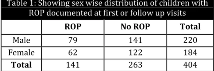
Table 1: Showing sex wise distribution of children with ROP documented at first or follow up visits
ROP No ROP Total
Male 79 141 220
Female 62 122 184
Total 141 263 404
A total of 141(34.9%) infants were found to have Retinopathy of Prematurity among the children who completed the follow up (n=404). Retinopathy of Prematurity was staged according to the ICROP guidelines.
Among the males who completed follow up (220), 68 (30.9%) had ROP requiring only regular follow up and not treatment (Type II according to Early Treatment of ROP guidelines – spontaneously resolved) and 11 (5%) had ROP requiring Laser treatment (Type I ROP according to ETROP guidelines). Among female infants who completed follow up (184), 54 (29.3%) had some ROP which required only regular follow up and 8 (4.3%) had retinopathy which required Laser treatment.
Low birth weight being the most significant risk factor known for ROP, presence or absence of ROP was compared with low birth weight.
Table 2: Distribution of screened infants according to birth weight and ROP status
Sex <2000 >2000 Total
Cases (%) ROP (%) Cases (%) ROP (%) Cases (%) ROP (%)
Male 178(44.1) 74(18.3) 42(10.3) 5(1.2) 220 (54.5) 79 (19.6)
Female 158(39.1) 59(14.6) 26 (6.4) 3(0.7) 184 (45.5) 62(15.3)
Total 336(83.2) 133(32.9) 68(16.7) 8(1.9) 404(100) 141(34.9)
Students test was applied for comparing mean birth weights of infants between groups with
and without ROP The mean birth weight and the S.D of the group without ROP was 1830.26g + 431.32g and the mean weight and S.D of the group with ROP was 1498.79 + 347.03, with a p value < 0.001 the association between low birth weight and ROP is significant.
Presence or absence of ROP was then compared with period of gestation.
Table 3: Distribution of screened infants according to Gestational age and ROP status
Sex < 34 wks >34 wks Total
Cases (%) ROP (%) Cases (%) ROP (%) Cases (%) ROP (%)
Male 153(37.9) 68(16.8) 67(16.5) 11(2.7) 220 (54.5) 79 (19.6)
Female 128(31.6) 54(13.4) 56(13.9) 08(1.9) 184 (45.5) 62(15.3)
J of Evolution of Med and Dent Sci/ eISSN- 2278-4802, pISSN- 2278-4748/ Vol. 3/ Issue 43/ Sep 11, 2014 Page 10669
Students test was applied for comparing mean period of gestation of infants between groups
with and without ROP. The mean POG and the S.D of the group without ROP was 33.9 + 2.48 weeks and the mean POG and S.D of the group with ROP was 31.82 + 2.24, with a p value < 0.001 the association between period of gestation and ROP is significant.
The mean birth weight and the mean gestational age of babies with ROP was similar to another Indian urban study done among a relatively large number of heavy infants n= , 10 year retrospective data) by Vinekar A, et al, where mean birth weight was 1533.9 g and the mean period of gestation was 30.9 weeks for newborns found to have ROP.15
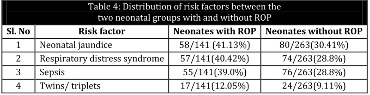
Of all the other known risk factors present in the infants with ROP (141) neonatal jaundice was presents in 57 children, 58 had respiratory distress syndrome and 55 had sepsis Some infants had more than one of the above risk factor, although there was no statistically significant association between the risk factors and ROP. Among the 19 infants who progressed to treatment threshold ROP, 4 infants had prematurity as the sole risk factor.
Table 4: Distribution of risk factors between the two neonatal groups with and without ROP
Sl. No Risk factor Neonates with ROP Neonates without ROP
1 Neonatal jaundice 58/141 (41.13%) 80/263(30.41%)
2 Respiratory distress syndrome 57/141(40.42%) 74/263(28.8%)
3 Sepsis 55/141(39.0%) 76/263(28.8%)
4 Twins/ triplets 17/141(12.05%) 24/263(9.11%)
Some of the other risk factors present were anemia, intraventricular hemorrhage, hypoglycemia, hyponatremia, hypercalcemia, NEC, birth asphyxia. Similar risk factors have been reported in studies conducted in NICU settings from India.16, 17,18
Of the 141 infants who were diagnosed to have ROP, 122 infants (86.5%) had disease that resolved spontaneously. Of these 122 infants, 56 (45.9%) had stage 1 ROP and 66 (54.1%) had stage 2 ROP. Of the 141 children with ROP, 19 infants (12.93% of infants with ROP) progressed to treatment threshold ROP and underwent laser photo ablation.
Laser was performed by a single surgeon (AV) using 532 nm Green Laser delivered using laser indirect ophthalmoscopy (LIO) at the NICU itself under topical anesthesia.12, 19 All treated
infants (100%) showed a favorable outcome following treatment as defined by the standard classification and guidelines.12,19
Of the 19 infants who underwent laser treatment 18 (36 eyes) of them were treated when they had reached Stage 2 with plus disease, whereas only 1 infant (5.3 %) had Aggressive Posterior ROP - APROP who had a birth weight of 1000g and delivered at 27 weeks of gestation. A higher proportion of APROP cases have been reported in Indian studies.15,18,20,21 The low incidence of severe
APROP in this study may be attributed to improved neonatal care, strict surveillance, and regular monitoring of the cases.
CONCLUSION: To the best of our knowledge, this is the largest study of a Tier 2 city Government managed district level hospital reporting ROP incidence thus far. A yield of disease in 141 cases out or
J of Evolution of Med and Dent Sci/ eISSN- 2278-4802, pISSN- 2278-4748/ Vol. 3/ Issue 43/ Sep 11, 2014 Page 10670
Proportion of severe ROP requiring laser treatment was 13.47% (n=141). Of the cases which required laser treatment only one case was Aggressive Posterior Retinopathy of Prematurity. The low incidence of severe APROP may be attributed to improved neonatal care and regular monitoring of the cases. It could also be a result of a significantly heavier cohort of infants being cared for in the rural NICU compared to their urban counterparts.
The study highlights that in the absence of risk factor stratification to predict which infants may progress to treatment requiring ROP, it is essential to include a larger inclusion criteria, especially in the setting of district level hospitals where the admission criteria may vary considerably. The screening cut-off of 2000 grams birth weight suggested by the recent National Neonatology Foundation (2010) guidelines21 seems to be validated in this study. With less than 2% of
infants weighting > 2000 grams who had ROP (Table 2), it seems that until more data is available, it would be useful to screen all infants < 2000 grams which allows the best chance to minimize missing
at-risk infants.
We also experienced, that being a rural cohort, we must rely more on the objective criteria of birth weight compared to the less reliable gestational age, as assessing this in the absence of stringent ante-natal care, ultrasonography and poor awareness of the last menstrual period is prone to error.
The study also reinforces what is recently published from rural Karnataka,10 wherein it
demonstrated that using Western-screening guidelines would result in a large number of potentially at risk infants not being screened.
This study being much larger and longer, adds credulous merit to this insight. Until, National level guidelines are evolved that would not only consider larger Level N)CU s in cities, but also Tier 2 and smaller centers that continue to serve a large number of infants who are premature, it would be prudent to screen all infants below 2000 grams at birth.
The study highlights that even at a Tier 2 city; improved neonatal care can tackle ROP blindness in a well-integrated ROP programme with a strong collaboration between Pediatrics, Ophthalmology and Community Medicine departments. Timely and appropriate screening strategy is necessary in the community to prevent ROP blindness in our rural infants.
REFERENCES:
1. Kanski JJ. Clinical Ophthalmology: A Systemic approach 6th Ed. Butterworth, Heinmann, Elsevier
2007; 606-611.
2. Gilbert C, Fielder A, Gordillo L, Quinn G, Semiglia R, Visintin P, Zin A; International NO-ROP group. Characteristics of infants with severe retinopathy of prematurity in countries with low, middle and high levels of development: implications for screening programs. Pediatrics 2005 May; 115 (5): e518-25. Epub 2005 Apr 1.
3. Rekha S, Battu RR, Chandrasekhara MK. Retinopathy of prematurity—a preliminary report. Indian Pediatr. 1992 May; 29 (5): 623-6.
4. Charan R, Dogra MR, Gupta A, Narang A. The incidence of retinopathy of prematurity in a neonatal care unit. Indian J Ophthalmol. 1995; 43: 123-126.
5. Gopal L, Sharma T, Ramchandran S. Retinopathy of Prematurity - a study. Indian J Ophthalmol 1995; 43: 59-61.
J of Evolution of Med and Dent Sci/ eISSN- 2278-4802, pISSN- 2278-4748/ Vol. 3/ Issue 43/ Sep 11, 2014 Page 10671
7. Maheshwari R, Kumar H, Paul VK, Singh M, Deorari AK, Tiwari HK. Incidence and risk factors of retinopathy of prematurity in a tertiary care new born unit in New Delhi. Natl Med J India. 1996 Sep-Oct; 9 (5): 211-4.
8. Varughese S, Jain S, Gupta N, Singh S, Tyagi V, Puliyel JM. Magnitude of the problem of Retinopathy of Prematurity. Experience in a large Maternity Unit with a medium size level - 3 nursery. Ind J Ophthalmol 2001; 49 (3): 187-188.
9. Gilbert C C, Foster A. Childhood blindness in the context of VISION 2020 – The Right to Sight. Bulletin of the World Health Organization, 2001, 79 (3); 227-32
10.Hungi B, Vinekar A, Datti N, Kariyappa P, Braganza S, Chinnaiah S, Donthi K, Shetty B. Retinopathy of Prematurity in a rural Neonatal Intensive Care Unit in South India--a prospective study. Indian J Pediatr. 2012 Jul; 79 (7): 911-5. Epub 2012 Feb 23.
11.Vinekar A, Trese MT, Capone A Jr. Photographic Screening for Retinopathy of Prematurity (PHOTO-ROP) Cooperative Group. Evolution of Retinal Detachment in Posterior Retinopathy of Prematurity: Impact on Treatment Approach. Am J Ophthalmol. 2008 Mar; 145 (3): 548-555. Epub 2008 Jan 22.
12.Pejaver RK. Vinekar A. Bilagi A. Research Issues in Retinopathy of Prematurity (Review Article): J of Neonatology 2009; 23 (4): 350-352.
13.International Committee for the Classification of Retinopathy of Prematurity. The International Classification of Retinopathy of Prematurity revisited. Arch Ophthalmol. 2005 Jul; 123 (7): 991-9. [Medline].
14.Early Treatment for Retinopathy of Prematurity Cooperative Group. Revised indications for the treatment of retinopathy of prematurity: results of the early treatment for retinopathy of prematurity randomized trial. Arch Ophthalmol. 2003 Dec; 121 (12): 1684-94.
15.Vinekar A, Dogra MR, Sangtam T, Narang A, Gupta A. Retinopathy of prematurity in Asian Indian babies weighing greater than 1250 grams at birth: ten year data from a tertiary care center in a developing country. Indian J Ophthalmol. 2007 Sep-Oct; 55 (5): 331-6.
16.Vinekar A, Hegde K, Gilbert C, Braganza S, Pradeep M, Shetty R, Shetty KB. Do platelets have a role in the pathogenesis of aggressive posterior retinopathy of prematurity? Retina. 2010 Apr; 30 (4 Suppl): S20-3.
17.Vinekar A, Avadhani K, Braganza S, Shetty B, Dogra M, Gilbert C. Outcomes of a protocol-based management for zone 1 retinopathy of prematurity: the Indian Twin Cities ROP Screening Program report number 2. Am J Ophthalmol. 2011 Oct; 152 (4): 712
18.Dogra MR, Vinekar A, Viswanathan K, Sangtam T, Das P, Gupta A, Dutta S. Laser treatment for retinopathy of prematurity through the incubator wall. Ophthalmic Surg Lasers Imaging. 2008 Jul-Aug; 39 (4): 350-2.
19.Sanghi G, Dogra MR, Das P, Vinekar A, Gupta A, Dutta S. Aggressive posterior retinopathy of prematurity in Asian Indian babies: spectrum of disease and outcome after laser treatment. Retina. 2009 Oct; 29 (9): 1335-9.
J of Evolution of Med and Dent Sci/ eISSN- 2278-4802, pISSN- 2278-4748/ Vol. 3/ Issue 43/ Sep 11, 2014 Page 10672
21.Pejaver RK, Bilagi AP, Vinekar A. National Neonatology Foundation s Evidence Based Clinical Practice Guidelines 2010. Retinopathy of Prematurity (NNF India, Guidelines) 2010; 253‐262.
AUTHORS:
1. Keerthi B. J. 2. P. Subhas Babu 3. Anand Vinekar 4. Nagaraja Goud 5. Asha Bullappa
PARTICULARS OF CONTRIBUTORS:
1. Assistant Professor, Department of Paediatrics, Mandya Institute of Medical Sciences, Mandya.
2. Assistant Professor, Department of Community Medicine, Mandya Institute of Medical Sciences, Mandya.
3. Associate Professor and HOD, Department of Ophthalmology (Paediatric Retina), Narayana Nethralaya PG Institute of Ophthalmology, Bangalore.
4. Assistant Professor and Statistician,
Department of Community Medicine, Mandya Institute of Medical Sciences, Mandya.
5. Assistant Professor, Department of
Community Medicine, Srinivasa Institute of Medial Sciences and Research Center, Suratkal.
NAME ADDRESS EMAIL ID OF THE CORRESPONDING AUTHOR:
Dr. P. Subhas Babu, Assistant Professor,
Department of Community Medicine, Mandya Institute of Medical Sciences, Mandya.
Email: [email protected]