Received: 2016 Apr 12; revised: 2016 Jul 17; rerevised: 2016 Jul 19; accepted: 2016 Jul 22; published online: 2017 Jan 05
Retinopathy of prematurity: results
from 10 years in a single neonatal
intensive care unit
Inês Coutinho1, Catarina Pedrosa1, Mafalda Mota1, Sofia Azeredo-Lopes2, Cristina Santos1, Graça Pires1, Susana Teixeira1, Manuel Cunha3
1Ophthalmology Department, Hospital Prof. Doutor Fernando Fonseca, Lisbon, Portugal
2Statistical Department, NOVA Medical School, Lisbon, Portugal
3Pediatrics Department, Hospital Prof. Doutor Fernando Fonseca, Lisbon, Portugal
Abstract
Introduction: Retinopathy of prematurity (ROP) is a vasoproliferative
disorder of the retina of preterm newborns and is an important and preventable cause of visual impairment in childhood. This study aimed to assess the incidence and main risk factors associated with the development of ROP in the last 10 years at Hospital Prof. Doutor Fernando Fonseca in Lisbon, Portugal.
Methods: Observational and retrospective study conducted between 2005
and 2014 at Hospital Prof. Doctor Fernando Fonseca. The study included newborns of gestational age < 32 weeks. We analyzed maternal, prenatal and neonatal factors associated with the development of ROP. Statistical analysis were performed with Statistical Package for Social Sciences (SPSS®) software. Univariate and multivariate analyses were performed and a multiple logistic regression model was carried out with a significance level α = 0.05.
Results: 527 premature infants with a gestational age < 32 weeks were
studied, of which 165 developed ROP. 60 of these patients needed treatment. In the univariate analysis, the risk factors for the development of ROP were maternal infection in pregnancy, low birth weight, low gestational age, low Apgar score at 5 minutes, need for oxygen therapy until the 28th day
of life, a high score on the CRIB and SNAPPE2 scales, use of surfactant, respiratory distress syndrome, persistence of patent ductus arteriosus, peri-intraventricular hemorrhage and neonatal sepsis. In the multiple logistic regression analysis, risk factors for ROP were the presence of neonatal sepsis, respiratory distress syndrome, persistence of patent ductus arteriosus and a high score on the neonatal SNAPPE2 scale.
Conclusions: We found a ROP incidence rate of 31.3%, with risk factors
similar to those observed in other studies.
Keywords
Prematurity, retinopathy, risk factor, incidence.
Corresponding author
Inês Coutinho, Ophthalmology Resident, Hospital Prof. Doutor Fernando Fonseca, Lisbon, Portugal; email: inescorga@gmail.com.
How to cite
Coutinho I, Pedrosa C, Mota M, Azeredo-Lopes S, Santos C, Pires G, Teixeira S, Cunha M. Retinopathy of prematurity: results from 10 years in a single neonatal intensive care unit. J Pediatr Neonat Individual Med. 2017;6(1):e060122. doi: 10.7363/060122.
Introduction
Retinopathy of prematurity (ROP) is a vaso- proliferative change secondary to inadequate vascularization of the retina of preterm newborns. It was first described as retrolental fibroplasia by Terry in 1942.
ROP is an important and preventable cause of poor vision in children, with 6-18% of cases of blindness occurring in developed countries being directly attributable to ROP [1, 2]. Despite the increased survival of premature infants with low gestational age and low birth weight, recent studies have shown a decrease in the incidence of ROP, which can be due to improved neonatal care and better understanding of the disease [3].
The pathophysiology of this disease is still not fully understood. Over the past 50 years, some studies have been performed in order to identify possible factors associated to the development of ROP. It is known that the retinal vasculature begins to develop from the optic disc to the periphery around the 16th week of gestation, with vascularization of
the nasal region occurring around 32-36 weeks and the temporal region at 40-42 weeks [4]. Thus, the degree of prematurity of the newborn determines the stage of retinal vasculature maturation and the affected zone [5].
Oxygen tension is low in utero and the
extra-uterine environment is hyperoxic for premature infants. After birth and until 30 weeks post con- ceptional age, retinal vascularization is inhib- ited because of hyperoxia and loss of growth factors provided at the maternal-fetal interface like IGF-1. However, as the newborn grows, the avascular retina becomes more metabolically ac- tive, which leads to tissue hypoxia, stimulating
pro-angiogenic factors (VEGF and IGF-1) and leading to retinal neovascularization between 32 and 34 weeks post-conceptional age [5, 6].
Several risk factors have been associated with the development of ROP, most commonly gestational age and birth weight. Other potential risk factors described in the literature are male gender, oxygen therapy, apnea, sepsis, peri-intraventricular hemorrhage, patent ductus arteriosus, anemia and blood transfusion, although the impact of these factors on the progression of the disease is not fully understood [1, 5, 7-9]. By contrast, preeclampsia [10] and lung maturation induced by prenatal steroids therapy have been suggested as protective factors.
Screening for ROP is critical to control visual sequelae. Infants with a birth weight < 1,500 g or gestational age < 32 weeks are groups considered at high risk, in which eye screening should be carried out. This screening can be extended to other children with co-morbidities associated with ROP as indicated by the neonatologist [8, 11-13]. Ophthalmologic screening occurs between 4-6 weeks of age or between 31-33 weeks post-conceptional age, with preference for a later date [8, 11-13].
The disease is classified according to the in- ternational classification (ICROP) (Tab. 1).
Current treatment criteria are summarized in Tab. 2 and are based on the CRYOP-ROP and ETROP
studies. Most cases of ROP resolve spontaneously without sequelae between 32 and 42 week of gestation [5, 6]. Currently, laser photocoagulation of the avascular retina is the treatment of choice, and various studies show that its functional and structural results are superior to cryotherapy [14].
The intravitreal injection of anti-VEGF appears to be another promising option for the treatment of ROP, and encouraging results have been presented, such as those in the BEAT-ROP study. It is a relatively quick and easy treatment to perform and it is associated with a lower rate of myopia than laser therapy, however more controlled studies are necessary to evaluate its long-term safety.
Its use, isolated or in combination with laser therapy, is currently restricted to cases of aggressive posterior ROP, ROP stage 3 in zone I, in the presence of local complications such as media opacity or poor dilation, in patients whose use of laser therapy is contraindicated because of clinical instability, in ROP stages 4 or 5 before a vitrectomy and in cases in which laser therapy fails [4, 15].
Table 1. International classification of retinopathy of prematurity.
Stage Localization Extension
Stage 1 – flat demarcation
line Zone I – circle area centered
on the optic nerve with a radius
twice the distance from the optic nerve to the macula
Evaluation in hours
Stage 2 – high crest between
the vascularized and non-vascularized retina
Stage 3 – fibrovascular
proliferation
Zone II – it extends from the end of Zone I to the nasal ora
serrata
Stage 4 – partial detachment of the retina
Zone III – it corresponds to the
growing remaining crescent
area
Stage 5 – full detachment of the retina
Plus disease
Vascular dilatation and tortuosity in the posterior pole Gravity signal and progression, which may arise at any stage Reflects an increase in VEGF1
Table 2. Treatment indications.
Threshold ROP
Classical indication for treatment according to the CRYO-ROP 1988 study
• Stage 3 in Zone II, at least 5 hours or 8 hours continuous extension interspersed in the presence of
plus disease in Zone I
Pre-threshold ROP Type 1
Indication for treatment according to the ET-ROP study
• Any stage of ROP in Zone I, with plus disease • Stage 3, Zone I, without plus disease • Stage 2 or 3, Zone II, with plus disease
Aggressive posterior ROP
Definition introduced in the international classification of 2005
• Unusual, severe and progresses rapidly
• Zone I or II with plus disease that does not follow a pattern according to development stage
timetable and procedures proposed by national and international guidelines for the screening of ROP.
The maternal variables analyzed were maternal age, multiple pregnancy, pre-eclampsia, infection (chorioamnionitis or TORCH) and prenatal use of steroids. Neonatal variables were gestational age, birth weight, sex, Apgar score at 5 minutes, respiratory distress syndrome, use of surfactant, oxygen therapy until the 28th day after birth,
peri-intraventricular hemorrhage, neonatal sepsis, CRIB and SNAPPE2 scores and persistence of patent ductus arteriosus.
Statistical analysis of the data was performed using SPSS®. Multiple logistic regression models were used, initially considering the explana- tory variables obtained by univariate analyses. Considering that the incidence and risk factors
for ROP vary from region to region, the present study aimed to analyze the situation in our hospital in the last 10 years.
Methods
Student’s t-test and the Chi-squared test were used when applicable or, alternatively, Fisher’s exact test was used. Mann-Whitney test was also used. Difference were considered statistically significant when α = 0.05.
Results
Between 2005-2014, 35,210 children were born, of whom 705 (2%) had a gestational age of less than 32 weeks. Of the 705 infants, 178 were excluded due to neonatal death or incomplete ophthalmic medical records. Thus, the sample of our study consisted of 527 infants with a
gestational age of less than 32 weeks. Of the 527 infants, 258 (49.0%) were male, mean gestational age was 29 weeks and the average birth weight was 1,137 g. The sample under study was divided into two groups with and without ROP; the main population characteristics are summarized in Tab. 3. The ROP group had a mean gestational age of
26.8 weeks and average birth weight of 882 g. In the group without ROP, mean gestational age was 30 weeks and average birth weight was 1,252 g.
The risk factors considered to be significant for ROP development in the univariate analyses (Tab. 3) were low birth weight, low gestational age, low
Apgar score at 5 minutes, need for oxygen therapy
Table 3. Sample characteristics and results of univariate logistic regression models.
Variable Preterm newborns with ROP
(n = 165)
Preterm newborns
without ROP (n = 362)
Univariate analysis
p-value OR
Maternal age (years), mean ± std. deviation 30.02 ± 5.86 29.74 ± 6.74 0.647 1.007
Pre-eclampsia, No / Yes 150 / 15 325 / 37 0.750 (ref = “No”)
0.903
Infection in pregnancy, No / Yes 150 / 15 346 / 16 0.032 (ref = “No”)2.219
Twin pregnancy, No / Yes 131 / 34 292/ 70 0.639 (ref = “No”) 1.116
Prenatal steroids, No / Partial / Complete 18 / 48 / 99 44 / 119 / 199 0.681
(ref = “No”)
0.986 1.169
Birth weight (g), mean ± std. deviation (min; max) 882 ± 263 (415; 1,835) 1,252 ± 287 (550; 2,000) < 0.001 0.995
Gestational age (days), mean ± std. deviation 26.8 weeks188 ± 14 30 weeks209 ± 14 < 0.001 0.909
Male / Female 78 / 87 180 / 182 0.790 (ref = “Female”) 0.951
Apgar at 5 min, median (min; max) 7.8 (2; 10) 8.38 (0; 10) < 0.001 0.763
Oxygen therapy until the 28th day of life, No / Yes 38 / 127 272 / 90 < 0.001 (ref = “No”) 8.194
Surfactant use, No / Yes 33 / 132 187 / 175 < 0.001 (ref = “No”)4.480
Respiratory distress syndrome, No / Yes 14 / 151 105 / 257 < 0.001 (ref = “No”)4.74
Persistence of ductus arteriosus, No / Yes 70 / 95 270 /92 < 0.001 (ref = “No”)4.115
Peri-intraventricular hemorrhage, No / Yes 97 / 68 277 / 85 < 0.001 (ref = “No”)2.342
Neonatal sepsis 118 143 < 0.001 (ref = “No”) 3.907
Gram positive 42 51
Gram negative 26 23
Fungal 11 6
Others 39 63
CRIB, median (min; max) 4 (0; 14) 1 (0; 18) < 0.001 1.358
SNAPPE2, median (min; max) 39 (0; 85) 20 (0; 101) < 0.001 1.036
until the 28th day after birth, higher value of CRIB and
SNAPPE2 score, peri-intraventricular hemorrhage, patent ductus arteriosus, use of surfactant, res- piratory distress syndrome, maternal infection during pregnancy and neonatal sepsis. There was no statistically significant difference between the two groups (with and without ROP) in terms of sex, maternal age, twin pregnancy, preeclampsia or the use of antenatal steroids.
It was also observed that birth weight and gestational age were correlated with a Spearman correlation coefficient of 0.773 (p < 0.001), so they should not be used together in the same model to avoid collinearity. Thus, when considered separately, each increase of 1 week in the gestational age provided a reduction of approximately 9% in the chance of developing ROP (OR = 0.909, p < 0.001). In turn, for each increase of 1 gram in birth weight, there was a decrease of about 0.5% in the risk of ROP (OR = 0.005, p < 0.001), i.e., for each increase of 10 g, there was a decrease in ROP risk of about 5%.
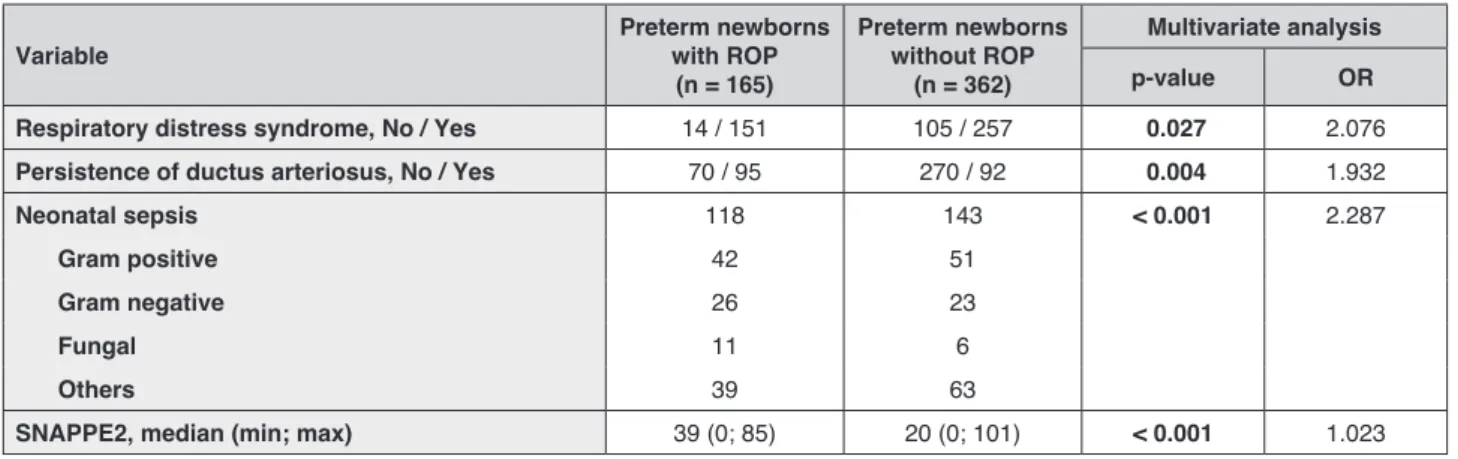
It was also observed that when the relationship between the occurrence of ROP and the variables weight and gestational age was adjusted by the effect of other covariates, the latter never exhibited statistically significant results. It can therefore be concluded that both gestational age and birth weight “cancel” statistical significance of other factors. Therefore, the effect of other possible factors were analysed when both weight and gestational age were not considered. The multivariate logistic regression model has found four risk factors: neonatal sepsis (OR = 2.287, p < 0.001), higher value of SNAPPE2 (OR = 1.023, p < 0.001), respiratory distress syndrome (OR = 2.076, p = 0.027) and persistence of ductus arteriosus (OR = 1.932, p = 0.004) (Tab. 4). Although
multivariate method is the preferred method to study this multifactorial disease, the results can be unstable on many occasions with the influence of one variable on others [3].
The incidence of ROP during this time period was 31.3% (n = 165). As for the stage, 59 infants (35.76%) developed ROP at stage 1, 61 newborns (36.97%) at stage 2, 43 infants (26.06%) at stage 3 and 2 newborns (1.21%) at stage 4. There were no cases of ROP stage 5 (Tab. 5). The association
between different parameters and the severity of ROP is described in Tab. 5. The severe stages of
ROP (stages 3, 4 and 5) were associated with lower birth weight, lower gestational age and increased oxygen therapy. The average birth weight and gestational age associated with severe ROP were 761 g and 27 weeks, respectively.
Of the 165 infants with ROP, 60 (36.36%) required treatment, with spontaneous remission of the disease in the other cases. Laser therapy was performed on 50 patients, while 10 patients received bevacizumab injection.
Discussion
In our study the incidence of ROP was 31.3%, a result that is similar to many other studies with similar selection criteria [16-18] and performed in developed countries. However, some studies showed lower incidence rates [19-23].
The risk factors we observed were similar to those described in the literature [9, 13, 22].
A low value of the Apgar score at 5 minutes and a high value of the CRIB and SNAPPE2 scores are indicators by themselves of morbidity and mortality in infants. It is known that ROP mainly affects the weakest newborns, although it is not fully understood if the most severe stages
Table 4. Sample characteristics and results of the multiple logistic regression models.
Variable Preterm newborns with ROP
(n = 165)
Preterm newborns
without ROP (n = 362)
Multivariate analysis
p-value OR
Respiratory distress syndrome, No / Yes 14 / 151 105 / 257 0.027 2.076 Persistence of ductus arteriosus, No / Yes 70 / 95 270 / 92 0.004 1.932
Neonatal sepsis 118 143 < 0.001 2.287
Gram positive 42 51
Gram negative 26 23
Fungal 11 6
Others 39 63
Parameter Stage 1+2 (n = 120)
Stage 3+4+5
(n = 45) p-value
Pre-eclampsia 0.241
No 111 39
Yes 9 6
Infection in pregnancy 0.076
No 38 8
Yes 82 37
Birth weight < 0.001
< 1,000 g 98 43
1,000-1,499 g 21 2
1,500-2,499 g 1 0
> 2,500 g 0 0
Gestational age 0.004
< 189 days (< 27 weeks) 64 35
190-224 days (27-32 weeks) 56 10
Apgar at 5 min, mean ± std. deviation 7.82 ± 1.39 7.73 ± 1.56 0.977
Oxygen therapy until the 28th of life 0.007
No 36 4
Yes 84 41
Surfactant use 0.080
No 28 5
Yes 92 40
Respiratory distress syndrome 0.355
No 12 2
Yes 108 43
Persistence of ductus arteriosus 0.493
No 52 17
Yes 68 28
Periventricular hemorrhage 0.220
No 74 23
Yes 46 22
Neonatal sepsis 0.076
No 38 8
Yes 82 37
SNAPPE2, mean ± std. deviation 38.11 ± 21.99 41.45 ± 18.60 0.457
Table 5. ROP incidence by stage of disease and results of the Chi-square test (or Fisher’s exact test) and Mann-Whitney test.
of the disease are associated with the severity of co-morbidities resulting from prematurity or therapeutic interventions necessary to maintain life [24, 25].
The presence of peri-intraventricular hemor- rhage, persistence of patent ductus arteriosus, maternal infection, oxygen therapy until the 28th day
after birth and respiratory distress syndrome relate to and allow ischemia to occur, therefore increasing the need for supplemental oxygen and leading to injury of immature capillaries, generation of
development of ROP (the risk of ROP in a child that received surfactant was about 4.5 times higher that of a child not given surfactant; OR = 4.480, p < 0.001, Tab. 3). This could be attributed to the
greater clinical instability of newborns who were treated with surfactant [29]. Moreover, the use of surfactant did not have a significant impact in reducing the severity of ROP (Tab. 5).
Neonatal sepsis was observed to be as a risk factor, possibly in relation to its inflammatory effect, which can stimulate retinal neovascularization [5, 30-32].
Unlike in other publications, male gender was not a risk factor for the development of ROP in the present study [24].
Another interesting fact is that prenatal steroid administration did not reduce the risk of ROP, which is concordant with other studies [9], despite having a positive impact on the surviv- al of premature infants and reducing the inci- dence of respiratory distress syndrome. Moreover, preeclampsia was not a protective factor in the current cohort although it has been previously described as protective against ROP in other studies [10].
As the present study was of retrospective design, it was not possible to analyze other causes described as important risk factors to ROP.
Conclusions
The pathogenesis of ROP is multifactorial. In this study the most significant risk factors to development of ROP were low birth weight, low gestational age, low Apgar score at 5 minutes, need for oxygen therapy until the 28th day after birth, a high score on CRIB and
SNAPPE2 scales, use of surfactant, respiratory distress syndrome, persistence of patent ductus arteriosus, peri-intraventricular hemorrhage, ma- ternal infection and neonatal sepsis.
The incidence of ROP was 31.3% and severe ROP (stage 3 or greater) was 27.27%. A treatment was performed in 36.36% of ROP cases.
ROP remains a major complication in pre- mature newborns despite all the advances that have been made in recent years.
Excellence in pre- and neonatal care, screening and early treatment of ROP are keys to prevent vision loss induced by this disease. It is mandatory to ensure that these newborns have regular ophthalmologic support, as they are more likely to have other ocular complications such as refractive
defects (myopia, astigmatism and anisometropy), oculomotor balance disorders (strabismus) and amblyopia [5, 6, 30].
Acknowledgements
A special thanks to the neonatology staff at Hospital Prof. Doutor Fernando Fonseca for all assistance provided.
Declaration of interest
The Authors have no conflicts of interest relevant to this article.
References
1. Abdel HÁ, Mohamed GB, Othman MF. Retinopathy of prematurity:
a study of incidence and risk factors in NICU of Al-Minya University Hospital in Egypt. J Clin Neonatol. 2012;1(2):76-81.
2. Vieira BC, Nascimento M, Ribeiro I, Caralho R, Martins JN.
Resultados de 12 anos de rastreio da retinopatia da prematuridade no Hospital Pedro Hispano. Oftalmologia. 2013;37(3):199-204.
3. Horbar JD, Carpenter JH, Badger GJ, Kenny MJ, Soll RF, Morrow
KA, Buzas JS. Mortality and neonatal morbidity among infants 501 to 1500 grams from 2000 to 2009. Pediatrics. 2012;129(6): 1019-26.
4. Paysse EA. Retinopathy of Prematurity. In: Garcia-Prats JA,
Saunders RA, Armsby C (Eds.). UpToDate. Available at: http:// www.uptodate.com/contents/retinopathy-of-prematurity, updated on: July 27, 2015, last access: July 2015.
5. Hellstörn A, Smith LE, Dammann O. Retinopathy of prematurity.
Lancet. 2013;382;1445-57.
6. Chawla D, Agarwal R, Deorari A, Paul VK, Chandra P, Azad RV.
Retinopathy of prematurity. Indian J Pediatr. 2012;79(4):501-9.
7. Filho JB, Eckert GU, Valiatti FB, Costa MC, Bonomo PP,
Procianoy RS. Prevalência e fatores de risco para a retinopatia da prematuridade: estudo com 450 pré-termos de muito baixo peso. Rev Bras Oftalmol. 2009;68(1):22-9.
8. Conselho Brasileiro de Oftalmologia Sociedade Brasileira de
Pediatria. Retinopatia da Prematuridade. Projeto Diretrizes – Associação Médica Brasileira e Conselho Federal de Medicina, 2011. Available at: http://diretrizes.amb.org.br/_BibliotecaAntiga/ retinopatia_da_prematuridade.pdf, last access: August 2016.
9. Henriques G, Brito C, Clemente F, Breda J, Teixeira S. Retinopatia da
Prematuridade. In: Marques Valido A, Guimarães H, Videira Amaral JM, Januário L, Carrapato R, Tomé T, Martins V (Eds.). Consensos Nacionais em Neonatologia. Coimbra: Secção de Neonatologia, Sociedade Portuguesa de Pediatria, 2004, pp. 101-3.
10. Tomé VA, Vieira JF, Oliveira LB, Pinto RM, Abdallah VO. Estudo da retinopatia da prematuridade em um hospital universitário. Rev Bras Oftalmol. 2011;74(4):279-82.
Association of Certified Orthoptists. Screening examination of premature infants for retinopathy of prematurity. Pediatrics. 2013;131(1):189-95.
12. Wilkinson AR, Haines L, Head K, Fielder AR. UK retinopathy of prematurity guideline. Early Hum Dev. 2008;84(2):71-4. 13. Teixeira S. Retinopatia da prematuridade. Porto: Monografia da
Sociedade Portuguesa de Oftalmologia, 2006.
14. Shalev B, Farr AK, Repka MX. Randomized comparison of diode laser photocoagulation versus cryotherapy for threshold retinopathy of prematurity: seven-year outcome. Am J Ophthalmol. 2001;132(1):76-80.
15. Mintz-Hittner HA, Kennedy KA, Chuang AZ; BEAT-ROP Cooperative Group. Efficacy of intravitreal bevacizumab for stage 3+ retinopathy of prematurity. N Engl J Med. 2011;364(7): 603-15.
16. Shinsato RN, Paccola L, Gonçalves WA, Barbosa JC, Martinez FE, Rodrigues Mde L, Jorge R. [Frequency of retinopathy of prematurity in newborns at the Clinical Hospital, Ribeirão Preto Medical School, University of São Paulo]. [Article in Portuguese]. Arq Bras Oftalmol. 2010;73(1):60-5.
17. Isaza G, Arora S, Bal M, Chaudhary V. Incidence of retinopathy of prematurity and risk factors among premature infants at a neonatal intensive care unit in Canada. J Pediatr Ophthalmol Strabismus. 2013;50(1):27-32.
18. Campo-Gesto A, Garcia S. Incidencia y gravedad de la retinopatía del premature. Rev Esp Inv Oftal. 2013;3(4):193-9.
19. Graziano RM, Leone CR, Cunha SL, Pinheiro AC. [Prevalence of retinopathy of prematurity in very low birth weight infants]. [Article in Portuguese]. J Pediatr (Rio J). 1997;73(6):377-82. 20. van Sorge AJ, Termote JU, Kerkhoff FT, van Rijn LJ, Simonsz
HJ, Peer PG, Schalij-Delfos NE. Nationwide inventory of risk factors for retinopathy of prematurity in the Netherlands. J Pediatr. 2014;164(3):494-8.
21. Kumar P, Sankar MJ, Deorari A, Azad R, Chandra P, Agarwal R, Paul V. Risk factors for severe retinopathy of prematurity in preterm low birth weight neonates. Indian J Pediatr. 2011;78(7):812-6.
22. Moinho R, Morais S, Monteiro M, Mimoso G. Retinopatia da Prematuridade numa Unidade de Cuidados Intensivos Neonatais: Experiência de Oito Anos. Acta Pediatrica Portuguesa. 2015;46(3):198-204.
23. Yu XD, Branch DW, Karumanchi SA, Zhang J. Preeclampsia and retinopathy of prematurity in preterm births. Pediatrics. 2012;130(1):e101-7.
24. Yang MB, Donovan EF, Wagge JR. Race, gender, and clinical risk index for babies (CRIB) score as predictors of severe retinopathy of prematurity. J AAPOS. 2006;10(3):253-61.
25. Fortes Filho JB, Dill JC, Ishizaki A, Aguiar WW, Silveira RC, Procianoy RS. Score for Neonatal Acute Physiology and Perinatal Extension II as a predictor of retinopathy of prematurity: study in 304 very-low-birth-weight preterm infants. Ophthalmologica. 2009;223(3):177-82.
26. Watts P, Adams GG, Thomas RM, Bunce C. Intraventricular haemorrhage and stage 3 retinopathy of prematurity. Br J Ophthalmol. 2000;84(6):596-9.
27. González Viejo I, Ferrer Novella C, Pueyo Royo V, García Martín E, Rite Gracia S, Caballero Pérez V, Romera Santa Bárbara B, Royo Pérez D. [Is patent ductus arteriosus a risk factor for retinopathy of prematurity?]. [Article in Spanish]. An Pediatr (Barc). 2011;74(1):25-30.
28. Saugstad OD. Oxygen and retinopathy of prematurity. J Perinatol. 2006;26(Suppl 1):S46-50; discussion S63-4.
29. Chen YH, Lien RI, Tsai S, Chang CJ, Lai CC, Chao AN, Chen KJ, Hwang YS, Wang NK, Chen YP, Chen TL, Wu WC. Natural history of retinopathy of prematurity: two-year outcomes of a prospective study. Retina. 2015;35(1):141-8.
30. Mitra S, Aune D, Speer CP, Saugstad OD. Chorioamnionitis as a risk factor for retinopathy of prematurity: a systematic review and meta-analysis. Neonatology. 2014;105(3):189-99.
31. Lee J, Dammann O. Perinatal infection, inflammation, and reti- nopathy of prematurity. Semin Fetal Neonatal Med. 2012;17(1):26-9. 32. Mantagos IS, VanderVeen DK, Smith LEH. Risk factors for