Iranian Journal of Basic Medical Sciences
ijbms.mums.ac.ir
Colchicine protects rat skeletal muscle from ischemia/reperfusion
injury by suppressing oxidative stress and inflammation
Liangrong Wang
1, Yuanlu Shan
1, Lei Chen
1, Bi Lin
1, Xiangqing Xiong
1, Lina Lin
1, Lida Jin
1*
1 Department of Anesthesiology, The First Affiliated Hospital of Wenzhou Medical University, No. 2, Fuxue Road, Wenzhou 325000, Zhejiang Province, People s Republic of China.
A R T I C L E I N F O A B S T R A C T
Article type:
Original article Objective(s):muscle injury. Neutrophils play an important role in ischemia/reperfusion (IR) induced skeletal Microtubules are required for neutrophil activation in response to various stimuli. This study aimed to investigate the effects of colchicine, a microtubule-disrupting agent, on skeletal muscle IR injury in a rat hindlimb ischemia model.
Materials and Methods: Twenty-one Sprague-Dawley rats were randomly allocated into three groups:IR group, colchicine treated-IR (CO) group and sham operation (SM) group. Rats ofboth the IR and CO groups were subjected to 3 hr of ischemia by clamping the right femoral artery followed by 2 hr of reperfusion. Colchicine (1 mg/kg)was administrated intraperitoneally prior to hindlimb ischemia in the CO group. After 2 hr of reperfusion, we measured superoxide dismutase (SOD) and myeloperoxidase (MPO) activities, and malondialdehyde (MDA), tumor necrosis factor (TNF)- and interleukin )L- levels in the muscle samples. Plasma creatinine kinase CK and lactate dehydrogenase (LDH)levelswere measured. We also evaluated the histological damage score and wet/dry weight (W/D) ratio.
Results: The histological damage score, W/D ratio, MPO activity, MDA, TNF- and )L- levels in muscle tissues were significantly increased, SOD activity was decreased, and plasma CK and LDH levels were remarkably elevated in both the IR and CO groups compared to the SM group (P<0.05). Colchicine treatmentsignificantly reduced muscle damage and edema, oxidative stress and levels of the inflammatory parameters in the CO group compared to the IR group (P<0.05). Conclusion:Colchicine attenuates IR-induced skeletal muscle injury in rats.
Article history: Received: Aug 15, 2015 Accepted: Mar 3, 2016
Keywords: Colchicine Inflammation Muscle
Reperfusion injury Skeletal
►
Please cite this article as:Wang L, Shan Y, Chen L, Lin B, Xiong X, Lin L, Jin L. Colchicine protects rat skeletal muscle from ischemia/reperfusion injury by suppressing oxidative stress and inflammation. Iran J Basic Med Sci 2016; 19:670-675.
Introduction
A timely and effective restoration of blood flow, i.e. reperfusion, is essential for ischemic limb recovery, but reperfusion can also aggravate the initial ischemia-induced injury, which is known as ischemia/reperfusion (IR) injury (1). IR injury not only exacerbates the damage of the local ischemic tissues, but also causesremote organ injury or even multiple organ dysfunction syndromes (2-4). The pathogenesis of IR injury is complex and multifactorial, involving an intrinsic intracellular injury process during the ischemic phase and an induced inflammatory response during the reperfusion phase. Importantly, neutrophils were found to play an important role in IR injury (5-7).
Microtubules are filamentous intracellular structures that are responsible for a variety of movements in all eukaryotic cells, and the microtubule network plays a crucial role in the development and maintenance of cellular polarity.
Therefore, compromising microtubule function in neutrophils is theoretically effective in inhibiting neutrophil migration and limiting neutrophil-dependent injuries (8). Colchicine, a microtubule-disrupting agent, has an anti-inflammatory feature and is currently used in the treatment of inflammation-linked diseases including acute gout,
Behcet s disease, Mediterranean fever, recurrent pericarditis, and secondary amyloidosis (9-11). Mechanistically, colchicine was shown to limit neutrophil-mediated inflammation and decrease the levels of inflammatory mediators in several settings (12-16). However, whether colchicine can ameliorate or prevent IR-induced skeletal muscle injury remains unknown.
The present study aimed to investigate if colchicine could protect skeletal muscle against IR-induced injury in a rat ischemic hindlimb model. We also aimed to study the associated molecular basis of
colchicine s protective mechanism. We hypothesized
*Corresponding author:Lida Jin. Department of Anesthesiology, The First Affiliated Hospital of Wenzhou Medical University, No. 2, Fuxue Road, Wenzhou
that colchicine attenuates skeletal muscle damage in response to IR via suppressing oxidative stress and inflammatory response. Our findings point to a potential clinical application of colchicine in treatment of IR injury.
Materials and Methods
Group allocation
This study was approved by the Animal Care and Use Committee of Wenzhou Medical University, and all protocols were in accordance with the Guidelines of the National Institutes of Health for Care and Use of Laboratory Animals. Twenty-one male adult Sprague-Dawley rats weighing 250-300 g were obtained from the Animal Center of Wenzhou Medical University and randomly allocated into three groups: IR group, colchicine treated-IR (CO) group, and sham operation (SM) group (7 rats per group). The rats inboth the IR and CO groups were subjected to3 hr of ischemia followed by 2 hr of reperfusion in the right hindlimb. Colchicine (1 mg/kg, Sigma Chemical, St. Louis, MO, USA) or an equal volume of saline was intraperitoneally administrated immediately before induction of hindlimb ischemia in the CO or IR group, respectively. The rats in theSM group underwentsham operation.
Experimental protocol
The rats were deprived of food but not water for 12 hr prior to the surgical procedure. Rats were administered an intraperitoneal injection of 80 mg/kg ketamine and 5 mg/kg diazepam and placed in a supine position on a heating pad to maintain body temperature at 36-37 °C. The right femoral artery was then isolated and clamped, and after limb exsanguination, a rubber band was applied on the right greater trochanter for 3 hr to ensure interruption of arterial blood supply to the hindlimb in both the IR and CO groups. After 3 hr of ischemia, the arterial clamps and rubber bands were removed and an additional 2 hr were allowed for reperfusion before sampling. In the SM group, the femoral artery was exposed but not occluded, and the rubber band was in place without being inflated. At the end of reperfusion, rats were euthanized, and the gastrocnemius muscles and blood samples from abdominal aortas were collected for subsequent analysis.
Histological examination
Muscle tissue samples were placed in 10% buffered formaldehyde for 48 hr, dehydrated in an ascending series of ethanol, cleared in xylene and embedded in paraffin wax. 5 μm thick sections were stained with hematoxylin and eosin (H&E) and examined under a light microscope by a pathologist who was blinded to the experimental groups. The histological damage score was used to
assess the extent of skeletal muscle injury. Muscle fiber degeneration, disorganization, and inflammatory cell infiltration were scored using previously describedmethods (17).
Measurement of wet/dry weight (W/D) ratio of muscle tissues
Gastrocnemius specimens were weighed immediately after euthanasia to obtain the wet weight, then dried in an 80°C ventilated oven for 48 hr and weighed again to obtain dry weight. The wet/dry weight (W/D) ratio was then calculated to compare muscle edema between the three groups.
Determination of oxidative stress and inflammatory parameters in muscle tissues
Muscle tissues were cut into small pieces, homogenized in ice-cold Tris-HCl buffer solution (pH 7.4, 0.2 mmol/l) and then centrifuged at 3,500 ×g for 60 min to obtain supernatant for the subsequent analyses. Superoxide dismutase (SOD) and myelo-peroxidase (MPO) activity and malondialdehyde (MDA) levels in the supernatants were measured with a spectrophotometer using commercially available kits (Jiancheng Bioengineering Institute, Nanjing, China). MDA levels were measured at wavelength 532 nm using the thiobarbituric acid reaction method. Quantification of thiobarbituric acid reactive substances (TBARS) was established comparing the absorption of 1, 1, 3, 3-tetraethoxypropane. The xanthine method employs the xanthine-oxidase system to produce superoxide radicals that react with 2‑(4‑iodophenyl)‑3‑ (4‑
nitrophenyl)‑5‑phenyltetrazolium chloride to form a purple formazan at wavelength of 550 nm, and one unit of SOD was described as the amount causing 50% inhibition of this reaction at 37 °C and expressed as U/mg total tissue protein. MPO activity was measured using o-dianisidine, as the chromogenic substrate, and H2O2 to initiate the
reaction. MPO activity was calculated based on light absorbance at 460 nm. Levels of inflammatory cytokines including tumor necrosis factor (TNF)-and interleukin (IL)- in injured muscles were measured using commercially available enzyme-linked immunosorbent assay kits (Westang Biotechnology Co. Ltd, Shanghai, China) according to
manufacturer s instructions.
Detection of circulating creatinine kinase and lactate dehydrogenase levels
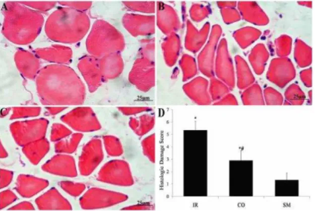
Figure 1. Representative images (×400) of H&E staining of skeletal muscle tissues: A, IR group; B, colchicine treatment-IR (CO) group; C, sham operation (SM) group. D: Histological damage scores of muscle tissues in three groups. * P<0.05 compared to SM; # P<0.05 compared to IR
Statistics analyses
Statistical analyses were performed with SPSS version 13.0 software. Continuous and normally distributed data were presented as mean±standard deviation, and the one-way analysis of variance (ANOVA) followed by the post-hoc Bonferroni
method were used to compare differences among groups. A P-value <0.05 was considered statistically significant.
Results
Colchicine significantly reduced skeletal muscle damage by IR
First, we used microscopic examination and histological damage scoring system to evaluate the degree of IR-induced muscle injury in the SM, IR and CO groups.Skeletal muscle cell edema, muscle fiber degeneration, necrosis, sarcoplasm dissolution, interstitial vessel hemorrhage, and neutrophil infiltration were commonly observed in skeletal muscles subjected to IR injury in both the IR and CO groups but not in the SM group (Figure 1A - C). However,colchicine treatment significantly reduced this damage, which was accompanied by a decrease in structural damage and scattered neutrophil infiltration (Figure 1, compare A and B). Figure 1D shows higher histological damage scores in both the IR and CO groups compared to the SM group (P<0.05
vs SM); however, the CO group exhibited a significantly reduced damage score compared to the IR group (P<0.05 vs. IR).
Colchicine significantly diminished the IR-induced edema in skeletal muscles
W/D ratio was usedas an indicator of edema in gastrocnemius muscles. As shown in Figure 2, IR-induced injury led to a significant increase in muscle edema, as evidenced by higher W/D ratios in the IR and CO groups compared to the SM group (P<0.05 vs SM); however, colchicine treatment greatly decreased theW/D ratio in theIR- injured
Figure 2. The degrees of muscle edema in the IR group,
colchicine treated-IR (CO) group and sham operation (SM) group. * P<0.05 compared to SM; #P<0.05 compared to IR
muscles compared to the IR group (P <0.05 vs. IR).
Colchicine attenuated the IR-elicited oxidative stress and inflammatory response in skeletal muscles
Next, we evaluated oxidative stress in the IR-damaged skeletal muscle. We measured MPO activity, which represents the number of infiltrated neutrophils, SOD activity, MDA levels, and the inflammatory markers TNF- and )L- . As expected, IR significantly increased MPO activity as well as MDA, TNF- and )L- levels in muscle tissues, but decreased SOD activity in both the IR and CO groups compared to the SM group (P<0.05 vs. SM; Figure 3). However, colchicine treatment greatly improved MPO activity, MDA, TNF- and )L- levels, and SOD activity compared to the IR group (P <0.05 vs. IR; Figure 3).
Colchicine reduced the levels of circulating CK and LDH in IR-injured rats
Plasma CK and LDH levels are considered systemic biomarkers of muscle injury. As shown in Figure 4, plasma CK and LDH levels in both the IR and CO groups were significantly increased compared to the SM group (P<0.05 vs. SM). As expected, colchicine treatment significantly lowered the levels of circulating CK and LDH compared to the IR group (P<0.05 vs. IR).
Discussion
Our study showed that the gastrocnemius muscle was damaged following IR, as evidenced by pathological changes, increased edema and systemic muscle injury biomarkers. Furthermore, our study also revealed that treatment with colchicine prior to ischemia significantly attenuated IR-induced skeletal muscle damage.
Our study showed an increase in MPO activity in the muscle tissues subjected to IR. MPO, a heme protein, is most abundantly expressed in neutrophils, and increased activity is therefore an indicator of neutrophil activation. Local
Figure 3. Measurement of superoxide dismutase (SOD) activity (A), malondialdehyde (DMA) level (B), myeloperoxidase (MPO) activity (C), tumor necrosis factor- (TNF- ) level (D) and interleukin- (IL- ) level (E) in skeletal muscle tissues of rats from the three groups. * P<0.05 compared to SM; # P<0.05 compared to IR
feature of IR-induced injury, and neutrophil depletion decreases oxidative stress and attenuates IR-induced skeletal muscle injury (6, 7). Recruited and activated neutrophils are associated with abundant production of reactive oxygen species (ROS) as well as proinflammatory cytokines including TNF- and )L- (1, 18-20). ROS is increased during the early stage of reperfusion and induces expression of adhesive molecules on the microvessel endothelium and facilitates transendothelial migration of leukocytes, which in turn causes further production of ROS (20, 21). The interaction between ROS and membrane lipids leads to an increase in membrane permeability, consequently causing cell death (17, 22, 23). MDA is one of the most commonly used biomarkers of lipid peroxidation and reflects the extent of cellular oxidative injury, while SOD is an important enzyme that catalyzes the conversion of superoxide radical into hydrogen peroxide. As demonstrated in our study, the increased MDA level and decreased SOD activity in the muscle tissues after IR suggests that increased oxidative stress might play a role in IR-induced skeletal muscle injury, which is consistent with previous findings (24, 25).
Increased levels of proinflammatory cytokines, including TNF- and )L- , are also implicated in muscle injury induced by IR (4). In agreement
with these findings, we observed significant increases in TNF- and )L- levels in the injured
Figure 4. The Circulating levels of biomarkers lactate
dehydrogenase (LDH, A) and creatinine kinase (CK, B) in rats from the IR group, colchicine treated-IR (CO) group and sham operation (SM) group. * P<0.05 compared to SM; # P<0.05 compared to IR
muscle tissues. TNF- has been shown to activate polymorphonuclear neutrophils and trigger neutron-phil infiltration. Moreover, TNF- induces chemo-tactic factors and adhesion molecules in vascular endothelial cells and facilitates leukocytes infiltra-tion, which contributes to excessive inflammatory processes (26, 27). Similarly, IL- up-regulates endothelial cell adhesion molecules and induces endothelial cells to release IL-8 which also contributes to excessive inflammatory responses (28). Conversely, polyclonal antibodies against
and )L-1, or soluble TNF- receptor and )L-1 receptor antagonists, reduce neutrophil chemotaxis and sequestration and provide significant protection against IR-induced limb injury (4).
and colchicine has been shown to attenuating lipid peroxidation and stabilizing membranes (38). Consistent with these observations, our study demonstrated that 1 mg/kg colchicine in rats significantly decreased MDA, TNF- and )L- levels, and increased SOD activity in IR-injured muscle tissues, indicating that colchicine treatment decreases levels of lipid peroxidation and inflammatory cytokines. However, further studies are needed to: 1) determine the dose-effect relationship and the functional interactions of colchicine with other agents, 2) evaluate the efficacy of colchicine treatment in IR injury in a clinical setting, and 3) investigate the effects of colchicine on remote organ dysfunction following IR injury in skeletal muscle.
Conclusion
IR leads to skeletal muscle damage, and colchicine treatment prior to ischemia attenuates IR-triggered skeletal muscle injury in part by suppressing the inflammatory response and oxidative stress. Therefore, colchicine may serve as a potential alternative strategy to prevent or treat skeletal muscle injury induced by IR.
References
. Menger MD, Vollmar B. Pathomechanisms of ischemia-reperfusion injury as the basis for novel preventive strategies: is it time for the
introduction of pleiotropic compounds?
Transplant Proc 2007; 39:485-488.
2. Lin L, Wang L, Bai Y, Zheng L, Zhao X, Xiong X, et al. Pulmonary gas exchange impairment following tourniquet deflation: a prospective, single-blind clinical trial. Orthopedics 2010; 33:395.
3. Zhang X, Jizhang Y, Xu X, Kwiecien TD, Li N, Zhang Y, et al. Protective effects of remote
ischemic conditioning against
ischemia/-reperfusion-induced retinal injury in rats. Vis Neurosci 2014; 31:245-252.
4. Seekamp A, Warren JS, Remick DG, Till GO, Ward PA. Requirements for tumor necrosis factor-alpha and interleukin-1 in limb ischemia/reperfusion injury and associated lung injury. Am J Pathol 1993; 143:453-463.
5. Clanton TL, Zuo L, Klawitter P. Oxidants and
skeletal muscle function: physiologic and
pathophysiologic implications. Proc Soc Exp Biol Med 1999; 222:253-262.
6. Carden DL, Granger DN. Pathophysiology of ischaemia-reperfusion injury. J Pathol 2000; 190:255-266.
7. Iwahori Y, Ishiguro N, Shimizu T, Kondo S, Yabe Y, Oshima T, et al. Selective neutrophil depletion
with monoclonal antibodies attenuates
ischemia/reperfusion injury in skeletal muscle. J Reconstr Microsurg 1998; 14:109-116.
8. Altinor S, Ozturkcan S, Hah MM. The effects of colchicine on neutrophil function in subjects with recurrent aphthous stomatitis. J Eur Acad Dermatol Venereol 2003; 17:469-470.
9. Paschke S, Weidner AF, Paust T, Marti O, Beil M, Ben-Chetrit E. Technical advance: Inhibition of neutrophil chemotaxis by colchicine is modulated through viscoelastic properties of subcellular compartments. J Leukoc Biol 2013; 94:1091-1096. 10. Lidar M, Livneh A. Familial Mediterranean
fever: clinical, molecular and management
advancements. Neth J Med 2007; 65:318-324. 11. Schlesinger N, Schumacher R, Catton M, Maxwell L. Colchicine for acute gout. Cochrane Database Syst Rev 2006; 18:CD006190.
12. Caner JE. Colchicine inhibition of chemotaxis. Arthritis Rheum 1965; 8:757-764.
13. Malawista SE, Bodel PT. The dissociation by colchicine of phagocytosis from increased oxygen consumption in human leukocytes. J Clin Invest 1967; 46:786-796.
14. Goldfinger SE, Howell RR, Seegmiller JE. Suppression of metabolic accompaniments of phagocytosis by colchicine. Arthritis Rheum 1965; 8:1112-1122.
15. Reibman J, Haines KA, Rich AM, Cristello P, Giedd KN, Weissmann G. Colchicine inhibits ionophore-induced formation of leukotriene B4 by human neutrophils: the role of microtubules. J Immunol 1986; 136:1027-1032.
16. Gillespie E, Lichtenstein LM. Histamine release from human leukocytes: studies with deuterium oxide, colchicine, and cytochalasin B. J Clin Invest 1972; 51:2941-2947.
17. Erkanli K, Kayalar N, Erkanli G, Ercan F, Sener
G, Kirali K. Melatonin protects against
ischemia/reperfusion injury in skeletal muscle. J Pineal Res 2005; 39:238-242.
18. Kozower BD, Kanaan SA, Tagawa T, Suda T, Grapperhaus K, Daddi N, et al. Intramuscular gene transfer of interleukin-10 reduces neutrophil recruitment and ameliorates lung graft ischemia-reperfusion injury. Am J Transplant 2002; 2:837-842.
19. Strieter RM, Kunkel SL, Bone RC. Role of tumor necrosis factor-alpha in disease states and inflammation. Crit Care Med 1993; 21:S447-463. 20. Li C, Jackson RM. Reactive species mechanisms of cellular hypoxia-reoxygenation injury. Am J Physiol Cell Physiol 2002; 282:C227-241.
21. Daddi N, Suda T, D'Ovidio F, Kanaan SA, Tagawa T, Grapperhaus K, et al. Recipient intramuscular cotransfection of naked plasmid transforming growth factor beta1 and interleukin 10 ameliorates lung graft ischemia-reperfusion injury. J Thorac Cardiovasc Surg 2002; 24:259-269.
22. Sadoshima J. The role of autophagy during ischemia/reperfusion. Autophagy 2008; 4:402-403.
23. Ma JQ, Ding J, Zhang L, Liu CM. Protective effects of ursolic acid in an experimental model of liver fibrosis through Nrf2/ARE pathway. Clin Res Hepatol Gastroenterol 2015; 39:188-197.
24. Bolcal C, Yildirim V, Doganci S, Sargin M, Aydin A, Eken A, et al. Protective effects of antioxidant medications on limb ischemia reperfusion injury. J Surg Res 2007; 139:274-279.
ischemia-reperfusion injury in rat skeletal muscle. J Surg Res 2014; 186:240-245.
26. Zhou Z, Liu Y, Miao AD, Wang SQ. Protocatechuic aldehyde suppresses TNF-alpha-induced ICAM-1 and VCAM-1 expression in human umbilical vein endothelial cells. Eur J Pharmacol 2005; 513:1-8.
27. Chai H, Wang Q, Huang L, Xie T, Fu Y. Ginsenoside Rb1 inhibits tumor necrosis factor-alpha-induced vascular cell adhesion molecule-1 expression in human endothelial cells. Biol Pharm Bull 2008; 31:2050-2056.
28. Strieter RM, Kunkel SL, Showell HJ, Remick DG, Phan SH, Ward PA, et al. Endothelial cell gene expression of a neutrophil chemotactic factor by TNF-alpha, LPS, and IL-1 beta. Science 1989; 243:1467-1469.
29. Yurttutan S, Ozdemir R, Canpolat FE, Oncel MY, Uysal B, Unverdi HG, et al. Protective effects of colchicine in an experimental model of necrotizing enterocolitis in neonatal rats. J Surg Res 2013; 183:156-162.
30. Forrat R, Sebbag L, Ferrera R, Hadour G, Canet E, Tabib A, et al. Effect of colchicine on circulating and myocardial neutrophils and on infarct size in a canine model of ischemia and reperfusion. J Cardiovasc Pharmacol 1996; 27:876-883.
31. Pratt J, Roux M, Henneguelle E, Stutzmann JM, Laduron PM. Neuroprotective effects of colchicine in the gerbil model of cerebral ischaemia. Neurosci Lett 1994; 169:114-118.
32. Kurt RK, Dogan AC, Dogan M, Albayrak A, Kurt SN, Eren F, et al. Protective effect of colchicine on ovarian ischemia-reperfusion injury: an
experimental study. Reprod Sci 2015; 22:545-550. 33. Ouyang Y, Wang W, Bhuta S, Chang YH. Mechanism of action of colchicine. VI: Effect of colchicine on generation of leukotriene B4 by human polymorphonuclear leukocytes. Clin Exp Rheumatol 1989; 7:397-402.
34. Mikenberg I, Widera D, Kaus A, Kaltschmidt B, Kaltschmidt C. TNF-alpha mediated transport of NF-kappaB to the nucleus is independent of the cytoskeleton-based transport system in non-neuronal cells. Eur J Cell Biol 2006; 85:529-536. 35. Jackman RW, Rhoads MG, Cornwell E, Kandarian SC. Microtubule-mediated NF-kappaB activation in the TNF-alpha signaling pathway. Exp Cell Res 2009; 315:3242-3249.
36. Mollinedo F, Nieto JM, Andreu JM. Cytoplasmic microtubules in human neutrophil degranulation: reversible inhibition by the colchicine analogue 2- methoxy-5-(2',3',4'-trimethoxyphenyl)-2,4,6-cycloheptatrien-1- one. Mol Pharmacol 1989; 36:547-555.
37. Reibman J, Haines KA, Gude D, Weissmann G. Differences in signal transduction between Fc gamma receptors (Fc gamma RII, Fc gamma RIII) and FMLP receptors in neutrophils. Effects of
colchicine on pertussis toxin sensitivity and diacylglycerol formation. J Immunol 1991;
146:988-996.
38. Das D, Pemberton PW, Burrows PC, Gordon C,
Smith A, McMahon RF, et al. Antioxidant
properties of colchicine in acute carbon