RBCCV 44205-1617 DOI 10.5935/1678-9741.20140066
Thymoquinone protects end organs from abdominal
aorta ischemia/reperfusion injury in a rat model
Timoquinona protege órgãos terminais da isquemia/reperfusão da aorta abdominal em modelo com ratos
Mehmet Salih Aydin
1, MD; Aydemir Kocarslan
1, MD; Sezen Kocarslan
2, MD; Ahmet Kucuk
4, MD;
İrfan
Eser
4, MD; Hatice Sezen
5, MD; Evren Buyukirat
3, MD; Abdussemet Hazar
1, MD
1Harran Univercity Faculty of Medicine, Department of Cardiovascular
Sur-gery, Sanliurfa, Turkey.
2Harran Univercity Faculty of Medicine, Department of Pathology,
Sanli-urfa, Turkey.
3Harran Univercity Faculty of Medicine, Department of Anesthesiology and
Reanimation, Sanliurfa, Turkey.
4Harran Univercity Faculty of Medicine, Department of Thoracic Surgery,
Sanliurfa, Turkey.
5Harran Univercity Faculty of Medicine, Department of Biochemistry,
San-liurfa, Turkey.
No inancial support.
This study was carried out at Harran Univercity Faculty of Medicine, Department of Cardiovascular Surgery, Sanliurfa, Turkey.
Correspondence address: Mehmet Salih Aydın
Department of Cardiovascular Surgery, Harran Univercity Faculty of Medicine 63000 Sanliurfa, Turkey
E-mail: [email protected]
Article received on February 19th, 2014
Article accepted on March 31st, 2014 Abstract
Introduction: Previous studies have demonstrated that thy-moquinone has protective effects against ischemia reperfusion injury to various organs like lungs, kidneys and liver in different experimental models.
Objective: We aimed to determine whether thymoquinone has favorable effects on lung, renal, heart tissues and oxidative stress in abdominal aorta ischemia-reperfusion injury.
Methods: Thirty rats were divided into three groups as sham (n=10), control (n=10) and thymoquinone (TQ) treatment group (n=10). Control and TQ-treatment groups underwent abdomi-nal aorta ischemia for 45 minutes followed by a 120-min period of reperfusion. In the TQ-treatment group, thymoquinone was given 5 minutes. before reperfusion at a dose of 20 mg/kg via an intraperitoneal route. Total antioxidant capacity, total oxidative status (TOS), and oxidative stress index (OSI) in blood serum were measured and lung, kidney, and heart tissue histopatholo-gy were evaluated with light microscopy.
Results: Total oxidative status and oxidative stress index activity in blood samples were statistically higher in the con-trol group compared to the sham and TQ-treatment groups (P<0.001 for TOS and OSI). Control group injury scores were
statistically higher compared to sham and TQ-treatment groups (P<0.001 for all comparisons).
Conclusion: Thymoquinone administered intraperitoneally was effective in reducing oxidative stress and histopathologic injury in an acute abdominal aorta ischemia-reperfusion rat model.
Descriptors: Aorta, Abdominal. Ischemia-Reperfusion Injury. Oxidative Stress.
Resumo
Introdução: Estudos prévios demonstraram que a timoqui-nona tem efeitos protetores contra a lesão de isquemia/reperfusão em vários órgãos como pulmão, rins e fígado em diferentes mo-delos experimentais.
Objetivo: Determinar se timoquinona tem efeitos positivos em tecidos do pulmão, rim e coração e no estresse oxidativo em lesão de isquemia/perfusão da aorta abdominal.
INTRODUCTION
Acute abdominal aorta ischemia followed by reperfusion may be encountered in several clinical circumstances, such as abdominal aortic aneurysm or dissection repair, acute thromboembolism with aortic atherosclerosis, or trauma sur-gery being brought to the emergency room. Such clinical sce-narios are associated with high mortality and morbidity rates
due to a systemic inlammatory response and multiple organ
dysfunction occurring during the reperfusion phase. Reperfu-sion of an acutely ischemic aorta may, paradoxically, lead to
systemic complications that account for signiicant morbidity
and mortality[1,2]. Overproduction of reactive oxygen species (ROS) and proinlammatory molecules and the subsequent inlammatory response is one of the most crucial underlying
mechanisms[2] that initiates injury, especially in the lungs and
vital organs, such as kidney and heart, with a subsequent high morbidity[1-4].
Thymoquinone (TQ; 2-isopropyl-5-methyl-1, 4-benzoqui-none), the active constituent of Nigella sativa seeds, is a macologically active quinone that has been shown to have phar-macological actions, such as antibacterial[5], antihypertensive[6],
antidiabetic[7], neuroprotective[8], anti-inlammatory[9] and
antia-poptotic[10] as well as, in some studies, apoptotic[11,12].
It has been reported that TQ prevents oxidative injury in var-ious in vitro and in vivo studies[13,14]. TQ possesses strong
anti-oxidant properties through its ability to scavenge different free radicals[15,16]. It has also been reported that TQ attenuated several
organ injuries (lung, renal, hepatic) in different ischemia-reper-fusion (I/R) models (renal, hepatic). However, no studies have evaluated the protective effects of TQ in an aorta I/R model[17-20].
The purpose of this study was to determine the eficacy
of TQ in preventing injury in vital organs (lung, heart and kidney) in an acute abdominal aorta ischemia-reperfusion model in rats.
submetidos à isquemia da aorta abdominal durante 45 minu-tos, seguido por um período de 120 minutos de reperfusão. No grupo de tratamento com TQ, a timoquinona foi administrada 5 minutos antes da reperfusão, dose de 20 mg/kg através da via intraperitoneal. A capacidade total antioxidante, estado oxida-tivo total (TOS) e o índice de estresse oxidaoxida-tivo (OSI) no soro
Abbreviations, acronyms & symbols
I/R Ischemia-Reperfusion OSI Oxidative Stress Index ROS Reactive Oxygen Species TAC Total Antioxidant Capacity TOS Total Oxidant Status
TQ Thymoquinone
METHODS
The experimental study was performed on a total of 30 three-month-old Wistar-albino rats weighing 200–250 g. All animals were maintained under standard conditions and treat-ed in compliance with National Institutes of Health guidelines. They were housed on a 12-h dark/light cycle schedule with lights on at 06.00 h. Rats were deprived of food, though not water, for 12 hours before surgery. Experiments were done in the Harran University Experimental Research Center. The rats were randomly assigned to three experimental groups: sham operation, control (I/R; non-treated), and TQ-treated I/R. Rats were anesthetized using ketamine hydrochloride (0.2mL/100 g) in all experiments. The abdomen was explored through a midline incision after shaving and disinfection. In the sham group, only laparotomy was performed. In the control group, I/R injury was induced by clamping the aorta under renal vas-cular pedicles for 45 minutes, followed by 2 hours of reper-fusion. In the TQ-treated I/R group, I/R injury was also in-duced by clamping the aorta under renal vascular pedicles for 45 minutes and TQ was given 5 minutes before reperfusion at a dose of 20 mg/kg via the intraperitoneal route, and again reperfusion was established for 2 hours. Heparin was not used due to possibility of affecting histopathological or biochemical
results. At the end of the procedures, the rats were sacriiced
after blood sampling, and then kidney, lung, and heart tissues were obtained from all rats.
TQ were purchased from Sigma–Aldrich (St. Louis, MO).
The purity (GC) of TQ was ≥98.5% as per the manufacturer’s speciication and was dissolved in dimethyl sulphoxide.
Biochemical Analyses
Measurement of Total Antioxidant Capacity
TAC of supernatant fractions was determined using a novel automated measurement method developed by Erel[21].
do sangue foram medidos, e a histopatologia dos tecidos do pul-mão, rim e coração foram avaliados com microscopia de luz.
Resultados: Estado oxidativo e índice de estresse oxidativo to-tal em amostras de sangue foram estatisticamente mais altos no grupo controle em relação aos grupos sham e tratamento com TQ (P<0,001 para TOS e OSI). Escores de lesões no grupo controle foram estatisticamente mais altos em relação aos grupos sham e tratamento com TQ (P<0,001 para todas as comparações).
Conclusão: A timoquinona administrada por via intraperitoneal foi eicaz na redução do estresse oxidativo e lesão histopatológica em modelo de rato de isquemia/reperfusão aguda da aorta abdominal.
Hydroxyl radicals, the most potent biological radicals, are produced in this method. In the assay, the ferrous ion solution present in Reagent 1 is mixed with hydrogen peroxide, which is present in Reagent 2. The subsequently produced radicals, such as brown-colored dianisidinyl radical cations produced by the hydroxyl radicals, are also potent radicals. Using this method, the antioxidative effect of the sample was measured against the potent-free radical reactions initiated by the pro-duced hydroxyl radicals. The assay has excellent precision,
with values lower than 3%. The results are expressed as nmo -lTrolox Equiv./mg protein.
Measurement of Total Oxidant Status
TOS of supernatant fractions was determined using a novel automated measurement method developed by Erel[22].
Oxidants present in the sample oxidize the ferrous ion–o-di-anisidine complex to ferric ion. The oxidation reaction is enhanced by glycerol molecules, which are abundant in the reaction medium. The ferric ion produces a colored complex with xylenol orange in an acidic medium. The color intensity, which can be measured spectrophotometrically, is related to the total amount of oxidant molecules present in the sample. The assay was calibrated with hydrogen peroxide, and the re-sults are expressed in terms of nmol H2O2 Equiv/mg protein.
Oxidative Stress Index
The percent ratio of TOS level to TAC level was deined
as OSI. OSI values were calculated according to the follow-ing formula[23]:
OSI (arbitrary unit) = TOS (nmol H2O2 Equiv/mg pro-tein)/TAC (nmolTroloxEquiv/mg protein).
Histopathological Evaluation
The kidney, lung, and heart of each animal were ob-tained for histological evaluation. Samples of these organs were placed in formalin and embedded in wax according to standard protocols. They were subsequently sectioned
at 5 μm slice thickness and stained with hematoxylin and
eosin. Magniication of × 20 was used (Olympus BX51
TF, USA). Samples were then graded histologically ac-cording to the severity of injury using a predetermined scoring system[24]. The predetermined scoring system, from
Solez et al.[24], included tubular necrosis, interstitial edema,
loss of brush border, and cast formation, in which the score was 0 for absent; 1 for mild to moderate; and 2 for marked renal involvement. The histological parameters for lung evaluation were alveolar congestion, intra-alveolar
hem-orrhage, and interstitial-perivascular iniltration of neutro -phils, in which the assessment score was 0 for absent; 1 for mild focal; 2 for moderate focal; and 3 for severe marked
lung involvement. Interstitial edema, inlammatory cell in
-iltration, and coagulation necrosis were assessed for heart
examination, in which the score was 0 for absent; 1 for mild focal; 2 for moderate focal; and 3 for severe marked heart involvement. Histological analysis was performed by a blinded expert.
Statistical Analysis
Statistical analyses were performed using SPSS 11.5 (SPSS for Windows 11.5, Chicago, IL). Continuous data are expressed as mean±SD whereas categorical variables are presented as number (count) and percentage. Distribution of continuous variables was assessed with one-sample Kolmog-orov-Smirnov test and indicated that all variables were ab-normally distributed. Therefore, nonparametric independent group comparisons were made: for multiple comparisons, the Kruskal-Wallis test was used, and for comparisons between groups, the Mann-Whitney test was used if any statistical
signiicance was found. A two-sided P value of <0.05 was
considered statistically signiicant.
RESULTS
All animals survived through the experimental protocol.
TAC activity in blood samples were signiicantly higher in
the sham group than in the treatment and control groups
Table 1. Oxidative and antioxidative parameters and histopathological evaluation in Sham, Control and TQ + I/R rats.
TAC (nmolTroloxEqv./mg protein) TOS (nmol H2O2 Eqv./ mg protein) OSI (arbitrary units)
Renal Pathology score Lung Pathology score Heart Pathology score
Sham (n=10) 1.39±0.18*
28.3±8.5 2.0±0.44 1.7±1.25 1.7±1.15 0.3±0.48
Control (n=10) 0.53±0.12 44.1±8.1+ 8.35±1.23+
4.4±0.69+ 4.6±0.51+ 1.5±0.52+
TQ+I/R (n=10) 0.65±0.12
25.8±2.3 1.30±0.41
2.1±1.37 3.0±1.24 0.9±0.73
P P<0.001
P<0.001
P<0.001
P<0.001
P<0.001
P<0.001
TAC=Total Antioxidant Capacity; TOS=Total Oxidant Status; OSI=Oxidative Stress Index
P<0.05 was considered as statistically signiicant.
Fig. 1 - TAC levels for sham, control, and thymoquinine groups. * P<0.001 (for all comparisons) compared with I/R and I/R+TQ
Fig. 2 - TOS levels in sham, control, and thymoquinine groups. + P<0.001 (for all comparisons) compared with sham and thymoquinine groups
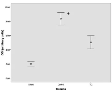
Fig. 3 - OSI levels in sham, control, and thymoquinine groups. +P<0.001 (for all comparisons) compared with sham and thymoquinine groups
(P<0.001; for all comparisons) but there were no
statistical-ly signiicant differences between the treatment group and
control group for TAC activity (P>0.05). TOS and OSI ac-tivity in blood samples were statistically higher in the control group than in the sham and thymoquinone group (P<0.001 for all comparisons). Histopathologic injury scores of renal, lung and heart tissues are summarized in Table 1. Control group injury scores were statistically increased compared to sham and thymoquinone groups (P<0.001 for all compari-sons). The results are summarized in Figures1, 2, and 3.
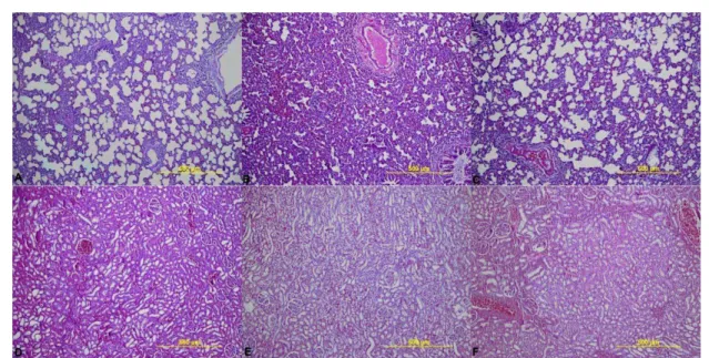
Upon histopathological evaluation, renal, lung and heart tissues were found to be normal with no pathological chang-es in the sham group (Figurchang-es 4A and 4D). Histopathologi-cal examination of the tissues in the control group revealed severe lesions, such as tubular damage characterized by cast formation, the loss of brush border and interstitial edema in the kidney. Histopathological examination of the tissues in
the control group revealed neutrophil and leukocyte iniltra -tion with alveolar conges-tion in the lung. Histopathological examination of the tissues in the control group revealed inter-stitial edema in the heart (Figures 4B and 4E). In rats receiv-ing TQ intraperitoneally, these lesions were less severe than in the control group (Figures 4C and 4F).
DISCUSSION
In our experimental study, we hypothesized that abdominal aorta ischemia for 45 minutes followed by reperfusion for 2 hours would cause renal, lung, and heart pathology and we have found that (i) abdominal aorta ischemia for 45 minutes followed
by reperfusion for 2 hours caused signiicant pathology in lung,
renal, and heart tissues (ii) TOS and OSI levels were increased in the control group and (iii) TOS, OSI, and histopathological injury scores were decreased in sham and TQ+IR groups.
It has been recognized that multiple organ dysfunction syndrome is a major cause of morbidity and mortality after abdominal aortic surgery and contributes to
approximate-ly 25% of all deaths in elective abdominal aorta repair. It
endotoxin across the intestinal mucosal barrier, leading to the
systemic release of reactive oxygen species (ROS) and inlam -matory cytokines, which not only damage the gut itself but also harm distant organs, including heart, kidney, and lung[25].
Nigella sativa (NS), also known as black seed or black cumin, has long been used in folk medicine. NS contains
more than 30% of a ixed oil and 0.40-0.45 w/w of a vola
-tile oil. The vola-tile oil has been shown to contain 18-24% thymoquinone (TQ) and 46% monoterpenes[7]. NS has been reported to exhibit anti-inlammatory, immunomodulatory,
and anti-neoplastic effects in many experimental and clinical studies[26-28]. TQ, the active constituent of Nigella sativa seeds
similar to NS, also showed favorable effects with respect to
oxidative stress and inlammation. Thus, TQ has attracted
the attention of scientists to investigate its molecular mech-anisms and potential use in the treatment of different
diseas-es. It has been shown to have antioxidant/anti-inlammatory
effects in several diseases, including experimental allergic encephalomyelitis, colitis, arthritis encephalomyelitis, diabe-tes, asthma, and carcinogenesis[10]. TQ attenuated lipid
per-oxidation and increased antioxidant enzyme activities. It has been reported to have strong antioxidant potential through its ability to scavenge different free radicals, its scaveng-ing power bescaveng-ing as effective as SOD against superoxide anions[16-18]. It acts as a scavenger of superoxide, hydroxyl
radicals and singlet molecular oxygen[29]. Furthermore, recent
studies have demonstrated that TQ supplementation increases
Fig. 4 - A and D show renal and lung tissues samples of the sham group and there were no pathological changes. B)
shows lung tissues samples of control group and neutrophil and leukocyte iniltration with alveolar congestion were
observed. E) shows renal tissues samples of control group and tubular damage characterized by cast formation; the loss of brush border and interstitial edema were observed. C and F show lung and renal tissues samples of thymoquinine treated group and there were less severe lesions than in the control group.
the expression of antioxidant genes, SOD, catalase and gluta-thione peroxidase in rat liver. Thus, TQ may reduce oxidative stress through a direct antioxidant effect as well as through the induction of endogenous antioxidant enzymes[30].
TQ also inhibited inducible nitric oxide synthase mRNA ex-pression in rat lipopolysaccharide-stimulated peritoneal macro-phage cells[31,32], which has been attributed to its ability to reduce oxidative stress-induced inlammation leading to the prevention
of inducible NOS (nitric oxide synthase) upregulation.
Several studies reported protective effects in the lung in different situations with different mechanisms. Suddek et al. showed that TQ produces a protective mechanism against cysplatin-induced pulmonary damage with anti-oxidant and
anti-inlammatory properties and, in addition, TQ has been found to have potential antiibrotic effects besides its antiox
-idant activity, which could be through NF-κB inhibition, in
bleomycin-induced oxidative stress of rat lungs[20,33]. Renal
protective effects of TQ have also been discussed in several studies, including vancomycin induced nephrotoxicity, inor-ganic mercury intoxication, and gentamicin-induced acute renal toxicity. These studies highlight the importance of reac-tive oxygen species in renal pathophysiology and the intrigu-ing possibility of TQ play a role in the prevention of and/ or protection from renal injury in humans[17,34-36]. Myocardial
has also been widely studied in different ischemia reperfu-sion models and reported to have favorable effects with dif-ferent potential mechanisms, including primarily antioxidant mechanisms[18,40]. In this study we also found protective
ef-fects of TQ in the lung, kidney, and heart with
histopatholog-ic evaluation. Signihistopatholog-icant oxidative stress in the control group
compared to sham and TQ groups also emphasizes that the anti-oxidant properties of TQ might be the probable protec-tive mechanism in the acute abdominal aorta ischemia-reper-fusion model in the rat.
We believe that there are suficient preclinical research
results with a considerable amount of information about TQ
regarding its molecular antioxidant, anti-inlammation, anti -cancer activity, drug toxicity, bioavailability and pharmaco-kinetics, and novel drug delivery approaches, to encourage the use of TQ in clinical settings[41]. However, the clinical
im-plications and appropriate pathophysiological mechanisms
of the indings of the present study remain to be elucidated
with further large-scale clinical studies.
Several limitations of this study should be considered. One of the potential limitations is the absence of oral ad-ministration of TQ versus an intraperitoneal route. Another limitation is the absence of biochemical analysis of different biochemical markers, including urea, creatinine, creatinine phosphokinase and creatinine kinase MB for the heart. Fur-ther studies focusing on IR injury of oFur-ther end organs, such as intestine, brain and medulla spinalis injury are needed.
CONCLUSION
In conclusion, TQ administered intraperitoneally was effec-tive in reducing oxidaeffec-tive stress and histopathologic injury in an acute abdominal aorta I/R rat model. Oxidative stress indices and
tissue injuries might be modiied with TQ treatment in different
clinical settings. However, further large scale studies are needed
to deine the possible favorable effects of TQ in clinical settings.
Authors’ roles & responsibilities
MSA Analysis and /or interpretation of data, statistical analysis, i -nal approval of the manuscript conception and study design, conduct of procedures, and/or experiments, writing of the manuscript or review of its content
AK Analysis and/or interpretation of data, inal approval of manuscript
SK Statistical analysis, conception and study design, conduct of procedures, and/or experiments
AK Conduct of procedures and/or experiments, writing of the manuscript or review of its content
IE Statistical analysis, inal approval of manuscript
HS Statistical analysis, inal approval of the manuscript, conception and study design, conduct of procedures, and/or experiments EB Statistical analysis, inal approval of the manuscript, drafting
the manuscript or revising it critically for its content AH Drafting of the manuscript or review of its content
REFERENCES
1. Yassin MM, Harkin DW, Barros D’Sa AA, Halliday MI, Rowlands
BJ. Lower limb ischemia-reperfusion injury triggers a systemic
inlammatory response and multiple organ dysfunction. World J
Surg. 2002;26(1):115-21.
2. Carvalho AC, Guillaumon AT, Cintra EdeA, Figueiredo LC, Moreira MM, Araújo S. Plasmatic vasopressin in patients undergoing con-ventional infra-renal abdominal aorta aneurysm repair. Rev Bras Cir Cardiovasc 2011;26(3):404-12
3. Harkın DW, Barros D’sa AA, Mccallion K, Hoper M, Hallıday
MI, Campbell FC. Circulating neutrophil priming and systemic
inlammation in limb ischaemia-reperfusion injury. IntAngiol
2001;20(1):78-89.
4. Groeneveld AB, Raıjmakers PG, Rauwerda JA, Hack CE. The in
-lammatory response to vascular surgery-associated ischaemia and
reperfusion in man: effect on postoperative pulmonary function. Eur J Vasc Endovasc Surg 1997;14(5):351-9.
5. Hanafy MS, Hatem ME. Studies on the antimicrobial activity of Nigella sativa seed (black cumin). J Ethnopharmacol. 1991;34(2-3):275-8.
6. el-Tahir K, Ashour M, al-Harbi M. The cardiovascular actions of the volatile oil of the black seed (Nigella sativa) in rats: elucidation of the mechanism of action. Gen Pharmacol 1993;24(5):1123-31.
7. Kanter M. Effects of Nigella sativa and its major constituent, thy-moquinone on sciatic nerves in experimental diabetic neuropathy. Neurochem Res. 2008;33(1):87-96.
8. Al-Majed AA, Al-Omar FA, Nagi MN. Neuroprotective effects of thymoquinone against transient forebrain ischemia in the rat hippo-campus. Eur J Pharmacol. 2006;543(1-3):40-7.
9. Mutabagani A, El-Mahdy SA. Study of the anti-inlammatory ac -tivity of Nigella sativa L. and thymoquinone in rats. Saudi Pharm J. 1997;5(2-3):110-3.
10. Woo CC, Kumar AP, Sethi G, Tan KH. Thymoquinone: potential
cure for inlammatory disorders and cancer. Biochem Pharmacol.
2012;83(4):443-51.
11. Wirries A, Breyer S, Quint K, Schobert R, Ocker M. Thymoquinone hydrazone derivatives cause cell cycle arrest in p53-competent colorectal cancer cells. Exp Ther Med. 2010;1(2):369-75.
12. Roepke M, Diestel A, Bajbouj K, Walluscheck D, Schonfeld P, Roessner A, et al. Lack of p53 augments thymoquinone-induced apoptosis and caspase activation in human osteosarcoma cells. Cancer Biol Ther. 2007;6(2):160-9.
13. Suguna P, Geetha A, Aruna R, Siva GV. Effect of thymoquinone on ethanol and high fat diet induced chronic pancreatitis--a dose response study in rats. Indian J Exp Biol. 2013;51(4):292-302.
14. Rifaioglu MM, Nacar A, Yuksel R, Yonden Z, Karcioglu M, Zorba
-quinone in an acute pseudomonas prostatitis rat model. Urol Int. 2013;91(4):474-81.
15.Selçuk CT, Durgun M, Tekin R, Yolbas L, Bozkurt M, Akçay C, et al. Evaluation of the effect of thymoquinone treatment on wound healing in a rat burn model. J Burn Care Res. 2013;34(5):e274-81.
16. Badary OA, Taha RA, Gamal el-Din AM, Abdel-Wahab MH. Thy-moquinone is a potent superoxide anion scavenger. Drug Chem Toxicol. 2003;26(2):87-98.
17. Fouda AM, Daba MH, Dahab GM, Sharaf El-Din OA. Thymoqui-none ameliorates renal oxidative damage and proliferative response induced by mercuric chloride in rats. Basic Clin Pharmacol Toxicol. 2008;103(2):109-18.
18. Awad AS, Kamel R, Sherief MA. Effect of thymoquinone on hepa-torenal dysfunction and alteration of CYP3A1 and spermidine/sper-mine N-1-acetyl-transferase gene expression induced by renal isch-aemia reperfusion in rats. J Pharm Pharmacol. 2011;63(8):1037-42.
19. Abd El-Ghany RM, Sharaf NM, Kassem LA, Mahran LG, Heikal OA. Thymoquinone triggers anti-apoptotic signaling targeting death ligand and apoptotic regulators in a model of hepatic ischemia reperfusion injury. Drug Discov Ther. 2009;3(6):296-306.
20. Suddek GM, Ashry NA, Gameil NM. Thymoquinone attenuates
cyclophosphamide-induced pulmonary injury in rats. Inlammo -pharmacology. 2013;21(6):427-35.
21. Erel O. A novel automated method to measure total antioxidant response against potent free radical reactions. Clin Biochem. 2004;37(2):112-9.
22. Erel O. A new automated colorimetric method for measuring total oxidant status. Clin Biochem. 2005;38(12):1103-11.
23. Bolukbas C, Bolukbas FF, Horoz M, Aslan M, Celik H, Erel O. Increased oxidative stress associated with the severity of the liver disease in various forms of hepatitis B virus infection. BMC Infect Dis. 2005;5:95.
24. Solez K, Morel-Maroger L, Sraer JD. The morphology of “acute tubular necrosis” in man: analysis of 57 renal biopsies and a comparison with the glycerol model. Medicine (Baltimore). 1979;58(5):362-76.
25. Li C, Li YS, Xu M, Wen SH, Yao X, Wu Y, et al. Limb remote
ischemic preconditioning for intestinal and pulmonary protection during elective open infrarenal abdominal aortic aneurysm repair: a randomized controlled trial. Anesthesiology. 2013;118(4):842-52.
26. Ammar el-SM, Gameil NM, Shawky NM, Nader MA. Comparative
evaluation of anti-inlammatory properties of thymoquinone and
curcumin using an asthmatic murine model. Int Immunopharmacol. 2011;11(12):2232-6.
27. Keyhanmanesh R, Boskabady MH, Khamneh S, Doostar Y. Effect of thymoquinone on the lung pathology and cytokine levels of ov-albumin-sensitized guinea pigs. Pharmacol Rep. 2010;62(5):910-6.
28. Yildiz F, Coban S, Terzi A, Savas M, Bitiren M, Celik H, et al. Protective effects of Nigella sativa against ischemia-reperfusion injury of kidneys. Ren Fail. 2010;32(1):126-31.
29. Mansour MA, Nagi MN, El-Khatib AS, Al-Bekairi AM. Effects of thymoquinone on antioxidant enzyme activities, lipid peroxidation and DT-diaphorase in different tissues of mice: a possible mecha-nism of action. Cell Biochem Funct. 2002;20(2):143-51.
30. Ismail M, Al-Naqeep G, Chan KW. Nigella sativa thymoqui-none-rich fraction greatly improves plasma antioxidant capacity and expression of antioxidant genes in hypercholesterolemic rats. Free Radic Biol Med. 2010;48(5):664-72.
31. Nagi MN, Almakki HA, Sayed-Ahmed MM, Al-Bekairi AM. Thymoquinone supplementation reverses acetaminophen-induced oxidative stress, nitric oxide production and energy decline in mice liver. Food Chem Toxicol. 2010;48(8-9):2361-5.
32. El-Mahmoudy A, Matsuyama H, Borgan MA, Shimizu Y, El-Sayed MG, Minamoto N, et al. Thymoquinone suppresses expression of inducible nitric oxide synthase in rat macrophages. Int Immuno-pharmacol. 2002;2(11):1603-11.
33. El-Khouly D, El-Bakly WM, Awad AS, El-Mesallamy HO,
El-De-merdash E. Thymoquinone blocks lung injury and ibrosis by
attenuating bleomycin-induced oxidative stress and activation of nuclear factor Kappa-B in rats. Toxicology. 2012;302(2-3):106-13.
34. Basarslan F, Yilmaz N, Ates S, Ozgur T, Tutanc M, Motor VK, et al. Protective effects of thymoquinone on vancomycin-induced nephrotoxicity in rats. Hum Exp Toxicol. 2012;31(7):726-33.
35. Ragheb A, Attia A, Eldin WS, Elbarbry F, Gazarin S, Shoker A. The
protective effect of thymoquinone, an anti-oxidant and anti-inlam -matory agent, against renal injury: a review. Saudi J Kidney Dis Transpl. 2009;20(5):741-52.
36. Sayed-Ahmed MM, Nagi MN. Thymoquinone supplementation prevents the development of gentamicin-induced acute renal tox-icity in rats. Clin Exp Pharmacol Physiol. 2007;34(5-6):399-405.
37. Nagi MN, Mansour MA. Protective effect of thymoquinone against doxorubicin-induced cardiotoxicity in rats: a possible mechanism of protection. Pharmacol Res. 2000;41(3):283-9.
38. Nagi MN, Al-Shabanah OA, Hafez MM, Sayed-Ahmed MM. Thy-moquinone supplementation attenuates cyclophosphamide-induced cardiotoxicity in rats. J Biochem Mol Toxicol. 2011;25(3):135-42.
39. Randhawa MA, Alghamdi MS, Maulik SK. The effect of thymo-quinone, an active component of Nigella sativa, on isoproterenol induced myocardial injury. Pak J Pharm Sci. 2013;26(6):1215-9.
40. Gökçe A, Oktar S, Koc A, Gonenci R, Yalcinkaya F, Yonden Z, Duru M. Protective effect of thymoquinone in experimental testicular torsion. Urol Int. 2010;85(4):461-5.
41. Abukhader MM. Thymoquinone in the clinical treatment of cancer: