O xidative stre ss in the latissim us
do rsi m uscle o f diabe tic rats
1Laboratório de Fisiologia Cardiovascular, Departamento de Fisiologia,
Instituto de Ciências Básicas da Saúde, Universidade Federal do Rio Grande do Sul, Porto Alegre, RS, Brasil
2Unidade de Hipertensão e Divisão de Experimentação, and
3Divisão de Bioengenharia, Instituto do Coração, Faculdade de Medicina,
Universidade de São Paulo, São Paulo, SP, Brasil K.L.D. De Angelis1,
I.A. Cestari3, J. Barp1,
P. Dall’Ago1,
T.G. Fernandes1,
P.I. Homem de Bittencourt1,
A. Belló-Klein1, A.A. Belló1,
S. Llesuy1 and
M.C. Irigoyen1,2
Abstract
The purpose of the present study was to investigate the effects of experimental diabetes on the oxidant and antioxidant status of latissi-mus dorsi (LD) latissi-muscles of male Wistar rats (220 ± 5 g, N = 11). Short-term (5 days) diabetes was induced by a single injection of streptozo-tocin (STZ, 50 mg/kg, iv; glycemia >300 mg/dl). LD muscle of STZ-diabetic rats presented higher levels of thiobarbituric acid reactive substances (TBARS) and chemiluminescence (0.36 ± 0.02 nmol/mg protein and 14706 ± 1581 cps/mg protein) than LD muscle of normal rats (0.23 ± 0.04 nmol/mg protein and 7389 ± 1355 cps/mg protein). Diabetes induced a 92% increase in catalase and a 27% increase in glutathione S-transferase activities in LD muscle. Glutathione peroxi-dase activity was reduced (58%) in STZ-diabetic rats and superoxide dismutase activity was similar in LD muscle of both groups. A positive correlation was obtained between catalase activity and the oxidative stress of LD, as evaluated in terms of TBARS (r = 0.78) and by chemiluminescence (r = 0.89). Catalase activity also correlated in-versely with glutathione peroxidase activity (r = 0.79). These data suggest that an increased oxidative stress in LD muscle of diabetic rats may be related to skeletal muscle myopathy.
Co rre spo nde nce M.C. Irigoyen
Unidade de Hipertensão e Divisão de Experimentação Instituto do Coração, HC-FMUSP Av. Enéas de Carvalho Aguiar, 44 05043-000 São Paulo, SP Brasil
Fax: + 55+ 11-3069-5048
E-mail: hipirigoyen@ incor.usp.br
Presented at the XV Annual Meeting of the Federação de Sociedades de Biologia Experimental, Caxambu, MG, Brazil, August 23-26, 2000.
Research supported by CNPq, CAPES, and FAPERGS. Publication supported by FAPESP.
Received April 12, 2000
Accepted July 26, 2000
Ke y wo rds
·Streptozotocin-diabetes
·Latissimus dorsi muscle
·O xidative stress
·Catalase
·Glutathione
Reactive oxygen species (ROS) have been implicated in the pathophysiology of a large number of diseases and this role has been attributed to their high reactivity and delete-rious effects on cell structures (1,2).
Mechanisms that contribute to the high level of oxidative stress in diabetes may in-clude not only increased non-enzymatic cosylation (glycation) and auto-oxidative gly-cosylation but also metabolic stress in sorbi-tol pathway activity, changes in the levels of inflammatory mediators and in the status of
antioxidant defense systems (1). Although diabetes has profound effects on the bio-chemical, morphological and contractile properties of skeletal and cardiac muscle (3), the importance of oxidative stress and its antioxidant adaptations in skeletal muscle of diabetic rats has been less studied.
muscle degeneration and necrosis that com-promise contractile bioassist (4). However, selection criteria for this type of treatment do not exclude diabetic patients. The purpose of the present study was to investigate the effects of experimental streptozotocin-in-duced diabetes on the oxidant and antioxi-dant process in LD muscle.
Male Wistar rats from the Animal House of the Federal University of Rio Grande do Sul, Porto Alegre, RS, Brazil, weighing 190-250 g, had free access to food and water. One group (N = 6) of rats was made diabetic with a single iv injection of streptozotocin (STZ, 50 mg/kg) dissolved in citrate buffer, pH 4.5. Rats were fasted overnight before STZ injec-tion. A second group (N = 5) was injected with citrate buffer and maintained as a nor-mal control group.
After 5 days of STZ-induced diabetes, the animals were killed with a blow to the head, 1 ml of blood was collected into tubes containing a drop of heparin saline (Produtos Roche Químicos e Farmacêuticos, Rio de Janeiro, RJ, Brazil), and the LD muscles were isolated and removed. The blood samples were centrifuged for 10 min at 3000 g and plasma was collected for later glucose determination by a colorimetric enzymatic test (Boehring Institute, Mannheim, Ger-many).
The LD muscles were homogenized in an ultra-Turrax blender using 1 g of tissue per 5 ml of 150 mmol/l potassium chloride and 20 nmol/l phosphate buffer, pH 7.4, and the homogenates were centrifuged at 600 g for 10 min at -2o
C. Proteins were assayed by measurement with the Folin phenol reagent (5).
Aliquots of LD homogenates were added to thiobarbituric acid and butanol and used for malondealdehyde (MDA) determination according to the technique of Buege and Aust (6) for thiobarbituric acid reactive sub-stances (TBARS).
The chemiluminescence assay was car-ried out with an LKB Rack Beta Liquid
Scintillation Spectrometer 1215 (LKB Producter AB, Bromma, Sweden) in the out-of-coincidence mode at room temperature (25-27o
C). The assay consists of the addition of an organic synthetic hydroperoxide to tissue sample homogenates and the observa-tion of the response. Hydroperoxides are highly unstable. They can react with mem-brane lipids, initiating the lipid peroxidation process and yielding singlet oxygen and ex-cited carbonyls. These species are exex-cited and can stabilize by means of light emission. The supernatants were diluted in 140 mM KCl and 20 mM phosphate buffer, pH 7.4, and added to glass tubes which were placed in scintillation vials; 3 mM tert -butylhydro-peroxide was added and chemiluminescence was determined up to the maximal level of emission (7).
Catalase (CAT) activity was measured by monitoring the decrease in H2O2
concen-tration spectrophotometrically over time. Aliquots of the samples were added to 50 mM phosphate buffer in a quartz cuvette. After zeroing the instrument, H2O2 was added
to a final concentration of 10 mM in 0.9 ml and absorbance was measured at 240 nm. The specific activity of each sample was calculated on the basis of the activity of pure CAT as described by Aebi (8).
Superoxide dismutase (SOD) activity was determined in the homogenates by measur-ing the inhibition of the rate of autocatalytic adrenochrome formation at 480 nm in a reaction medium containing 1 mM epineph-rine and 50 mM glycine-NaOH, pH 10.5 (9).
g-Glutamyl-cysteine-synthetase (g-GCS) was assayed in LD homogenized in buffer A (100 mM Tris-HCl, 150 mM KCl, 20 mM MgCl2, 2 mM EDTA, and 5 mM
ß-mercap-toethanol, pH 8.0) by coupling ATP hy-drolysis to NADH consumption via the pyru-vate kinase/lactate dehydrogenase reaction (10). The reaction (37o
phosphoenolpyruvate, 5 mM ATP, 1.85 U/ ml pyruvate kinase, and 2.13 U/ml lactate dehydrogenase, pH 8.0.
Glutathione peroxidase (GPx) activity was measured by the method of Del Maestro (11). Sample aliquots were added to the assay mixture of 1 U/ml glutathione reduc-tase and 2 mM glutathione in 1 ml phosphate buffer and the mixtures were preincubated at 37ºC for 30 min. Subsequently NADPH and tert-butylhydroperoxide were added to final concentrations of 155 and 580 µM, respec-tively, and the change in absorbance at 340 nm was recorded at regular intervals over a period of 4 min. One enzyme unit was de-fined as the amount of GPx required to oxi-dize 1 µmol NADPH in 1 min at 25ºC and calculated on the basis of molar absorbance for NADPH at 340 nm.
Glutathione S-transferase (GST) was quantified in muscle homogenates that were assayed (37o
C) in a buffer containing 0.2 M sodium phosphate, pH 6.5, 20 mM reduced glutathione and 20 mM 1-Cl-2,4-dinitroben-zene. The formation of 2,4 dinitrophenyl-S-glutathione by GST was monitored spectro-photometrically at 340 nm (12).
Data are reported as mean ± SEM and the values obtained for the two groups were compared by the unpaired Student t-test. Correlations were determined by linear re-gression analysis.
All rats given STZ developed severe
hy-perglycemia (106 ± 15 vs 306 ± 19 mg/dl for the diabetic group, P = 0.001) accompanied by a 20% decrease in body weight. At the beginning of the experiments diabetic and control rats presented similar body mass (213 ± 4 g vs 220 ± 11 g).
Homogenates of LD muscle used to evalu-ate in vitro lipoperoxidation presented higher oxidative stress in diabetic than in control rats. TBARS, which reflect MDA produc-tion, in LD muscle showed an increase of 56% in STZ rats as compared to normal rats (Table 1). Chemiluminescence, that reflects the balance between the pro-oxidants and antioxidants, was twice higher in diabetic than in control LD muscles (Table 1).
The activity of antioxidant enzymes in LD muscle presented different alterations in response to diabetic oxidative stress, as can be observed in Table 1. The activity of CAT, an enzyme that catalyzes the reduction of H2O2 to H2O and O2, was greatly increased
(92%) in LD muscle of STZ rats. The SOD enzyme, that catalyzes the dismutation of superoxide into less toxic hydrogen perox-ide, presented similar activity in the LD muscle of the groups studied. Diabetic LD muscle showed higher g-GCS activity (17.7 ± 1 µmol min-1
mg protein-1
) than control LD muscle (12.6 ± 1.4 µmol min-1
mg protein-1
), indicating increased glutathione synthesis. Diabetes induced an increase (27%) in GST activity and a decrease (58%) in GPx
activ-Table 1 - Determination of pro-oxidants (thiobarbituric acid reactive substances (TBARS) and chemilumines-cence) and antioxidants (catalase, superoxide dismutase, glutathione S-transferase and glutathione peroxi-dase activities) in latissimus dorsi muscle of control and diabetic rats.
Data are reported as mean ± SEM . * P<0.05 compared to control (Student t-test). NS, Not significant.
Control Diabetic P
TBARS (nmol/mg protein) 0.23 ± 0.04 0.36 ± 0.02* 0.03
Chemiluminescence (cps/mg protein) 7389 ± 1355 14706 ± 1581* 0.03
Catalase (pmol min-1 mg protein-1) 8.7 ± 1 16.7 ± 1.5* 0.03
Superoxide dismutase (U/mg protein) 7.2 ± 1.1 5.3 ± 0.3 NS
Glutathione S-transferase (nmol min-1 mg protein-1) 72.7 ± 6 92.5 ± 5* 0.03
ity. These enzymes catalyze the detoxifica-tion of peroxides and the reducdetoxifica-tion of H2O2
by reduced glutathione, respectively (Table 1).
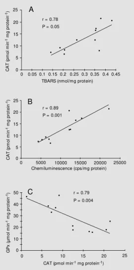
A positive correlation was obtained by linear regression between oxidative stress and CAT activity in LD muscle from the groups studied, with higher CAT activity at higher levels of oxidative stress, as evalu-ated by TBARS (r = 0.78, P = 0.05) (Figure 1A) or by chemiluminescence (r = 0.89, P = 0.001) (Figure 1B). CAT activity was also inversely correlated with GPx activity in LD muscle from all groups (r = 0.79, P = 0.004) (Figure 1C).
The major findings of the present experi-ments are that LD muscle from 5-day STZ-diabetic rats showed increased oxidative
stress, indicated by TBARS and chemilumi-nescence, and adaptations in antioxidant en-zymes.
This is the first experiment carried out to study oxidative stress in LD muscle. The present results are in agreement with previ-ous studies showing increased levels of oxi-dative stress in red blood cells, kidney, liver, brain, and heart in diabetes (1,2,13,14). A probable mechanism explaining these changes is related to the increased glucose concentration. Several diabetic complications including myopathy have been attributed to hyperglycemia stimulation of the polyol path-way (1-3). Moreover, it has been demon-strated that aldose reductase inhibitor (ARI) treatment has a marked beneficial effect on skeletal muscle contractile performance in experimental diabetes, thus implicating the polyol pathway as a causative factor in dia-betic myopathy. The reduction of tetanic tension output in the extensor digitorum lon-gus muscle was related to polyol pathway activity, since it was prevented by ARI treat-ment. Combined insulin and ARI therapy produced an improvement of the contractile properties of soleus muscle that was more marked than the improvement caused by each agent alone (3). Furthermore, Cameron and Cotter (15) demonstrated a decrease in ROS damage in STZ-diabetic rats treated with ARI, confirming that hyperglycemia is related to the activation of the polyol path-way leading to increased oxidative stress.
The causes of the increased pro-oxidant activity in diabetes are multifactorial and not completely understood. Probably, hypergly-cemia can lead to both a rise in ROS produc-tion and to the attenuaproduc-tion of free radical scavenging compounds (2). In a ROS re-view, Baynes (1) concluded that diabetes with complications is associated with in-creased chemical modification of protein and lipids. Hyperglycemia leads to protein glycation, glucose auto-oxidation and fatty acid oxidation, which may contribute to in-creased ROS generation. Antioxidant
en-C A T ( p m o l m in
-1 m
g
p
ro
te
in
-1) 25
r = 0.78 P = 0.05
A 20 15 10 5 0
0 0.05 0.1 0.15 0.2 0.25 0.3 0.35 0.4 0.45 TBARS (nmol/mg protein)
r = 0.89 P = 0.001
B 25 20 15 10 5 0 C A T ( p m o l m in
-1 m
g p ro te in -1)
0 5000 10000 15000 20000 25000
Chemiluminescence (cps/mg protein)
50 40 30 20 10 0 G P x ( µ m o l m in
-1 m
g p ro te in -1)
0 5 10 15 20 25
CAT (pmol min-1 mg protein-1)
C r = 0.79
P = 0.004 Figure 1 - Positive correlation
zymes appear to be important for cell de-fense against oxidative damage. Changes in antioxidant concentration occur according to the tissue studied, and these alterations may be related to the capacity of these tis-sues to adapt to oxidative stress (2). Kakkar et al. (13) observed high levels of TBARS in pancreas, heart and blood of diabetic rats. Catalase activity was increased in liver, heart and blood, but not in kidneys. The GPx enzyme presented higher activity in pan-creas and kidneys of STZ animals and SOD activity was increased in liver, heart and pancreas. The above results are an example of differences in the adaptive responses of tissues to the diabetic process. Moreover, there are time-course changes in antioxidant enzymes in the same tissue (14).
In the present experiments we observed higher CAT activity in LD muscle from short-term diabetic rats compared to normal rats. The increase in muscle CAT, located in microperoxisomes, may reflect increased lev-els of fatty acyl CoA oxidase that initiates the ß-oxidation of fatty acids in peroxisomes (16,17). Reinforcing the importance of CAT as an antioxidant tool to block the increase in skeletal muscle diabetic oxidative stress, we observed a positive correlation between TBARS levels and CAT activity (r = 0.78) and also between chemiluminescence and CAT activity (r = 0.89), suggesting that oxi-dative stress induced by H2O2 leads to
eleva-tion of CAT activity.
In the present study there was no change in SOD activity in LD muscle from diabetic rats, suggesting a less intense effect of diabe-tes on SOD than on CAT activity. Similar data, increased CAT and unchanged SOD, were observed in skeletal muscles of 80-day STZ-diabetic rats (17). These results may indicate an important role of peroxisomes in the adaptation of LD muscle to hyperglyce-mia. Other data showing an increase or a decrease in SOD activity may be due to
differences in experimental design or in the tissue evaluated (2).
The increase in the formation of glutathi-one, evidenced by higher g-GCS activity, suggests a role for glutathione in the defense of cells against oxidative stress. In fact, the activity of GST, a nonspecific peroxide scav-enger, was increased in LD muscle, prob-ably helping with cell detoxification. How-ever, GPx activity was reduced in muscle from diabetic rats. A similar reduction in GPx activity was observed in most studies on experimental diabetes (2). The reduction in GPx activity associated with enhanced oxidative stress in diabetic muscle observed here may be related to increased H2O2 levels
(18). Although we did not quantify H2O2
production, the increase in CAT activity, a specific H2O2 scavenger, may indicate an
increase in H2O2 formation in diabetic LD
muscle. The inverse correlation between CAT and GPx activities (r = 0.79) reinforces the occurrence of this possibility in skeletal muscle of diabetic rats.
The increase in oxidative stress has been associated with inactivation of voltage-de-pendent calcium channels and of plasma membrane Ca2+
ATPase (19) and Na+
K+
ATPase (3). Moreover, the increase in type and frequency of skeletal muscle mitochon-drial DNA deletions in diabetic patients may be related to oxidative damage by ROS (20). All these data seem to indicate that oxidative stress plays an important role in diabetic myopathy.
We have previously observed that re-duced tetanic force in LD muscle of short-term diabetic rats (21) was associated with reduced Na+
K+
ATPase and Ca2+
Re fe re nce s
1. Baynes JW (1991). Role of oxidative stress in development of complications in diabetes. Diabetes, 40: 405-412. 2. Van Dam PS, Van Asbeck BS, Erkelens W,
M arx JJM , Gispen WH & Bravenboer B (1995). The role of oxidative stress in neu-ropathy and other diabetic complications.
Diabetes M etabolism Review s,11:
181-192.
3. Cameron NE, Cotter M A & Robertson S (1990). Changes in skeletal muscle con-tractile properties in streptozotocin-in-duced diabetic rats and role of polyol path-w ay and hypoinsulinemia. Diabetes, 39: 460-465.
4. Chachques JC, M arino JP, Lajos P, Zegdi R, D’Attellis N, Fornes P, Fabiani JN & Carpentier A (1997). Dynamic cardiomyo-plasty: clinical follow -up at 12 years.
Euro-pean Journal of Cardio-Thoracic Surgery,
12: 560-568.
5. Low ry OH, Rosebrough NJ, Farr AL & Randall RJ (1951). Protein measurement w ith the Folin phenol reagent. Journal of
Biological Chemistry, 193: 265-275.
6. Buege JA & Aust SD (1978). M icrosomal lipid peroxidation. M ethods in Enzymol-ogy,52: 302-310.
7. Gonzalez Flecha B, Llesuy S & Boveris A (1991). Hydroperoxide-initiated chemilu-minescence: an assay for oxidative stress in biopsies of heart, liver, and muscle.
Free Radical Biology and M edicine, 10:
93-100.
8. Aebi H (1984). Catalase in vitro. M ethods
in Enzymology, 105: 121-126.
9. M isra HP & Fridovich I (1972). The role of superoxide anion in the autooxidation of epinephrine and simple assay for super-oxide dismutase. Journal of Biological
Chemistry, 247: 3170-3175.
10. Seelig GF & M eister A (1984). Glutathi-one biosynthesis; g-glutamylcysteine syn-thetase from rat kidney. Journal of
Bio-logical Chemistry, 259: 379-390.
11. Del M aestro R (1985). Oxidative enzymes in tissue homogenates. In: Greenw ald RA (Editor), CRC Handbook of M ethods for
Oxygen Radical Research. CRC Press Inc.,
Boca Raton, FL, 294-296.
12. M annervik B & Guthenberg C (1981). Glu-tathione transferase (human placenta).
M ethods in Enzymology, 77: 231-237.
13. Kakkar R, Kalra J, M antha SV & Prasad K (1995). Lipid peroxidation and antioxidant enzymes in diabetic rats. M olecular and
Cellular Biochemistry, 151: 113-119.
14. Kakkar R, M antha SV, Kalra J & Prasad K (1996). Time course study of oxidative stress in aorta and heart of diabetic rat.
Clinical Science, 91: 441-448.
15. Cameron NE & Cotter M A (1997). M eta-bolic and vascular factors in the pathogen-esis of diabetic neuropathy. Diabetes,46: S31-S37.
16. Christie KN (1979). Catalase in skeletal
muscle fibers. Journal of Histochemistry
and Cytochemistry,27: 814-819.
17. Lammi-Keefe CJ, Sw an PB & Hegarty PVJ (1984). Evidence for increased peroxida-tive activity in muscles from streptozoto-cin-diabetic rats. Proceedings of the Soci-ety for Experimental Biology and M edi-cine,176: 27-31.
18. Arnaiz SL, Travacio M , M onserrat AJ, Cutrín JC, Llesuy S & Boveris A (1997). Chemiluminescence and antioxidant lev-els during peroxisome proliferation by fenofibrate. Biochim ica et Biophysica Acta,1360: 222-228.
19. Viner RI, Hühmer AFR, Bigelow DJ & Schöneich C (1997). The oxidative inacti-vation of sarcoplasmatic reticulum Ca2+ ATPase by peroxynitrite. Free Radical
Re-search,24: 243-259.
20. Liang P, Hughes V & Fukagaw a NK (1997). Increased prevalence of mitochondrial DNA deletions in skeletal muscle of older individuals w ith impaired glucose toler-ance: possible marker of glycemic stress.
Diabetes,46: 920-923.