an Antimicrobial Peptide
Stephanie J. Soscia1,2, James E. Kirby3, Kevin J. Washicosky1, Stephanie M. Tucker1, Martin Ingelsson4, Bradley Hyman1,5, Mark A. Burton6,7, Lee E. Goldstein6,7, Scott Duong3, Rudolph E. Tanzi1,5*, Robert D. Moir1,5
1Genetics and Aging Research Unit, Mass General Institute for Neurodegenerative Disease and Department of Neurology, Massachusetts General Hospital, Charlestown, Massachusetts, United States of America,2Department of Anatomy and Neurobiology, Boston University School of Medicine, Boston, Massachusetts, United States of America,3Department of Pathology, Beth Israel Deaconess Medical Center, Boston, Massachusetts, United States of America,4Department of Public Health/Geriatrics, Uppsala University, Uppsala, Sweden,5Harvard Medical School, Boston, Massachusetts, United States of America,6Molecular Aging and Developmental Laboratory, Photonics Center, College of Engineering, Boston University School of Medicine, Boston University, Boston, Massachusetts, United States of America,7Boston University Alzheimer’s Disease Center, Boston University, Boston, Massachusetts, United States of America
Abstract
Background:The amyloidb-protein (Ab) is believed to be the key mediator of Alzheimer’s disease (AD) pathology. Abis most often characterized as an incidental catabolic byproduct that lacks a normal physiological role. However, Abhas been shown to be a specific ligand for a number of different receptors and other molecules, transported by complex trafficking pathways, modulated in response to a variety of environmental stressors, and able to induce pro-inflammatory activities.
Methodology/Principal Findings:Here, we provide data supporting anin vivofunction for Abas an antimicrobial peptide (AMP). Experiments used establishedin vitroassays to compare antimicrobial activities of Ab and LL-37, an archetypical human AMP. Findings reveal that Ab exerts antimicrobial activity against eight common and clinically relevant microorganisms with a potency equivalent to, and in some cases greater than, LL-37. Furthermore, we show that AD whole brain homogenates have significantly higher antimicrobial activity than aged matched non-AD samples and that AMP action correlates with tissue Ablevels. Consistent with Ab-mediated activity, the increased antimicrobial action was ablated by immunodepletion of AD brain homogenates with anti-Abantibodies.
Conclusions/Significance:Our findings suggest Ab is a hitherto unrecognized AMP that may normally function in the innate immune system. This finding stands in stark contrast to current models of Ab-mediated pathology and has important implications for ongoing and future AD treatment strategies.
Citation:Soscia SJ, Kirby JE, Washicosky KJ, Tucker SM, Ingelsson M, et al. (2010) The Alzheimer’s Disease-Associated Amyloidb-Protein Is an Antimicrobial Peptide. PLoS ONE 5(3): e9505. doi:10.1371/journal.pone.0009505
Editor:Ashley I. Bush, Mental Health Research Institute of Victoria, Australia
ReceivedNovember 12, 2009;AcceptedJanuary 20, 2010;PublishedMarch 3, 2010
Copyright:ß2010 Soscia et al. This is an open-access article distributed under the terms of the Creative Commons Attribution License, which permits unrestricted use, distribution, and reproduction in any medium, provided the original author and source are credited.
Funding:This work was supported by grants from the Cure Alzheimer’s Disease Fund (http://www.curealzfund.org/). The funder had no role in study design, data collection and analysis, decision to publish, or preparation of the manuscript.
Competing Interests:Dr Tanzi is a consultant to and holds stock options in Prana Biotechnology.
* E-mail: [email protected]
Introduction
The past 25 years has witnessed the accrual of a large body of data concerning the physiochemistry and biological activities of the amyloid b-peptide (Ab), the main component of b-amyloid deposits in the brains of Alzheimer’s disease (AD) patients [1]. Ab, which is generated in the brain and peripheral tissues, is widely believed an incidental catabolic byproduct of the amyloid b protein precursor (APP) with no normal physiological function. However, Ab has been shown to be a ligand for a number of different receptors and other molecules [2,3,4], transported by complex trafficking pathways between tissues and across the blood brain barrier [1,5], modulated in response to a variety of environmental stressors, and able to induce pro-inflammatory activities [6,7]. Despite these clues, the normal physiological role of Ab remains unknown. We have observed that many of the physiochemical and biological properties previously reported for
Ab are similar to those of a group of biomolecules collectively known as ‘‘antimicrobial peptides’’ (AMPs) which function in the innate immune system. AMPs (also called ‘‘host defense peptides’’) are potent, broad-spectrum antibiotics that target Gram-negative and Gram-positive bacteria, mycobacteria, enveloped viruses, fungi, protozoans and in some cases, transformed or cancerous host cells. AMPs are also potent immunomodulators that mediate cytokine release and adaptive immune responses (see review by Zaiou, 2007 [8]).
infections [12]. Conversely, high levels of LL-37 are associated with the pathology of several presumably non-infectious diseases [13], including plaques in atherosclerosis [14]. We have observed that LL-37 exhibits striking similarities to Ab, including a propensity to form cytotoxic soluble oligomers [15,16,17,18] and insoluble fibrils that demonstrate congophilia and birefringence [19], two classical histochemical properties of tinctorial amyloid. While the microbiocidal activity of LL-37 has been well characterized [20], the activity of Abagainst microbial organisms has not been tested.
Here we show that Abis active against at least eight common and clinically relevant microorganisms. Thein vitroantimicrobial activity of Abmatched, and in some cases, exceeded, that of LL-37, an archetypical human AMP. Furthermore, anti-Ab immu-noreactive material in AD whole brain homogenates is active against Candida albicans, the pathogen we identified as most sensitive to synthetic Ab. Most strikingly, temporal lobe samples from AD brain contained significantly higher antimicrobial activity than material from the same brain area of aged-matched, non-AD subjects. Consistent with an Ab-mediated action, cerebellum samples with lowb-amyloid loads from the same set of affected and unaffected subjects were not significantly different with regards to antimicrobial activity. Our findings show Ab possesses antimicrobial activity and may function in vivo as an AMP and, thus, play a role as an effector molecule of innate immunity.
Results
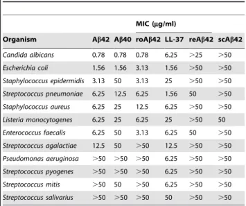
Antimicrobial activity against a particular microorganism is measuredin vitroby a peptide’s minimal inhibitory concentration (MIC), which is defined as the lowest concentration able to visibly inhibit growth overnight. We compared the MICs of synthetic LL-37, Ab40, and Ab42, against a panel of clinically relevant organisms (Table 1). The antimicrobial activity of Ab peptides was equivalent to or greater than LL-37 for seven of the pathogens tested. These data indicate that Ab is a bona fide AMP with potencies similar to, or, in some cases surpassing those of LL-37. The synthetic Abpeptides demonstrated antibiotic activity against Gram-negative and Gram-positive bacteria and the yeast C. albicans. Activity was isoform-specific for six organisms with Ab42 showing greater potency compared to Ab40. Equivalent findings were observed for recombinant Ab42, material that is free of the potentially toxic contaminants associated with conventional solid-phase peptide synthesis (data not shown). Rodent Ab42 also demonstrated antimicrobial activity. However, microbial growth was not inhibited by reverse (rAb42) or scrambled (scAb42) negative control peptides, thus confirming the antimicrobial action is peptide-specific.
AMPs, including LL-37 [21], can be bacteriostatic or bactericidal depending on peptide concentration, ionic strength, and the type of stressor a colony has previously encountered. The growth curves for E. faecalis in the presence of Ab42 suggest a predominantly bacteriostatic action for the peptide against this organism under our incubation conditions (Figure 1A). Consis-tent with previous studies, LL-37 showed poConsis-tent bactericidal activity againstE. faecalis. Microbial growth resumed at later time points, most likely due to degradation of LL-37 and Ab by protective bacterial proteases (Figure 1B).
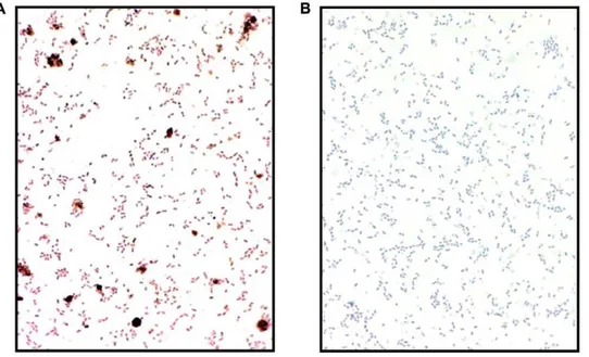
The capacity to associate with microbial lipid bilayers is considered a definitive feature of AMPs [22]. Most antimicrobial peptides are cationic to facilitate binding to anionic bacterial membranes. However, Ab peptides are anionic under physiolog-ical conditions [23]. Nonetheless, data from light microscopic
examination of immunostained bacteria pre-incubated with Ab confirm that the peptide binds to the surface of bacterial cells (Figure 2). Binding of Ab to bacterial membranes is consistent with previous studies showing that Ab readily binds and disrupts negatively charged synthetic lipid bilayers [24,25] and anionic mitochondria membranes [26,27,28], believed to have been originally derived from bacterial membranes.
In the next experiments we tested if the antimicrobial activity observed for synthetic peptides in vitro could be identified in temporal lobe and cerebellum from human brain. Typically b -amyloid load is high in AD temporal lobe and low in cerebellum. Tissue taken from AD (n = 32) or age matched control subjects (n = 13) were homogenized and normalized for protein. Ab40 and Ab42 levels in brain homogenates were determined by ELISA. Homogenates were then diluted into culture broth and inoculated withCandida albicans. Growth ofC. albicanswas determined using a fluorescence-based alamar blue microplate assay previously described for following cell viability with this organism [29]. AD temporal lobe homogenates inhibited the growth of C. albicans
significantly more (p = 0.0048) than non-demented control sam-ples (Figure 3A). Consistent with an Ab-mediated antimicrobial activity in AD temporal lobe homogenates, a significant difference inC. albicans growth was not observed with cerebellum samples, which carry a considerably lower Ab load. Also consistent with Ab-mediated antimicrobial activity,C. albicansgrowth significantly correlated with Ab concentration in temporal lobe homogenates (Figure 3B) but not in cerebellum samples with Pearson’s correlation coefficients (r) of 20.484, p = 0.0012 and 20.091, p = 0.56, respectively. In addition, the increased antimicrobial activity of AD temporal lobe samples could be significantly attenuated (p = 0.0007) by immunodepletion of homogenates with anti-Abantibodies (Figure 4A), consistent with an Ab-mediated antimicrobial activity in AD brain. Analysis of immunodepleted
Table 1.Abpeptides possess antimicrobial activity.
MIC (mg/ml)
Organism Ab42 Ab40 roAb42 LL-37 reAb42 scAb42
Candida albicans 0.78 0.78 0.78 6.25 .25 .50
Escherichia coli 1.56 1.56 3.13 1.56 .50 .50
Staphylococcus epidermidis 3.13 50 3.13 25 .50 .50
Streptococcus pneumoniae 6.25 12.5 6.25 1.56 50 .50
Staphylococcus aureus 6.25 25 12.5 6.25 .50 .50
Listeria monocytogenes 6.25 25 6.25 25 .50 50
Enterococcus faecalis 6.25 50 3.13 6.25 50 .50
Streptococcus agalactiae 12.5 50 .50 12.5 .50 .50
Pseudomonas aeruginosa .50 .50 .50 6.25 .50 .50
Streptococcus pyogenes .50 .50 .50 6.25 .50 .50
Streptococcus mitis .50 50 .50 6.25 .50 .50
Streptococcus salivarius .50 .50 .50 50 .50 .50
The antimicrobial activity of synthetic Ab1-42 (Ab42), Ab1-40 (Ab40), LL-37 ( LL-37), reverseAb42-1(rAb42), or scrambled Ab42 (scAb42) peptides were determined as minimal inhibitory concentrations (MIC) against 12 microorganisms. Antimicrobial activity was assayed by broth microdilution susceptibility test on 96-well plates with microbial growth in wells determined by visual inspection following an overnight incubation. Inhibition of growth in plate wells was confirmed by alamar blue cell viability assay and by surface plating of incubants on agar and counting CFU. Inoculums contained mid-logarithmic phase cells. Consistent with antimicrobial activity specific to the Ab sequence, inhibition was not observed for reverse and scrambled peptides. doi:10.1371/journal.pone.0009505.t001
homogenates confirmed Ab levels were attenuated in samples incubated with rabbit anti-Abantibody (Figure 4B). Additional experiments confirmed that antimicrobial activity in AD temporal lobe homogenates is also attenuated following immunodepletion with the anti-Abmouse monoclonal antibody 6E10 (Figure S1).
Discussion
Ab peptides inhibited the growth of eight of 12 clinically important pathogens screened (Table 1), including the bacteriaS. pneumoniae, which is a leading cause of bacterial meningitis [30], andC. albicans, the most common cause of neurocandidiasis [31]. If the normal function of Abis to function as an AMP, then an absence of the peptide may result in increased vulnerability to infection. Such an association has been shown for LL-37 and the disorder morbus Kostmann in which patients deficient in this AMP cannot mount an effective defense against pathogens [32]. To our knowledge a relationship between human immunodefi-ciency and low Ab levels has not been investigated. However, knockout mice that lack the proteases that generate endogenous rodent Ab appear to have increased susceptibility to pathogens [33]. BACE1 knockout (KO) mice that generate low levels of Ab and BACE1- and BACE2-deficient double KO mice, which do
not express Ab, have mortality rates of 40 and 60 percent, respectively. Housing the animals in a pathogen-free environment restores survival rates to that of wild-type mice (.95 percent). The etiology of the immunodeficiency has been investigated but not identified. Adaptive immune responses to vesicular stomatitis virus are the same for BACE-KO and wild-type mice. In addition, markers for adaptive immune system function are normal in BACE-KO mice, including leukocytes migration into the peri-toeum following thioglycolateacute-induced acute peritonitis and T-cell cytotoxiocity towards non-host cells. More recently, in a clinical trial of the Ab42- lowering agent tarenflurbil patients receiving the drug have significantly increased rates of infection [34]. Increased pathogen susceptibility of apparently adaptive immunocompetent BACE-KO mice and AD patients with suppressed Ab expression is consistent with our finding that Ab may have a normal protective function as an antimicrobial peptide of the innate immune system.
The immunostatus of APP knockout (APP-KO) mice has yet to be characterized. APP is a member of a larger protein family that includes the amyloid protein precursor-like proteins 1 and 2 (APLP1 and APLP2) [35,36]. APP and APLP proteins appear to have overlapping and partially redundant functions [37,38,39] and share processing pathways, including BACE-mediated generation
Figure 1. Growth of E. faecalisis inhibited by Ab42. E. faecaliswere cultured alone (circle) with 25mg/ml of Ab42 (triangle) or LL-37 (diamond).Panel A; Bacterial growth with time was monitored by inoculation of agar with diluted incubants and counting CFU. Representative data from six experiments is shown as mean signal of four replicates6s.e.m.Panel BIncubants were monitored for Ab42 and LL-37 by Western blot with mAb 6E10 or anti-LL-37. The figure shows representative signal for Ab42 (odd lanes) or LL-37 (even lanes) incubants from six replicate experiments.
of APLP-derived peptides analogous to Ab [37,38,39,40]. It is unclear to what degree APLP-derived proteins may compensate for deficiencies associated with low APP expression. Mice lacking both APP and APLP proteins (triple APP/APLP KO-mice) show early postnatal mortality with severe developmental abnormalities [41,42,43,44,45]. Interestingly, local cortical dysplasias can be infection-mediated and are observed in 68% of triple APP/APLP KO-mice [45]. Partial penetrance is also suggestive of an environmental component in this ectopia.
Recent studies have shown that while the adaptive immune system has limited access to the brain, the CNS can still mount a robust response to invading pathogens via antimicrobial peptides and the innate immune system. Numerous innate immune molecules with potent antimicrobial activity are found in brain, including the recently identified chromogranins [46], neuropep-tides neurokinin-1, enkelytin and peptide B, neuropeptide Y, polypeptide tyrosine-tyrosine, and the peptide hormones a -melanocyte stimulating hormone, adenoregulin, adrenomedullin
Figure 2.E. faecalispre-incubated with Ab42 are mAb 6E10 immunoreactive.Bacteria were incubated (1 hr at 37uC) with (panel A) or without (panel B) Ab42 (25mg/ml). Following repeated washes, the bacteria were fixed onto glass slides and immunostained with the HRP conjugated anti-Abantibody (mAb 6E10-HRP).
doi:10.1371/journal.pone.0009505.g002
Figure 3. AD brain homogenates have increased antimicrobial activity againstC. albicans.AD and non-AD brain samples were tested for Ab-mediated inhibition ofC. albicans. Samples of temporal lobe (Temp. L.) and cerebellum (Cereb.) from AD (n = 32) and age-matched control subjects (n = 13) were homogenized in culture broth.Panel A; Homogenates were inoculated with log-phaseC. albicansand microbial growth determined by alamar blue viability assay. Data is shown as percentage of signal forC. albicansalone (average of four replicates)6s.e.m.Panel B; Homogenates were assayed for Ab40 and Ab42 by commercially available ELISA. Graph shows Absignal (sum of Ab40 and Ab42) againstC. albicans growth for temporal lobe homogenates from combined AD and non-demented cohorts (n = 42). Probability analysis used unpaired two-tailed t-tests (p). Correlation was determined by calculating the Pearsonrcorrelation coefficient (r).
doi:10.1371/journal.pone.0009505.g003
and proadrenomudullin, corticostatin RK-1, neurotensin, and bradykinin [47]. Consistent with an antimicrobial role for brain generated Ab, we found AD temporal lobe homogenates contain an average of 24% greater activity againstC. albicansthan samples from non-AD subjects (Figure 3A). Furthermore, higher Ablevels in temporal lobe samples correlated with increased inhibition ofC. albicans(Figure 3B) while immunodepletion of Abfrom AD brain homogenates restored antimicrobial activity to levels equivalent to those of control homogenates (Figures 4A and S1). Immunoblot analysis confirmed attenuated Ab levels in anti-Ab antibody immunodepleted samples and lowb-amyloid load in cerebellum tissue (Figure 4B). These data support a protective role for Ab under the conditions found in the brain milieu even thoughin vivo
concentrations of soluble peptide are substantially lower than levels in experiments using synthetic peptide [48]. Several factors may contribute to this apparent discrepancy. First, synergistic AMP interactions in vivo potentiate antimicrobial activity [49]. This effect has been demonstrated for CRAMP (rodent LL-37), for which peptide levels in rodent CNS do not approach concentra-tions that are needed to obtain positive signals inin vitro assays. However, rat brain extracts depleted of CRAMP have substan-tially attenuated antimicrobial activity [50]. Moreover, mutant mice lacking CRAMP are more susceptible to CNS infection by
meningococcal meningitis[11]. Second, AD brain contains a large pool of neurotoxic oligomeric Ab species [51,52]. Oligomerization plays a key role in the targeting and permeabilization of bacterial membranes by AMPs [19,53,54]. Neurotoxic oligomeric Ab species present in AD brain may enhance the antimicrobial activity of homogenates beyond that predicted from in vitro
experiments, which add synthetic monomeric peptides to micro-bial cultures.
A large body of data supports a central role for neuroinflamma-tion in AD neuropathology [55]. A number of studies have proposed Ab as the source of AD-associated inflammation [56]. However, a re-evaluation of the role of Abin inflammation may now be warranted in view of these data suggesting that the peptide functions as an AMP in tissues. Inflammatory response in the immunologically privileged CNS is mediated by the innate immune system. Rather than Ab acting as a sole independent initiator of neuroinflammation, our data raise the possibility that the peptide may be part of a response mounted by the innate immune system. Thus, Abmay be one of a family of AMPs known to contribute pro-inflammatory activities under disease conditions. At least one other disease has been shown to involve deposition of an AMP as amyloid, corneal amyloidosis. In corneal amyloidosis the widespread and well-characterized antimicrobial protein lactoferrin accumulates in the subepithelium as insoluble amyloid [57,58]. Semenogelin-derived antimicrobial peptides [59] are also deposited as seminal vesicle amyloid [60] in a common sub-clinical pathology found in elderly men [61]. Based on our current findings, we postulate that stimulation of the innate immune system may initially trigger Ab generation and the b-amyloid cascade that leads tob-amyloid deposition. Along these lines at least three pathogenic mechanisms could conceivably lead to Ab generation and accumulation in the CNS via stimulation of an innate immune response. First, persistent sub-acute CNS infection may drive chronic activation of the innate immune system. A number of studies have reported that the CNS of AD patients is
Figure 4. Immunodepletion of Abfrom AD brain homogenates attenuatesC. albicansinhibition.Homogenates of temporal lobe (Temp. L.) and cerebellum (Cereb.) were prepared from AD (n = 32) or non-demented (n = 13) subjects. AD (AD) or non-demented (non-AD) homogenates were pooled and then incubated with Magno-beads pre-loaded with rabbit IgG (IgG) or a polyconal rabbit anti-Abantibody (a-Ab). Following bead removal samples were analyzed for Absignal by Western blot and assayed forC. albicansgrowth by alamar blue viability assay.Panel AshowsC. albicansgrowth in treated homogenates as a percentage of signal in culture broth alone. Immunodepletion of AD temporal lobe homogenates with a-Abrestored microbial growth to levels equivalent to non-demented control samples. Graph shows average of five replicates6s.e.m.Panel B; Untreated and immunodepleted homogenates (1:16 dilution) were Western blotted and probed with the Ab-specific mAb 4G8 antibody. Analysis confirmed Absignal was reduced in temporal lobe homogenate incubated with anti-amyloidb-peptide antibody (Lane 1) compared to sample incubate alone (Lane 2) or with rabbit IgG (Lane 3). Abin dilutions of cerebellum homogenate is below the level of detection for our experimental conditions (Lanes 4–6). Blots included synthetic Ab42 (Ab42) standard (Lane 7). Statistical probability analysis (p) of data used unpaired two-tailed t-test.
infected with pathogens including Chlamydia pneumoniae [62],
Borrelia spirochetes [63], Helicobacter pylori [64], and HSV [65]. Deposition of b-amyloid has also been reported for acquired immunodeficiency syndrome patients with brain HIV infection [66]. Given the known genetic influence on Ab accumulation, genetic factors may contribute to activation of the innate immune system by regulating Abproduction and clearance. At one of the end of the spectrum of known AD genes, highly penetrant mutations such as those in the early-onset familial AD genes,APP,
PSEN1, and PSEN2, would constitutively trigger cerebral Ab accumulation with no need for activation of the innate immune system [67]. At the other end of the spectrum, consistent with the increase risk of AD associated with the e4 variant of the apolipoprotein E gene [68], carriers of thee4allele are reported to have higher rates of CNS infection for several of these pathogens [69]. Finally, in a recent family-based genome-wide association scan for late-onset AD, one of four genes achieving genome-wide significance for association with AD was a homologue of CD33, a lectin involved in the innate immune system [70].
In a second potential pathogenic mechanism, a transient infection may lead to a self-perpetuating innate immune response. Transient triggers may include pathogens reported to be present in AD brain. And in a third mechanism, an inappropriate inflammatory response by the innate immune system to transient or persistent non-infectious insults could also trigger a self-perpetuating innate immune response. While dozens of diseases have been suggested to involve immune abnormalities, for most, the underlying cause of the aberrant immunoresponse remains unclear. For AD, traumatic brain injury [71], stroke [72] and certain forms of inhalant anesthetics [73] have been linked to increased cerebral Ab levels. Thus, while an infection-mediated pathological mechanism for AD is certainly one possibility for triggering an innate immune response in the CNS and subsequent production of antimicrobially active Ab, other non-microbial factors may also be involved. Interestingly, peptides containing the microtubule binding sites on tau proteins have also been shown to harbor antimicrobial properties [74].
The capacity to associate with lipid bilayers is considered a definitive feature of AMPs, and the peptides usually affect their antimicrobial activity by membrane permeabilization [75]. Membrane disruption is also thought to be a mechanism for Ab -mediated cytotoxicity [24,26]. Our finding that bacterial mem-branes stain positive for Abfollowing incubation with the peptide (Figure 2) is consistent with a mechanism that involves association with microbial lipid bilayers. While most AMPs are cationic, Ab peptides are anionic. Repulsive electrostatic forces between anionic peptides and electronegative phospholipids in bacterial membranes potentially limit antimicrobial activity of this class of AMP. However, in addition to our data, previous studies have conclusively shown that Ab readily binds and disrupts both synthetic anionic lipid bilayers [24] as well as mitochondrial membranes [26]. Interestingly, mitochondria are thought to be of endosymbiont origin and have anionic membranes that resemble the lipid bilayers of bacteria. A number of AMPs, including LL-37, appear to target and disrupt the mitochondrial membranes of parasitic protozoans [8]. Recent studies have also identified a number of anionic mammalian peptides with antimicrobial activity, including CNS neuropeptides [76] and peptide hormones [47]. Structural studies on the important epithelial anionic AMP dermicidin have shown that an overall positive charge is not a prerequisite for binding of bacterial membranes [77]. Rather, the key modulators of lipid bilayers/peptide association are the peptides charge distribution and secondary conformation.
Collec-tively, these data indicate that AMP activity is not limited to cationic species and that anionic peptides such as Abcan readily bind bacterial membranes and act as potent antimicrobial agents. In E. faecalis cultures, Ab was more resistant to bacterial-mediated degradation than LL-37 (Figure 1B). Bacterial defense mechanisms secrete proteases that target positively charged peptides. Anionic AMPs are believed to be, at least in part, a host counter measure to bacterial resistance mechanisms [78]. Oligomerization is also thought to protect AMPs from microbially-mediated degradation, and Aboligomers have been shown to be highly protease resistant. An anionic charge and propensity to oligomerize may therefore help render Ab resistant to bacterial attack.
AMPs cytotoxicity is usually highly specific for microbes. However, AMPs can also be cytotoxic to select host cells under physiological conditions. Host cell cytotoxicity has been shown for LL-37 [14] which, like Ab [79], is cytotoxic towards vascular smooth muscle cells. AMP host cell cytoxicity often involves disruption of mitochondrial function, an activity reported for both LL-37 [80,81] and Ab [27]. Thus, neurotoxicity that has been shown for Abis consistent with AMP behavior. The role of AMP host cell cytoxicity in disease and defense is unclear. LL-37 cytotoxicity has been implicated in disease pathology [14] but may also have a normal function in antibody-dependent cell cytotox-icity, a host mechanism for the clearance of virus-infected and transformed cells [81]. At present Ab’s host cell cytotoxicity is only associated with disease. Identification of Abas an AMP raises the possibility that host cell cytotoxicity, or at least a component of this activity, may also have a role in innate immunity.
In summary, our finding that Abis an antimicrobial peptide is the first evidence that the species responsible for amyloidosis may have a normal function. This stands in stark contrast to current models, which assumeb-amyloid deposition to be an accidental process resulting from the abnormal behavior of an incidental product of catabolism. Our data suggest increased Abgeneration, and resulting AD pathology, may be a mediated by a response of the innate immune system to a perceived infection. This model has important implications for current and future AD treatment strategies. First, it raises the possibility of preventing amyloidosis from initiating by pre-emptive targeting of pathogens/insults that stimulate the brain’s innate immune system. Second, our model identifies the inflammatory pathways of the innate immune system as targets for modulating Abgeneration/accumulation. The target pathways implicated here are downstream of the inflammatory trigger. Thus, this approach would likely be useful independently of the involvement of infectious agents in AD pathology.
Materials and Methods
Synthetic Peptides
Experiments used Ab1-40 (Ab40), Ab1-42 (Ab42), scrambled Ab(scAb42), Ab42-1 (rAb42), LL-37, and scrambled LL-37 (scLL-37) peptides. Aband LL-37 peptides were prepared and purified by Dr. James I. Elliott at Yale University (New Haven, CT) using solid-phase peptide synthesis. Scrambled LL-37 peptide was from AnaSpec (San Jose, CA). Recombinant human Ab42 (recAb42) and rodent Ab42 (roAb42) were purchased from rPeptide (Bogart, GA) and Calbiochem (Gibbstown, NJ) respectively. Findings for recombinant and SPPS prepared peptides were equivalent in all experiments.
Brain Samples
at 270uC and the other hemisphere was fixed in formalin for histological examination. The clinical diagnosis of AD was confirmed by subsequent histological evidence of amyloid plaques and neurofibrillary tangles. Samples were provided by the Neurobiology Tissue Bank at the Mass General Institute for Neurodegenerative Disease and Massachusetts General Hospital and included temporal lobe and cerebellum from 32 AD patients and 13 non-demented age-matched control subjects.
Cell Cultures
Bacteria were from the American Type Culture Collection (ATCC, Manassas, VA) and included Candida albicans ATCC 10231,Escherichia coliATCC 25922,Staphylococcus epidermidisATCC 12228, Streptococcus pneumoniae ATCC 49619, Staphylococcus aureus
ATCC 25923, Listeria monocytogenes ATCC 19112, Enterococcus faecalis ATCC 29212, Streptococcus agalactiae ATCC 12386,
Pseudomonas aeruginosa ATCC 27853, Streptococcus pyogenes ATCC 19615, Streptococcus mitis ATCC 6249, and Streptococcus salivarius
ATCC 13419. Bacteria were cultured aerobically in Mueller-Hinton broth (MHB), Brain and Heart Infusion broth (BHIB), or BHIB supplemented with 1% lysed horse blood and plated on Tryptone Soy Agar (TSA) plates containing 5% defibrinated sheep blood. C. albicans was grown in RPMI-1640 medium (Hyclone, Logan, UT) with 2% glucose buffered (pH 7.0) and 0.165 M MOPS and surface plated on sabouraud dextrose agar plates. Culture conditions for each organism are included inTable S1. Organisms were subcultured for 2 hrs to generate mid-logarithmic growth cultures for use as inoculates in experiments. Media reagents were obtained from Becton, Dickinson and Company (Sparks, MD).
Preparation of Inoculum Containing Mid-Logarithmic Phase Cells
Colonies from agar were transferred by sterile loop to growth media and incubated for 2 hrs at 37uC to achieve a McFarland density of 0.5. Bacteria inoculum cell densities were normalized to 56105 cells/ml immediately before use photometrically and subsequently confirmed by colony count. Inoculum ofC. albicans
contained a cell density of 2.56103CFU/ml.
Peptide Pre-Treatment and Preparation of Stock Solutions
Bulk powdered peptides were first dissolved in 30% trifluor-oethanol (TFE) at 1 mg/ml. Five hundred microliter aliquots of the stock solutions were lyophilized and stored under nitrogen at 220uC. Stock solutions at 2 mg/ml were prepared the day of experimentation from the peptide films by solubilizing a second time in either water or 20% TFE. Abstocks prepared in water were sonicated and insoluble peptide aggregates pelleted by centrifugation (10 min616,000g). Peptide concentrations in stock solutions were determined immediately before use by bicincho-ninic acid (BCA) protein assay. The validity of BCA for assaying Ab peptides has been established previously [82]. For MIC experiments, peptides were serially diluted into growth media. For other experiments stocks were diluted into required working buffers. Experiments included controls for peptide buffer vehicle alone.
MIC Determination
Peptide antimicrobial activity was determined as minimal inhibitory concentration (MIC) [83]. Experiments identified peptide MIC by broth microdilution susceptibility test in conjunction with CFU and alamar blue assays. Inoculum
containing mid-logarithmic phase cells was dispensed into the wells of polypropylene 96-well plates (Fisher, Pittsburgh, PA) containing seven two-fold dilutions of test peptide in growth media. Plates were then incubated aerobically overnight (12 to 18 hrs) at 37uC. Peptide MIC was taken as the lowest concentration able reduce cell growth by CFU and alamar blue assays by at least two-fold and which correlated with the visible loss of a growth button on the bottom of microtiter wells. Experiments were repeated a minimum of three times for each organism, and tests included at least three replicates for each assay condition. Experiments included control serial dilutions of buffer vehicle alone.
Note on radial diffusion assays (RDAs). RDAs have been widely used in previous studies to assess AMP antimicrobial activity. However, in our experiments RDAs proved unreliable for testing Ab antimicrobial activity because the peptide failed to diffuse away from the point of application (data not shown). Ab solutions are prone to aggregation, particularly in the presence of even trace amounts of metal, and interaction with contaminates or the media matrix may lead to rapid precipitation of the peptide within the agar. Abpeptides also appear to irreversibly absorb to the cellulose disks often used as sample reservoirs in RDAs.
CFU Assay
Serial dilutions of incubants were prepared and streaked onto the surface of agar. The agar plates were then incubated overnight at 37uC and colonies forming units counted.
Alamar Blue Cell Viability Assay
Microbial growth was determined by following the reduction of the synthetic metabolic substrate resazurin (alamar blue) to a fluorescent product by respiratory enzymes in living cells [84]. Alamar blue assay is used in high throughput screens for antimicrobial agents [85] and is available commercially in kit form from Invitrogen. Microbial growth in experiments was assayed with alamar blue kits according to the manufacturer’s instructions. Briefly, resazurin reagent was added to microbial cultures (1:10) and samples incubated for 30 or 60 minutes. Fluorescence signal was measured at excitation of 530 nm and emission at 590 nm. Signal was blanked on sterile media. For experiments with brain homogenates, blank wells contained all components as tests but were not inoculated withC. albicans. Anti-Ab Imunostaining of Bacteria
E. faecalissmears were air-dried on Superfrost/Plus microscope slides (Fisher Scientific, Pittsburgh, PA) and then heated to kill and fix bacterial cells. Fixed cells were incubated with 3% methanolic hydrogen peroxide for 30 minutes at room temperature to inhibit endogenous peroxidase activity, passed through graded alcohol, and rinsed three times in deionized water and phosphate-buffered saline (PBS). Slides were then incubated with a 1:2,000 dilution of the anti-Abmonoclonal antibody (mAb) 6E10 (Covance, Prince-ton, NJ) in TBST. Following washing, slides were incubated with goat anti-mouse IgG-coupled to HRP (1:200). Detection and localization steps were performed using Vectastain ABC kit and DAB Substrate Kit (Vector Laboratories, Burlingame, CA).
(Figure S1). Samples were then inoculated (2.56103 CFU/ml) with mid-logarithmic growth culture ofC. albicansand incubated aerobically for 3 hrs at 37uC in 96-well microplates (100ml/well).
Alamar blue reagent was added to wells (10ml) and fluorescence
measured after 30 and 60 minutes incubation. Signal from test wells was blanked on samples incubanted without C. albicans. Signal from homogenate blanks was equivalent to uninoculated media alone (data not shown). Samples were assayed in quadruplicate.
Assaying Abin Tissue Homogenates
Ab40 and Ab42 in samples were determined using commer-cially available ELISA kits (Covance, Princeton, NJ). Brain homogenates were assayed according to the manufacturer’s instructions.
Immunodepletion of Brain Homogenates
MagnaBind goat anti-rabbit IgG beads (Pierce, IL) were pre-incubated overnight with the Ab specific rabbit anti-amyloid b -peptide antibody (Invitrogen, CA) or rabbit IgG then washed repeatedly. Pooled samples were prepared from temporal lobe (30 AD and 12 non-AD) or cerebellum (32 AD and 13 non-AD) homogenates. The pooled brain homogenates were incubated alone or with the antibody loaded beads at 4uC for 2 hrs. Final incubation conditions were 5mg of antibody per mg of original
tissue (w/w). Beads were pelleted and soluble fraction removed. Fractions were immunoblotted and probed with the Ab specific mAb 4G8 (Covance, Princeton, NJ). Analysis confirmed anti-Ab antibody treated homogenates were depleted of Ab(Figure 4B). Soluble fractions were then tested for antimicrobial activity against
C. albicansby alamar blue assay. Immunoblotting (Western Blotting)
Samples were first resolved by electrophoresis on SDS-PAGE (4–12% Bis-Tris gels) and then transferred to polyvinylidene fluoride membrane. Membranes were blocked with bovine serum albumin (10%) then probed with mAb 4G8 (1:200), mAb 6E10 (1:2,000), or mAb anti-LL-37 (1:200) (Hycult Biotechnology, Uden, The Netherlands). Following washing, membranes were incubated with goat anti-mouse IgG-coupled to HRP. Blots were developed with chemiluminescence reagent (Pierce, Rockford IL) and signal captured using a VersDoc digital imaging system (BioRad, Hercules, CA). Blot incubations used Tris buffered saline, pH 8 containing 0.1% Tween (TBST).
Statistical Analysis
Association coefficients between Ablevels in brain homogenate and C. albicansgrowth were calculated using Pearson correlation test and linear regression. Experimental groups were compared by unpaired two-tailed t-test with a nominal alpha criterion level of
0.05. Antimicrobial signal in AD and non-AD cohorts passed a D’Agostino-Pearson test for normality (alpha = 0.05) with p values of 0.077 and 0.24, respectively. Variances of signal from AD and non-AD cohorts were not significantly different (p = 0.18). Alternative non-parametric statistical analysis of antimicrobial activity in temporal lobe homogenates by two-tailed Mann-Whitney U test also returned a significant difference between AD and non-AD cohorts (p = 0.018). Statistical analysis used Graph-Pad Prism software package (La Jolla, CA).
Supporting Information
Figure S1 Ab-mediated inhibition of C. albicans in AD brain homogenates is dose dependant. AD temporal lobe (Temp. L.) or cerebellum (Cereb.) were homogenized in phosphate buffer. Temporal lobe (n = 30.) or cerebellum (n = 32) homogenates were pooled and 1:16, 1:32, and 1:64 serial dilutions prepared in culture broth. Homogenate dilutions were incubated with mouse IgG (IgG) or anti-Ab mAb 6E10 (6E10) antibody immobilized on MagnaBind beads. Following pelleting of the beads incubants were inoculated with mid-logarithmic phaseC. albicansin 96-well plates. Microbial growth was determined by alamar blue cell viability assay. Graphs shows percentage signal ofC. albicansalone (average of five replicates) 6 s.e.m. Consistent with Ab-mediated antimicrobial activity, C. albicans growth is highest for samples with low Ablevels and increases with homogenate dilution. Found at: doi:10.1371/journal.pone.0009505.s001 (0.10 MB TIF)
Table S1 Experimental culture conditions for test organisms. The table shows test organisms used for peptide MIC determina-tion with Gram staining (Gram Stain) properties, American Type Culture Collection designation (ATCC No.), culture media (Growth Media), and growth period (Incub. hrs) used for broth microdilution susceptibility testing. Organisms were grown aerobically at 37uC in Mueller-Hinton broth (MHB), Brain and Heart Infusion broth alone (BHIB) or supplemented with 1% lysed horse blood (BHIB/LHB), or RPMI-1640 medium with 2% glucose (RPMI-1640).
Found at: doi:10.1371/journal.pone.0009505.s002 (0.05 MB PDF)
Acknowledgments
The authors wish to thank Lucius Chiaraviglio for expert technical assistance.
Author Contributions
Conceived and designed the experiments: SJS JEK RET RDM. Performed the experiments: SJS KJW SMT MAB. Analyzed the data: SJS JEK KJW LEG SD RET RDM. Contributed reagents/materials/analysis tools: MI BTH LEG. Wrote the paper: SJS JEK RET RDM.
References
1. Tanzi RE, Moir RD, Wagner SL (2004) Clearance of Alzheimer’s Abpeptide: the many roads to perdition. Neuron 43: 605–608.
2. Le Y, Gong W, Tiffany HL, Tumanov A, Nedospasov S, et al. (2001) Amyloid (b)42 activates a G-protein-coupled chemoattractant receptor, FPR-like-1. J Neurosci 21: RC123.
3. Koldamova RP, Lefterov IM, Lefterova MI, Lazo JS (2001) Apolipoprotein A-I directly interacts with amyloid precursor protein and inhibits Abaggregation and toxicity. Biochemistry 40: 3553–3560.
4. Maezawa I, Jin LW, Woltjer RL, Maeda N, Martin GM, et al. (2004) Apolipoprotein E isoforms and apolipoprotein AI protect from amyloid precursor protein carboxy terminal fragment-associated cytotoxicity. J Neurochem 91: 1312–1321.
5. Zlokovic BV, Yamada S, Holtzman D, Ghiso J, Frangione B (2000) Clearance of amyloidb-peptide from brain: transport or metabolism? Nat Med 6: 718–719.
6. Lee M, You HJ, Cho SH, Woo CH, Yoo MH, et al. (2002) Implication of the small GTPase Rac1 in the generation of reactive oxygen species in response tob -amyloid in C6 astroglioma cells. Biochem J 366: 937–943.
7. Paris D, Town T, Parker TA, Tan J, Humphrey J, et al. (1999) Inhibition of Alzheimer’sb-amyloid induced vasoactivity and proinflammatory response in microglia by a cGMP-dependent mechanism. Exp Neurol 157: 211–221. 8. Zaiou M (2007) Multifunctional antimicrobial peptides: therapeutic targets in
several human diseases. J Mol Med 85: 317–329.
9. Gudmundsson GH, Agerberth B, Odeberg J, Bergman T, Olsson B, et al. (1996) The human gene FALL39 and processing of the cathelin precursor to the antibacterial peptide LL-37 in granulocytes. Eur J Biochem 238: 325– 332.
10. Zanetti M (2004) Cathelicidins, multifunctional peptides of the innate immunity. J Leukoc Biol 75: 39–48.
11. Bergman P, Johansson L, Wan H, Jones A, Gallo RL, et al. (2006) Induction of the antimicrobial peptide CRAMP in the blood-brain barrier and meninges after meningococcal infection. Infect Immun 74: 6982–6991.
12. Ong PY, Ohtake T, Brandt C, Strickland I, Boguniewicz M, et al. (2002) Endogenous antimicrobial peptides and skin infections in atopic dermatitis. N Engl J Med 347: 1151–1160.
13. Von Haussen J, Koczulla R, Shaykhiev R, Herr C, Pinkenburg O, et al. (2008) The host defence peptide LL-37/hCAP-18 is a growth factor for lung cancer cells. Lung Cancer 59: 12–23.
14. Ciornei CD, Tapper H, Bjartell A, Sternby NH, Bodelsson M (2006) Human antimicrobial peptide LL-37 is present in atherosclerotic plaques and induces death of vascular smooth muscle cells: a laboratory study. BMC Cardiovasc Disord 6: 49.
15. Kirkitadze MD, Bitan G, Teplow DB (2002) Paradigm shifts in Alzheimer’s disease and other neurodegenerative disorders: the emerging role of oligomeric assemblies. J Neurosci Res 69: 567–577.
16. Wogulis M, Wright S, Cunningham D, Chilcote T, Powell K, et al. (2005) Nucleation-dependent polymerization is an essential component of amyloid-mediated neuronal cell death. J Neurosci 25: 1071–1080.
17. Walsh DM, Klyubin I, Shankar GM, Townsend M, Fadeeva JV, et al. (2005) The role of cell-derived oligomers of Abin Alzheimer’s disease and avenues for therapeutic intervention. Biochem Soc Trans 33: 1087–1090.
18. Oren Z, Lerman JC, Gudmundsson GH, Agerberth B, Shai Y (1999) Structure and organization of the human antimicrobial peptide LL-37 in phospholipid membranes: relevance to the molecular basis for its non-cell-selective activity. Biochem J 341(Pt 3): 501–513.
19. Sood R, Domanov Y, Pietia¨inen M, Kontinen VP, Kinnunen PK (2008) Binding of LL-37 to model biomembranes: insight into target vs host cell recognition. Biochim Biophys Acta 1778: 983–996.
20. Turner J, Cho Y, Dinh NN, Waring AJ, Lehrer RI (1998) Activities of LL-37, a cathelin-associated antimicrobial peptide of human neutrophils. Antimicrob Agents Chemother 42: 2206–2214.
21. Sieprawska-Lupa M, Mydel P, Krawczyk K, Wo´jcik K, Puklo M, et al. (2004) Degradation of human antimicrobial peptide LL-37 by staphylococcus aureus-derived proteinases. Antimicrob Agents Chemother 48: 4673.
22. Bechinger B, Lohner K (2006) Detergent-like actions of linear amphipathic cationic antimicrobial peptides. Biochim Biophys Acta 1758: 1529–1539. 23. Tomski SJ, Murphy RM (1992) Kinetics of aggregation of syntheticb-amyloid
peptide. Arch Biochem Biophys 294: 630–638.
24. Chauhan A, Ray I, Chauhan VP (2000) Interaction of amyloidb-protein with anionic phospholipids: possible involvement of Lys28 and C-terminus aliphatic amino acids. Neurochem Res 25: 423–429.
25. Chi E, Ege C, Winans A, Majewski J, Wu G, et al. (2008) Lipid membrane templates the ordering and induces the fibrillogenesis of Alzheimer’s disease amyloid-bpeptide. Proteins 72: 1–24.
26. Rodrigues CM, Sola´ S, Silva R, Brites D (2000) Bilirubin and amyloid-bpeptide induce cytochrome c release through mitochondrial membrane permeabiliza-tion. Mol Med 6: 936–946.
27. Eckert GP, Wood WG, Mu¨ller WE (2001) Effects of aging andb-amyloid on the properties of brain synaptic and mitochondrial membranes. Journal of Neural Transmission 108: 1051–1064.
28. Chen X, Stern D, Yan SD (2006) Mitochondrial dysfunction and Alzheimer’s disease. Curr Alzheimer Res 3: 515–520.
29. Repp KK, Menor SA, Pettit RK (2007) Microplate Alamar blue assay for susceptibility testing of Candida albicans biofilms. Med Mycol 45: 603–607. 30. Leib SL, Ta¨uber MG (1999) Pathogenesis of bacterial meningitis. Infect Dis Clin
North Am 13: 527–548, v–vi.
31. Chakrabarti A (2007) Epidemiology of central nervous system mycoses. Neurology India 55: 191–197.
32. Pu¨tsep K, Carlsson G, Boman HG, Andersson M (2002) Deficiency of antibacterial peptides in patients with morbus Kostmann: an observation study. Lancet 360: 1144–1149.
33. Dominguez D, Tournoy J, Hartmann D, Huth T, Cryns K, et al. (2005) Phenotypic and biochemical analyses of BACE1- and BACE2-deficient mice. J Biol Chem 280: 30797–30806.
34. Green RC, Schneider LS, Amato DA, Beelen AP, Wilcock G, et al. (2009) Effect of tarenflurbil on cognitive decline and activities of daily living in patients with mild Alzheimer disease: a randomized controlled trial. JAMA 302: 2557–2564. 35. Wasco W, Bupp K, Magendantz M, Gusella JF, Tanzi RE, et al. (1992) Identification of a mouse brain cDNA that encodes a protein related to the Alzheimer disease-associated amyloidbprotein precursor. Proc Natl Acad Sci USA 89: 10758–10762.
36. Wasco W, Brook JD, Tanzi RE (1993) The amyloid precursor-like protein (APLP) gene maps to the long arm of human chromosome 19. Genomics 15: 237–239.
37. Li Q, Su¨dhof TC (2004) Cleavage of amyloid-bprecursor protein and amyloid-b
precursor-like protein by BACE 1. J Biol Chem 279: 10542–10550. 38. Pastorino L, Ikin AF, Lamprianou S, Vacaresse N, Revelli JP, et al. (2004)
BACE (b-secretase) modulates the processing of APLP2 in vivo. Mol Cell Neurosci 25: 642–649.
39. Jacobsen KT, Iverfeldt K (2009) Amyloid precursor protein and its homologues: a family of proteolysis-dependent receptors. Cell Mol Life Sci 66: 2299–2318. 40. Minogue AM, Stubbs AK, Frigerio CS, Boland B, Fadeeva JV, et al. (2009)
gamma-secretase processing of APLP1 leads to the production of a p3-like
peptide that does not aggregate and is not toxic to neurons. Brain Res 1262: 89–99.
41. Zheng H, Jiang M, Trumbauer ME, Sirinathsinghji DJ, Hopkins R, et al. (1995)
b-Amyloid precursor protein-deficient mice show reactive gliosis and decreased locomotor activity. Cell 81: 525–531.
42. Chen Y, Tang BL (2006) The amyloid precursor protein and postnatal neurogenesis/neuroregeneration. Biochem Biophys Res Commun 341: 1–5. 43. Heber S, Herms J, Gajic V, Hainfellner J, Aguzzi A, et al. (2000) Mice with
combined gene knock-outs reveal essential and partially redundant functions of amyloid precursor protein family members. J Neurosci 20: 7951–7963. 44. Magara F, Mu¨ller U, Li ZW, Lipp HP, Weissmann C, et al. (1999) Genetic
background changes the pattern of forebrain commissure defects in transgenic mice underexpressing theb-amyloid-precursor protein. Proc Natl Acad Sci USA 96: 4656–4661.
45. Herms J, Anliker B, Heber S, Ring S, Fuhrmann M, et al. (2004) Cortical dysplasia resembling human type 2 lissencephaly in mice lacking all three APP family members. EMBO J 23: 4106–4115.
46. Shooshtarizadeh P, Zhang D, Chich J-F, Gasnier C, Schneider F, et al. (2009) The antimicrobial peptides derived from chromogranin/secretogranin family, new actors of innate immunity. Regulatory peptides: In Press.
47. Brogden KA, Guthmiller JM, Salzet M, Zasloff M (2005) The nervous system and innate immunity: the neuropeptide connection. Nat Immunol 6: 558–564. 48. Cherny RA, Legg JT, McLean CA, Fairlie DP, Huang X, et al. (1999) Aqueous dissolution of Alzheimer’s disease Abamyloid deposits by biometal depletion. J Biol Chem 274: 23223–23228.
49. McCafferty DG, Cudic P, Yu MK, Behenna DC, Kruger R (1999) Synergy and duality in peptide antibiotic mechanisms. Curr Opin Chem Biol 3: 672–680. 50. Bergman P, Terme´n S, Johansson L, Nystro¨m L, Arenas E, et al. (2005) The
antimicrobial peptide rCRAMP is present in the central nervous system of the rat. J Neurochem 93: 1132–1140.
51. Kuo YM, Emmerling MR, Vigo-Pelfrey C, Kasunic TC, Kirkpatrick JB, et al. (1996) Water-soluble Ab (N-40, N-42) oligomers in normal and Alzheimer disease brains. J Biol Chem 271: 4077–4081.
52. Roher AE, Chaney MO, Kuo YM, Webster SD, Stine WB, et al. (1996) Morphology and toxicity of Ab-(1-42) dimer derived from neuritic and vascular amyloid deposits of Alzheimer’s disease. J Biol Chem 271: 20631–20635. 53. Sal-Man N, Oren Z, Shai Y (2002) Preassembly of membrane-active peptides is
an important factor in their selectivity toward target cells. Biochemistry 41: 11921–11930.
54. Glukhov E, Stark M, Burrows LL, Deber CM (2005) Basis for selectivity of cationic antimicrobial peptides for bacterial versus mammalian membranes. J Biol Chem 280: 33960–33967.
55. Tuppo EE, Arias HR (2005) The role of inflammation in Alzheimer’s disease. Int J Biochem Cell Biol 37: 289–305.
56. Townsend KP, Obregon D, Quadros A, Patel N, Volmar Ch, et al. (2002) Proinflammatory and vasoactive effects of Ab in the cerebrovasculature. Ann N Y Acad Sci 977: 65–76.
57. Ando Y, Nakamura M, Kai H, Katsuragi S, Terazaki H, et al. (2002) A novel localized amyloidosis associated with lactoferrin in the cornea. Lab Invest 82: 757–766.
58. Araki-Sasaki K, Ando Y, Nakamura M, Kitagawa K, Ikemizu S, et al. (2005) Lactoferrin Glu561Asp facilitates secondary amyloidosis in the cornea. Br J Ophthalmol 89: 684.
59. Edstro¨m AML, Malm J, Frohm B, Martellini JA, Giwercman A, et al. (2008) The major bactericidal activity of human seminal plasma is zinc-dependent and derived from fragmentation of the semenogelins. J Immunol 181: 3413–3421. 60. Linke RP, Joswig R, Murphy CL, Wang S, Zhou H, et al. (2005) Senile seminal
vesicle amyloid is derived from semenogelin I. J Lab Clin Med 145: 187–193. 61. Kee KH, Lee MJ, Shen SS, Suh JH, Lee O-J, et al. (2008) Amyloidosis of
seminal vesicles and ejaculatory ducts: a histologic analysis of 21 cases among 447 prostatectomy specimens. Ann Diagn Pathol 12: 235–238.
62. Itzhaki RF, Wozniak MA, Appelt DM, Balin BJ (2004) Infiltration of the brain by pathogens causes Alzheimer’s disease. Neurobiol Aging 25: 619–627. 63. Miklossy J, Kis A, Radenovic A, Miller L, Forro L, et al. (2006)b-amyloid
deposition and Alzheimer’s type changes induced by Borrelia spirochetes. Neurobiol Aging 27: 228–236.
64. Kountouras J, Tsolaki M, Gavalas E, Boziki M, Zavos C, et al. (2006) Relationship between Helicobacter pylori infection and Alzheimer disease. Neurology 66: 938–940.
65. Itzhaki RF, Lin WR, Shang D, Wilcock GK, Faragher B, et al. (1997) Herpes simplex virus type 1 in brain and risk of Alzheimer’s disease. Lancet 349: 241–244.
66. Youngsteadt E (2008) Virology. Alzheimer’s risk factor also aids HIV. Science 320: 1577.
67. Tanzi RE, Kovacs DM, Kim TW, Moir RD, Guenette SY, et al. (1996) The gene defects responsible for familial Alzheimer’s disease. Neurobiol Dis 3: 159–168.
68. Strittmatter WJ, Saunders AM, Schmechel D, Pericak-Vance M, Enghild J, et al. (1993) Apolipoprotein E: high-avidity binding tob-amyloid and increased frequency of type 4 allele in late-onset familial Alzheimer disease. Proc Natl Acad Sci USA 90: 1977–1981.
70. Bertram L, Lange C, Mullin K, Parkinson M, Hsiao M, et al. (2008) Genome-wide association analysis reveals putative Alzheimer’s disease susceptibility loci in addition to APOE. Am J Hum Genet 83: 623–632.
71. Roberts GW, Gentleman SM, Lynch A, Graham DI (1991)bA4 amyloid protein deposition in brain after head trauma. Lancet 338: 1422–1423. 72. Tesco G, Koh YH, Kang EL, Cameron AN, Das S, et al. (2007) Depletion of
GGA3 stabilizes BACE and enhancesb-secretase activity. Neuron 54: 721–737. 73. Xie Z, Dong Y, Maeda U, Moir R, Inouye SK, et al. (2006) Isoflurane-induced apoptosis: a potential pathogenic link between delirium and dementia. J Gerontol A Biol Sci Med Sci 61: 1300–1306.
74. Kobayashi N, Masuda J, Kudoh J, Shimizu N, Yoshida T (2008) Binding sites on tau proteins as components for antimicrobial peptides. Biocontrol Sci 13: 49–56. 75. Bolintineanu D, Hazrati E, Davis HT, Lehrer RI, Kaznessis YN (2009) Antimicrobial mechanism of pore-forming protegrin peptides: 100 pores to kill E. coli. Peptides 31: 1–8.
76. Goumon Y, Lugardon K, Kieffer B, Lefevre J, Van Dorsselaer A, et al. (1998) Characterization of Antibacterial COOH-terminal Proenkephalin-A-derived Peptides (PEAP) in Infectious Fluids. Importance of enkelytin, the antibacterial PEAP209-237 secreted by stimulated chromaffin cells. J Biol Chem 273: 29847. 77. Steffen H, Rieg S, Wiedemann I, Kalbacher H, Deeg M, et al. (2006) Naturally processed dermcidin-derived peptides do not permeabilize bacterial membranes and kill microorganisms irrespective of their charge. Antimicrob Agents Chemother 50: 2608–2620.
78. Dennison SR, Morton LH, Brandenburg K, Harris F, Phoenix DA (2006) Investigations into the ability of an oblique alpha-helical template to provide the
basis for design of an antimicrobial anionic amphiphilic peptide. FEBS J 273: 3792–3803.
79. Subasinghe S, Unabia S, Barrow CJ, Mok SS, Aguilar MI, et al. (2003) Cholesterol is necessary both for the toxic effect of Abpeptides on vascular smooth muscle cells and for Ab binding to vascular smooth muscle cell membranes. J Neurochem 84: 471–479.
80. Risso A, Braidot E, Sordano MC, Vianello A, Macrı` F, et al. (2002) BMAP-28, an antibiotic peptide of innate immunity, induces cell death through opening of the mitochondrial permeability transition pore. Mol Cell Biol 22: 1926–1935. 81. Aarbiou J, Tjabringa GS, Verhoosel RM, Ninaber DK, White SR, et al. (2006)
Mechanisms of cell death induced by the neutrophil antimicrobial peptides alpha-defensins and LL-37. Inflamm Res 55: 119–127.
82. Huang X, Atwood CS, Moir RD, Hartshorn MA, Vonsattel JP, et al. (1997) Zinc-induced Alzheimer’s Ab1-40 aggregation is mediated by conformational factors. J Biol Chem 272: 26464–26470.
83. National Committee for Clinical Laboratory Standards (2009) Methods for determining bactericidal activity of antimicrobial agents; approved guideline. WaynePA: NCCLS Document M26-A.
84. Shiloh MU, Ruan J, Nathan C (1997) Evaluation of bacterial survival and phagocyte function with a fluorescence-based microplate assay. Infect Immun 65: 3193–3198.
85. Hamid R, Rotshteyn Y, Rabadi L, Parikh R, Bullock P (2004) Comparison of alamar blue and MTT assays for high through-put screening. Toxicol In Vitro 18: 703–710.