Prostanoids modulate inflammation
and alloimmune responses during
graft rejection
Serviço de Imunologia, Hospital Universitário Professor Edgard Santos, Faculdade de Medicina, Universidade Federal da Bahia, Salvador, BA, Brasil P.N. Rocha and
E.M. Carvalho
Abstract
Acute rejection of a transplanted organ is characterized by intense inflammation within the graft. Yet, for many years transplant re-searchers have overlooked the role of classic mediators of inflamma-tion such as prostaglandins and thromboxane (prostanoids) in alloim-mune responses. It has been demonstrated that local production of prostanoids within the allograft is increased during an episode of acute rejection and that these molecules are able to interfere with graft function by modulating vascular tone, capillary permeability, and platelet aggregation. Experimental data also suggest that prostanoids may participate in alloimmune responses by directly modulating T lymphocyte and antigen-presenting cell function. In the present paper, we provide a brief overview of the alloimmune response, of prosta-noid biology, and discuss the available evidence for the role of prostaglandin E2 and thromboxane A2 in graft rejection.
Correspondence P.N. Rocha Serviço de Imunologia Hospital Universitário Prof. Edgard Santos, UFBA Rua João das Botas, s/n, 5º andar 40110-160 Salvador, BA Brasil
Fax: +55-71-245-7110 E-mail:
Received May 4, 2005 Accepted August 12, 2005
Key words
•Thromboxane •Prostaglandins •Transplantation •Inflammation •Rejection •Corticosteroids
Introduction
Alloimmune response is a term that de-scribes the immune system’s reaction to al-loantigens (1). The main alal-loantigens in trans-plantation are the major histocompatibility complex (MHC) molecules present on the surface of graft cells. T lymphocytes of the host are capable of detecting foreign MHC through surface proteins called T cell recep-tors (TCRs). However, transplant immunolo-gists have long recognized that the mere binding of foreign MHC to the TCR - the so-called signal 1 - is not sufficient to induce an immune response. A second signal, deliv-ered by activated antigen-presenting cells (APCs), also known as co-stimulation, is required for the full activation of T cells (2).
In fact, in the absence of co-stimulation, alloantigen recognition by T lymphocytes promotes tolerance and not immunity. Since APCs are not antigen-specific and, thus, do not possess the capability of recognizing foreign MHC, other stimuli are required for APC activation.
example, experimental evidence has shown that inflammatory stimuli induce accumula-tion of MHC class II complexes on dendritic cells (4). Inflammation also up-regulates the expression of co-stimulatory molecules such as B7 members and CD40 on the surface of APCs (5). On this basis, perhaps kidneys from living donors are accepted more easily than those from cadavers because of the lower extent of damage to the graft.
Once activated, APCs can then home to local lymph nodes and efficiently present foreign MHC to T cells. Proper T cell activa-tion by alloantigens in the context of danger signals initiates a cascade of events culmi-nating in the generation of effector elements that mediate graft damage (6). Activated CD8+ T cells injure the graft by way of perforin- or fas-induced apoptosis. CD4+ T cells activate B cells by way of cell-to-cell interactions (such as CD40-CD40L) and Th2
cytokines; activated B cells mature into plasma cells and secrete alloantibodies that harm the graft by activating the complement cascade or antibody-dependent cellular cy-totoxicity (7). Finally, CD4+ T cells release several cytokines and chemokines that or-chestrate a delayed hypersensitivity-type re-sponse, attracting inflammatory cells such as neutrophils, eosinophils and macrophages to the graft. Activated macrophages produce soluble mediators that can regulate graft func-tion and modulate the alloimmune response. If left unchecked, the full-blown alloimmune response leads to acute graft rejection (Fig-ure 1), which is characterized by intense inflammation within the graft. In the ab-sence of immunosuppression, subjects with acute rejection present impaired graft func-tion along with erythema, swelling, tender-ness, and increased temperature at the graft site, the cardinal features of inflammation.
Therefore, inflammation is not only cen-tral to the initiation of the alloimmune re-sponse but also occurs as a result of the effector elements generated by this response. In this manuscript, we will focus on the role of the prostanoids, a group of classic soluble mediators of inflammation generated during acute allograft rejection (8). Prostanoids is the collective term used to describe the cy-clooxygenase (COX) metabolites of arachi-donic acid, prostaglandins and thromboxane A2 (TXA2). As we will show, these mol-ecules have several functions that are rel-evant to their ability to modulate the inten-sity of graft rejection, such as regulation of platelet aggregation, vascular tone and per-meability, as well as direct effects on T cells and APCs.
Overview of prostanoid biology
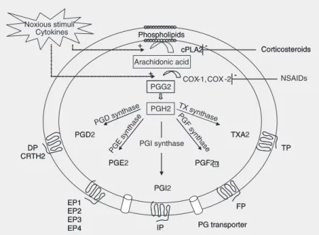
The breakdown of cell membrane phos-pholipids into the 20-carbon fatty acid ara-chidonic acid, the initial rate-limiting step in prostanoid production, is mediated by a group of cytosolic enzymes called phospholipases
(Figure 2). Several noxious stimuli and cy-tokines can up-regulate the activity of these enzymes, which increases the availability of the substrate arachidonic acid during inflam-matory states. Conversely, adrenal steroids inhibit phospholipases and, consequently, the production of prostanoids; this is one of the mechanisms behind the anti-inflamma-tory properties of corticosteroids.
Prostaglandin H (PGH) synthase, also known as COX, catalyzes the conversion of arachidonic acid into PGG2, an unstable prostaglandin that is peroxidized to PGH2. There are two known COX isoforms: COX-1 and COX-2. COX-COX-1 is constitutively ex-pressed in virtually every nucleated cell and is responsible for the basal production of prostanoids involved in physiologic func-tions. Conversely, COX-2 activity is not detectable in most tissues during physiologic states but is characteristically up-regulated by inflammatory stimuli. Therefore, COX-2 is responsible for the increased output of prostanoids during pathologic conditions such as arthritis, glomerulonephritis, and acute allograft rejection.
PGH2 is the intermediate prostaglandin that is metabolized by tissue-specific isom-erases to generate prostaglandins and TXA2. After synthesis, prostanoids exit the cell via poorly characterized prostaglandin transport-ers and are rapidly converted into inactive metabolites; therefore, these compounds have few systemic effects and must exert their actions on neighboring cells or on the very cells from which they were generated. The cellular actions of prostanoids are mediated via specific G-protein coupled receptors pres-ent in most tissues and particularly abundant in cells of the immune system. These recep-tors have been well characterized and the genes encoding them have been cloned. Mouse lines with targeted disruption of each prosta-noid receptor have been subsequently gener-ated, leading to a better understanding of the function of individual prostanoids in physiol-ogy and disease.
In transplantation, the function of two prostanoids, PGE2 and TXA2, has been best characterized. Since these molecules have very distinct roles in graft rejection, they will be analyzed separately.
PGE2 in transplantation
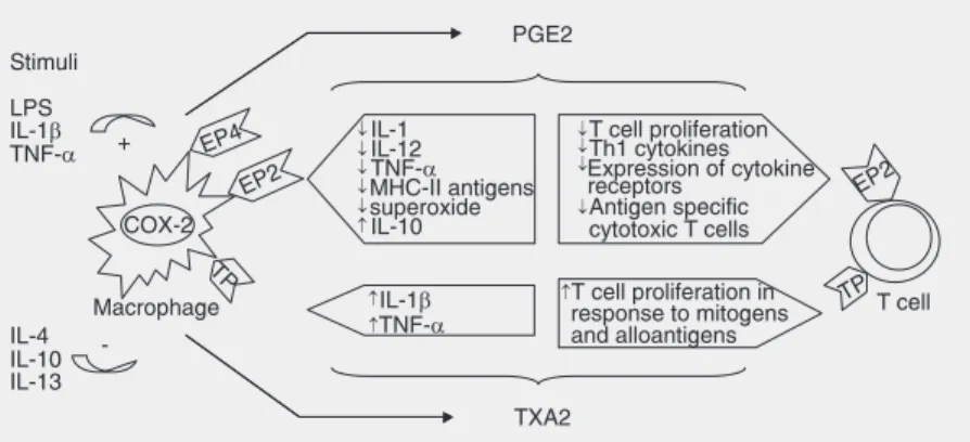
PGE2 is generated by the sequential me-tabolism of arachidonic acid by COX and PGE synthase. The protean biologic actions of this compound are mediated by four re-ceptors: EP1, EP2, EP3, and EP4. Mice with targeted deletions of each receptor subtype have been generated, contributing to a de-tailed understanding of the role of these receptors in physiology and disease. On the immune system, the actions of PGE2 result in suppression of T cell and APC functions, effects that are mediated via EP2 and EP4 receptors (9). Both receptor subtypes share
similar intracellular signaling pathways that involve binding to Gαs, stimulation of aden-ylate cyclase and generation of increased intracellular levels of cyclic AMP, a phe-nomenon typically associated with suppres-sion of immune cell function. In an elegant study, Nataraj et al. (9) demonstrated that the suppressive effects of PGE2 on T cells are mediated by EP2 receptors whereas in macrophages, both EP2 and EP4 receptors are involved.
In macrophages, PGE2 down-regulates the expression of MHC class II antigens (10), inhibits the production of 1 (11), IL-12 (IL-12,13), TNF-α (14), and superoxide (15)
and increases IL-10 production in response to LPS (12,16). This pattern of cytokine release by APCs favors the differentiation of naive T cells towards a Th2 phenotype and promotes B lymphocyte isotype switching to IgE. In addition, PGE2 has been shown to directly inhibit T cell proliferation in re-sponse to mitogens and alloantigens, an ef-fect that has been attributed to inhibition of IL-2 production (17-21).
In concert with their properties of inhib-iting cell-mediated immunity in vitro, PGE
analogues have been shown to prolong the survival of renal (22), cardiac (23), and small-intestinal (24) allografts in rats, as well as skin allografts in mice (25). Human trans-plantation studies also indicate a favorable effect of PGE analogues. In a double-blind, placebo-controlled study of 77 renal allograft recipients receiving a cyclosporine- and pred-nisone-based regimen, Moran et al. (26) ran-domized patients to receive additional treat-ment with misoprostol (N = 38) or placebo (N = 39) for the first 12 weeks; the drug or placebo were then discontinued and the pa-tients followed for an additional 4 weeks. At the end of follow-up, patients receiving mi-soprostol had significantly better renal func-tion, as judged by serum creatinine and creati-nine clearance. Moreover, misoprostol-treated patients experienced a 50% reduc-tion in the incidence of acute rejecreduc-tion (10/
38 vs 20/39, P = 002). Beneficial effects of
PGE have also been demonstrated in liver transplantation. Henley and co-workers (27) showed that, compared to placebo (N = 82), the intravenous infusion of a PGE1 analogue (N = 78) for the first 21 days after liver transplantation reduced the intensive care unit stay, the overall duration of hospitaliza-tion, and the need for renal support.
Thromboxane A2 in transplantation
TXA2 is generated by the metabolism of arachidonic acid by COX and TX synthase. TXA2 is widely known for its effects on blood vessels (vasoconstriction) and plate-lets (aggregation) but an increasing body of evidence indicates that TXA2 also has im-portant actions on the immune system. The cellular actions of TXA2 are mediated by a single thromboxane prostanoid (TP) recep-tor, which is highly expressed in immune cells; indeed, the mouse organs with the highest concentrations of TP receptors are the thymus and the spleen (28). The TP receptor signals through Gαq and promotes an increase in intracellular calcium concen-tration, a phenomenon associated with acti-vation of immune cells.
un-primed mouse spleen cells in the mixed lym-phocyte reaction. They demonstrated that the synthase inhibitor and receptor antago-nist similarly inhibited splenocyte prolifera-tion and that the use of both drugs in combi-nation did not promote any additional effect, indicating that TXA2 directly influences phoproliferative responses in the mixed lym-phocyte reaction. Recently, researchers in Dr. Thomas Coffman’s (31) laboratory gen-erated mice with a targeted deletion of TP receptors (TP-/-). In vitro assays
demon-strated that, compared to wild-type controls, both mitogen- and alloantigen-induced pro-liferative responses are substantially reduced in TP-/- spleen cells (32). Taken together, these results unequivocally confirm direct pro-inflammatory actions of TXA2 enhanc-ing cellular immune responses.
Additional studies have shown that TXA2 enhances cell-mediated immunity through distinct actions on T cells and APCs. In macrophages, TXA2 induces MHC class II molecule expression (10) and regulates
TNF-α and IL-1 synthesis (33). Moreover, pliminary data from our laboratory have re-vealed that, compared to wild-type cells, TP-/- macrophages stimulated with IFN-γ
express lower levels of the co-stimulatory molecules CD40 and CD86 (Rocha PN and Coffman TC, unpublished observations). TXA2 also has direct effects on T cells. We have shown that a population of highly puri-fied TP-/- T cells (>97% purity) exhibits a decreased proliferative response to plate-bound anti-CD3 antibody (32). In our stud-ies, the decreased proliferation observed in TP-/- lymphocytes was not associated with altered cytokine production or expression of co-stimulatory molecules. Rather, we found that when TP-/- cells were exposed to stimuli that triggered an exaggerated, antigen-inde-pendent calcium signal (i.e., phorbol 12-myristate 13-acetate + ionomycin), the pro-liferative defect was completely reversed (32). Our interpretation of these results is that TP receptors might contribute to the
intensity of the calcium signal set off by antigen engagement of the TCR.
A possible role for TXA2 in human trans-plant rejection was first proposed by the studies of Foegh et al. (34), who measured the levels of TXB2 (the stable metabolite of TXA2) in daily urine samples from 12 pa-tients after renal transplantation and demon-strated a several-fold increase in urinary TXB2 excretion during episodes of acute rejection. In the vast majority of cases, the increase in TXB2 excretion preceded the increase in serum creatinine and the clinical diagnosis of rejection. Because of this, sev-eral investigators have since advocated the use of urinary TXB2 levels as a noninvasive tool to aid in the diagnosis of acute renal allograft rejection (35-38). The sources of TX in this setting are the inflammatory cells infiltrating the graft, as the intensity of the infiltrate seems to correlate with the amount of prostanoid produced. Moreover, treatment of acute renal transplant rejection with cyclo-phosphamide has been shown to decrease the urinary excretion of TXB2 (39); as this drug does not interfere with any of the steps involved in TXA2 synthesis, the likely ex-planation for the reduction in TXB2 excre-tion in these studies is the resoluexcre-tion of the inflammatory infiltrate. Of the mononuclear cells typically encountered in the rejecting allograft, monocytes and macrophages are known to produce prostanoids in response to a variety of inflammatory stimuli, such as cytokines, bacterial products, and comple-ment components.
Given the in vitro actions of TXA2 on
allograft survival compared to wild-type con-trols; allografts harvested from TP knockout animals exhibited a marked attenuation in acute rejection scores (32). These effects were only seen when recipients were treated with small doses of cyclosporin A (CsA) during the initial week of transplantation. In a kidney transplantation model, however, we observed attenuation in acute rejection in TP-/- animals even in the absence of phar-macologic manipulation (Rocha PN and Coffman TC, unpublished observations).
The detrimental effects of TXA2 during acute rejection may extend beyond its ef-fects on immune cells. For example, the initial fall in glomerular filtration rate (GFR) during acute renal allograft rejection is much greater than expected based on the observed morphologic changes in the kidney (41-43). The rapid fall in GFR in this setting is likely to represent a vasoactive phenomenon me-diated by molecules such as TXA2 that are produced within the allograft being rejected. Similarly, the quick (and sometimes dra-matic) improvement in GFR observed after steroid treatment for acute rejection is due, at least in part, to reversal of TXA2-induced renal allograft vasoconstriction. The same rationale was used by Pierucci et al. (44) to explain the improvement in renal function observed after the acute infusion of a TXA2 antagonist in humans with lupus nephritis. It was later shown that chronic TXA2 block-ade profoundly altered the course of renal disease in lupus-prone mice, effects that go beyond vasodilation (45).
The role of TXA2 in vascular thrombosis is well established and has been elegantly re-demonstrated by Cheng et al. (46). Local production of this molecule during acute rejection may lead to intragraft thrombosis and irreversible dysfunction. TXA2 block-ade is also essential for the prevention of coronary events in high-risk patients. Since a large number of renal transplant recipients are at increased risk for coronary disease, prevention of myocardial infarction
consti-tutes another advantage of TXA2 blockade in this setting (47).
In summary, TXA2 produced by immune cells infiltrating the graft during acute rejec-tion might contribute to graft dysfuncrejec-tion and injury by: 1) exerting direct pro-inflammatory effects on T cells and APCs that enhance cell-mediated immunity and, consequently, the intensity of allograft rejection; 2) inducing vasoconstriction and reducing graft perfusion; 3) promoting platelet aggregation and intragraft thrombosis. Finally, TXA2 has also been im-plicated as a potential mediator of CsA neph-rotoxicity. Animal studies by Spurney et al. (48) showed that the production of TXA2 is increased during experimental chronic CsA toxicity and that TXA2 blockade is beneficial in this setting. To investigate the significance of these findings in humans, Smith et al. (49) administered the TX synthase inhibitor CGS 13080 in a 48-h intravenous infusion to kid-ney transplant recipients with renal impair-ment due to CsA toxicity. Renal function and plasma flow were measured before and 1 week after the infusion. At the end of the observa-tion, there was a 9% improvement in GFR and a 33% increase in renal plasma flow compared to baseline values. In a subsequent study, however, the oral administration of the TX synthase inhibitor CGS 12970 for 4 weeks did not improve renal function or plasma flow in patients with CsA nephrotoxicity (50).
with these findings, we have recently shown that TXA2 mediates CD4+CD8+ thymocyte apoptosis in vivo (53). Finally, Remuzzi and colleagues (54) demonstrated that administra-tion of a TP receptor antagonist during the peritransplant period completely abrogated the unresponsive state induced by intra-thymic injection of synthetic class II MHC allopeptides in the Wistar-Furth x Lewis rat strain combi-nation. In the same study, inhibition of TXA2 generation by aspirin or dexamethasone also abolished the induction of acquired thymic tolerance. These findings, if confirmed, indi-cate that future immunosuppressive regimens aiming to achieve acquired central tolerance to alloantigens should not include drugs that promote TXA2 blockade in the peritransplant period.
Effect of cyclooxygenase inhibition on the alloimmune responses
Prostanoids with opposing actions on immune cells are generated during an epi-sode of acute rejection (Figure 3). Whereas inhibition of TXA2 is at least theoretically advantageous, the same is not true for PGE2, which has well-described immunosuppres-sive properties. The COX inhibitors (selec-tive or non-selec(selec-tive) are capable of globally inhibiting the synthesis of all prostanoids. Despite the clinically relevant anti-inflam-matory properties of these agents, in vitro
studies indicate that COX blockade with indomethacin actually enhances cellular im-mune responses, as evidenced by prolifera-tion and cytotoxicity assays (29,32). One can speculate that these experiments reveal the dominant effects of immunosuppressive prostanoids such as PGE2 in this model. Alternatively, global COX blockade might shift eicosanoid production towards the li-poxygenase pathway and lead to increased production of pro-inflammatory leukotrienes. Regardless of the exact mechanism behind these apparent pro-inflammatory actions of indomethacin on immune cells, it is
reason-able to theorize that global COX inhibition could exert deleterious effects on allograft function. In kidney allografts, nonsteroidal anti-inflammatory drugs (NSAIDs) could also promote worsening renal function by blocking vasodilator prostanoids. Unfortu-nately, the effects of traditional NSAIDs on allograft function have not been well studied
in vivo. The availability of COX-deficient
mice and specific pharmacologic inhibitors will certainly facilitate this project (55,56). Such studies are urgently needed given that NSAIDs are amongst the most commonly used drugs on the market and transplantation has become the preferred form of therapy for many heart, kidney, and liver diseases.
A recent study by Ma et al. (57) showed that the administration of a selective COX-2 inhibitor doubled the survival of cardiac al-lografts in rats. This prolongation in allograft survival was associated with significant at-tenuation of histological changes at days 3 and 5, as well as a reduction in the number of
apoptotic cardiomyocytes. Taken together, these findings suggest that COX-2-derived prostanoids play a dominant pro-inflamma-tory role during allograft rejection.
Final considerations
We have discussed the intricate relation-ship between inflammation and the alloim-mune response. In the perioperative stages of transplantation, tissue damage and in-flammation set the stage for allorecognition to occur in the presence of danger signals; these signals activate quiescent APCs that, in turn, provide the critical co-stimulatory stimuli required to activate host’s T cells. During episodes of acute rejection, inflam-mation occurs because of the effector ele-ments generated by the alloimmune response. Prostanoid inflammatory mediators produced by activated mononuclear cells infiltrating the graft can modulate the intensity of the alloimmune response and directly regulate graft function by changes in blood flow (Sum-mary box). TXA2 promotes vasoconstric-tion and platelet aggregavasoconstric-tion and enhances cell-mediated immunity via direct actions on both APCs and T cells. These effects are deleterious to the allograft and acute
rejec-tion has been attenuated in animal models by inhibiting the actions of TXA2. In contrast, PGE2 is a vasodilator and powerful immu-nosuppressant and PGE analogues have been shown to ameliorate acute rejection in ani-mal and human transplantation. Global block-ade of prostanoid production with COX in-hibitors enhances alloimmune responses
in vitro, revealing a dominant effect of PGE2, but at present the relevance of COX inhibi-tion in animal models of transplantainhibi-tion is poorly understood. Steroids are the only drugs in the current immunosuppressive ar-mamentarium that possess anti-inflamma-tory properties and are capable of inhibiting the synthesis of prostanoids. Recently, there has been growing interest in steroid-free protocols. In most of these protocols, how-ever, steroids are still used during the times of inflammation described herein: the periop-erative stage of transplantation and episodes of acute rejection. Given the data on TXA2 and thymic tolerance, one might question if steroids are the ideal anti-inflammatory drugs for the perioperative period. Perhaps, in the future, we will have better drugs to down-regulate innate immunity at the time of trans-plantation to efficiently shut off the innate immune system and transform allorecognition into an event that generates tolerance instead of immunity. In addition, targeted anti-inflam-matory agents that can inhibit only the pro-inflammatory prostanoids (such as TXA2) and leave the anti-inflammatory agents (such as PGE2) untouched, might be of great value for the treatment of acute rejection.
Acknowledgments
The authors would like to thank Dr. David Howell (Department of Pathology, Duke University Medical Center) for kindly pro-viding the kidney allograft biopsy images. Summary Box
Prostanoids and Graft Rejection
·
Prostanoids are produced by inflammatory cells within the rejecting allograft·
The vasoactive properties of these compounds enable them to rapidlyinfluence graft function by altering blood flow
·
Prostanoid-induced changes in vascular permeability promote local swellingand facilitate the leakage of inflammatory cells into the interstitium of the graft
·
Platelet function is also regulated by prostanoids: TXA2 promotes whereasPGI2 inhibits aggregation
·
Prostanoids modulate the intensity of allograft rejection by direct effects onReferences
1. Arakelov A & Lakkis FG (2000). The alloimmune response and effector mechanisms of allograft rejection. Seminars in Nephrology, 20: 95-102.
2. Sayegh MH & Turka LA (1995). T cell costimulatory pathways: promising novel targets for immunosuppression and tolerance in-duction. Journal of the American Society of Nephrology, 6: 1143-1150.
3. Matzinger P (2002). The danger model: a renewed sense of self.
Science, 296: 301-305.
4. Cella M, Engering A, Pinet V et al. (1997). Inflammatory stimuli induce accumulation of MHC class II complexes on dendritic cells.
Nature, 388: 782-787.
5. Van Gool SW, Vandenberghe P, de Boer M et al. (1996). CD80, CD86 and CD40 provide accessory signals in a multiple-step T-cell activation model. Immunological Reviews, 153: 47-83.
6. Rocha PN, Plumb TJ, Crowley SD et al. (2003). Effector mechan-isms in transplant rejection. Immunological Reviews, 196: 51-64. 7. Rocha PN, Butterly DW, Greenberg A et al. (2003). Beneficial effect
of plasmapheresis and intravenous immunoglobulin on renal al-lograft survival of patients with acute humoral rejection. Transplan-tation, 75: 1490-1495.
8. Rocha PN, Plumb TJ & Coffman TM (2003). Eicosanoids: lipid mediators of inflammation in transplantation. Springer Seminars in Immunopathology, 25: 215-227.
9. Nataraj C, Thomas DW, Tilley SL et al. (2001). Receptors for prosta-glandin E(2) that regulate cellular immune responses in the mouse.
Journal of Clinical Investigation, 108: 1229-1235.
10. Snyder DS, Beller DI & Unanue ER (1982). Prostaglandins modu-late macrophage Ia expression. Nature, 299: 163-165.
11. Kunkel SL, Chensue SW & Phan SH (1986). Prostaglandins as endogenous mediators of interleukin 1 production. Journal of Immu-nology, 136: 186-192.
12. van der Pouw Kraan TC, Boeije LC, Smeenk RJ et al. (1995). Prostaglandin-E2 is a potent inhibitor of human interleukin 12 pro-duction. Journal of Experimental Medicine, 181: 775-779.
13. van der Pouw Kraan TC, Boeije LC, Snijders A et al. (1996). Regula-tion of IL-12 producRegula-tion by human monocytes and the influence of prostaglandin E2. Annals of the New York Academy of Sciences, 795: 147-157.
14. Scales WE, Chensue SW, Otterness I et al. (1989). Regulation of monokine gene expression: prostaglandin E2 suppresses tumor necrosis factor but not interleukin-1 alpha or beta-mRNA and cell-associated bioactivity. Journal of Leukocyte Biology, 45: 416-421. 15. Metzger Z, Hoffeld JT & Oppenheim JJ (1981). Regulation by PGE2
of the production of oxygen intermediates by LPS-activated macro-phages. Journal of Immunology, 127: 1109-1113.
16. Harizi H, Juzan M, Pitard V et al. (2002). Cyclooxygenase-2-issued prostaglandin e(2) enhances the production of endogenous IL-10, which down-regulates dendritic cell functions. Journal of Immunol-ogy, 168: 2255-2263.
17. Goodwin JS, Messner RP & Peake GT (1978). Prostaglandin sup-pression of mitogen-stimulated lymphocytes in vitro. Changes with mitogen dose and preincubation. Journal of Clinical Investigation, 62: 753-760.
18. Walker C, Kristensen F, Bettens F et al. (1983). Lymphokine regula-tion of activated (G1) lymphocytes. I. Prostaglandin E2-induced inhibition of interleukin 2 production. Journal of Immunology, 130: 1770-1773.
19. Chouaib S, Chatenoud L, Klatzmann D et al. (1984). The mechan-isms of inhibition of human IL-2 production. II. PGE2 induction of suppressor T lymphocytes. Journal of Immunology, 132: 1851-1857. 20. Chouaib S, Welte K, Mertelsmann R et al. (1985). Prostaglandin E2 acts at two distinct pathways of T lymphocyte activation: inhibition of interleukin 2 production and down-regulation of transferrin receptor expression. Journal of Immunology, 135: 1172-1179.
21. Minakuchi R, Wacholtz MC, Davis LS et al. (1990). Delineation of the mechanism of inhibition of human T cell activation by PGE2.
Journal of Immunology, 145: 2616-2625.
22. Strom TB & Carpenter CB (1983). Prostaglandin as an effective antirejection therapy in rat renal allograft recipients. Transplanta-tion, 35: 279-281.
23. Fabrega AJ, Blanchard J, Rivas PA et al. (1992). Prolongation of rat heart allograft survival using cyclosporine and enisoprost, a prosta-glandin E1 analog. Transplantation, 53: 1363-1364.
24. Koh IH, Kim PC, Chung SW et al. (1992). The effects of 16, 16 dimethyl prostaglandin E2 therapy alone and in combination with low-dose cyclosporine on rat small intestinal transplantation. Trans-plantation, 54: 592-598.
25. Anderson CB, Jaffee BM & Graff RJ (1977). Prolongation of murine skin allografts by prostaglandin E1. Transplantation, 23: 444-447. 26. Moran M, Mozes MF, Maddux MS et al. (1990). Prevention of acute
graft rejection by the prostaglandin E1 analogue misoprostol in renal-transplant recipients treated with cyclosporine and prednisone.
New England Journal of Medicine, 322: 1183-1188.
27. Henley KS, Lucey MR, Normolle DP et al. (1995). A double-blind, randomized, placebo-controlled trial of prostaglandin E1 in liver transplantation. Hepatology, 21: 366-372.
28. Namba T, Sugimoto Y, Hirata M et al. (1992). Mouse thromboxane A2 receptor: cDNA cloning, expression and Northern blot analysis.
Biochemical and Biophysical Research Communications, 184: 1197-1203.
29. Leung KH & Mihich E (1980). Prostaglandin modulation of develop-ment of cell-mediated immunity in culture. Nature, 288: 597-600. 30. Ruiz P, Rey L, Spurney R et al. (1992). Thromboxane augmentation
of alloreactive T cell function. Transplantation, 54: 498-505. 31. Thomas DW, Mannon RB, Mannon PJ et al. (1998). Coagulation
defects and altered hemodynamic responses in mice lacking recep-tors for thromboxane A2. Journal of Clinical Investigation, 102: 1994-2001.
32. Thomas DW, Rocha PN, Nataraj C et al. (2003). Proinflammatory actions of thromboxane receptors to enhance cellular immune re-sponses. Journal of Immunology, 171: 6389-6395.
33. Caughey GE, Pouliot M, Cleland LG et al. (1997). Regulation of tumor necrosis factor-alpha and IL-1 beta synthesis by thromboxane A2 in nonadherent human monocytes. Journal of Immunology, 158: 351-358.
34. Foegh ML, Winchester JF, Zmudka M et al. (1981). Urine i-TXB2 in renal allograft rejection. Lancet, 2: 431-434.
35. Winchester JF, Gelfand MC, Foegh ML et al. (1983). Early indicators of renal allograft rejection. Kidney International. Supplement, S-34-S-40.
36. Johnson BF, Wiley KN, Greaves M et al. (1994). Urinary thrombox-ane and 6-keto-prostaglandin F1 alpha are early markers of acute rejection in experimental pancreas transplantation. Transplantation, 58: 18-23.
B2 as an indicator of acute rejection in human liver transplantation.
Surgery Today, 26: 242-249.
38. Zhao Y, Katz NM, Lefrak EA et al. (1997). Urinary thromboxane B2 in cardiac transplant patients as a screening method of rejection.
Prostaglandins, 54: 881-889.
39. Coffman TM, Yarger WE & Klotman PE (1985). Functional role of thromboxane production by acutely rejecting renal allografts in rats.
Journal of Clinical Investigation, 75: 1242-1248.
40. Coffman TM, Ruiz P, Sanfilippo F et al. (1989). Chronic thrombox-ane inhibition preserves function of rejecting rat renal allografts.
Kidney International, 35: 24-30.
41. Gardner LB, Guttmann RD & Merrill JP (1968). Renal transplanta-tion in the inbred rat. IV. Alteratransplanta-tions in the microvasculature in acute unmodified rejection. Transplantation, 6: 411-418.
42. Hollenberg NK, Retik AB, Rosen SM et al. (1968). The role of vasoconstriction in the ischemia of renal allograft rejection. Trans-plantation, 6: 59-69.
43. Rosen SM, Truniger BP, Kriek HR et al. (1967). Intrarenal distribu-tion of blood flow in the transplanted dog kidney: effect of denerva-tion and rejecdenerva-tion. Journal of Clinical Investigation, 46: 1239-1253. 44. Pierucci A, Simonetti BM, Pecci G et al. (1989). Improvement of
renal function with selective thromboxane antagonism in lupus ne-phritis. New England Journal of Medicine, 320: 421-425.
45. Spurney RF, Fan PY, Ruiz P et al. (1992). Thromboxane receptor blockade reduces renal injury in murine lupus nephritis. Kidney International, 41: 973-982.
46. Cheng Y, Austin SC, Rocca B et al. (2002). Role of prostacyclin in the cardiovascular response to thromboxane A2. Science, 296: 539-541.
47. Averna M, Barbagallo CM, Ganci A et al. (2001). Determinants of enhanced thromboxane biosynthesis in renal transplantation. Kid-ney International, 59: 1574-1579.
48. Spurney RF, Mayros SD, Collins D et al. (1990). Thromboxane receptor blockade improves cyclosporine nephrotoxicity in rats.
Prostaglandins, 39: 135-146.
49. Smith SR, Creech EA, Schaffer AV et al. (1992). Effects of throm-boxane synthase inhibition with CGS 13080 in human cyclosporine nephrotoxicity. Kidney International, 41: 199-205.
50. Smith SR, Kubacki VB, Rakhit A et al. (1993). Chronic thromboxane synthase inhibition with CGS 12970 in human cyclosporine nephro-toxicity. Transplantation, 56: 1422-1426.
51. Nusing R, Lesch R & Ullrich V (1990). Immunohistochemical local-ization of thromboxane synthase in human tissues. Eicosanoids, 3: 53-58.
52. Ushikubi F, Aiba Y, Nakamura K et al. (1993). Thromboxane A2 receptor is highly expressed in mouse immature thymocytes and mediates DNA fragmentation and apoptosis. Journal of Experimen-tal Medicine, 178: 1825-1830.
53. Rocha PN, Plumb TJ, Robinson LA, Spurney R, Pisetsky D, Koller BH & Coffman TM (2005). Role of thromboxane A2 in the induction of
apoptosis of immature thymocytes by lipopolysaccharide. Clinical and Diagnostic Laboratory Immunology, 12: 896-903.
54. Remuzzi G, Noris M, Benigni A et al. (1994). Thromboxane A2 receptor blocking abrogates donor-specific unresponsiveness to renal allografts induced by thymic recognition of major histocom-patibility allopeptides. Journal of Experimental Medicine, 180: 1967-1972.
55. Langenbach R, Loftin CD, Lee C et al. (1999). Cyclooxygenase-deficient mice. A summary of their characteristics and susceptibili-ties to inflammation and carcinogenesis. Annals of the New York Academy of Sciences, 889: 52-61.
56. Langenbach R, Morham SG, Tiano HF et al. (1995). Prostaglandin synthase 1 gene disruption in mice reduces arachidonic acid-in-duced inflammation and indomethacin-inacid-in-duced gastric ulceration.
Cell, 83: 483-492.