Research Article
Caffeic Acid Phenethyl Ester: Consequences of
Its Hydrophobicity in the Oxidative Functions and
Cytokine Release by Leukocytes
Luana Chiquetto Paracatu,
1Carolina Maria Quinello Gomes Faria,
1Camila Quinello,
2Camila Rennó,
2Patricia Palmeira,
2Maria Luiza Zeraik,
3Luiz Marcos da Fonseca,
1and Valdecir Farias Ximenes
1,41Department of Clinical Analysis, School of Pharmaceutical Sciences, S˜ao Paulo State University (UNESP),
14801-902 Araraquara, SP, Brazil
2Department of Pediatrics, Medical School, University of S˜ao Paulo, 05403-900 S˜ao Paulo, SP, Brazil
3Department of Organic Chemistry, Institute of Chemistry, S˜ao Paulo State University (UNESP), 14800-900 Araraquara, SP, Brazil 4Department of Chemistry, Faculty of Sciences, S˜ao Paulo State University (UNESP), 17033-360 Bauru, SP, Brazil
Correspondence should be addressed to Valdecir Farias Ximenes; [email protected]
Received 22 April 2014; Revised 23 July 2014; Accepted 24 July 2014; Published 28 August 2014
Academic Editor: Rafaele Capasso
Copyright © 2014 Luana Chiquetto Paracatu et al. his is an open access article distributed under the Creative Commons Attribution License, which permits unrestricted use, distribution, and reproduction in any medium, provided the original work is properly cited.
Numerous anti-inlammatory properties have been attributed to cafeic acid phenethyl ester (CAPE), an active component of propolis. NADPH oxidases are multienzymatic complexes involved in many inlammatory diseases. Here, we studied the importance of the CAPE hydrophobicity on cell-free antioxidant capacity, inhibition of the NADPH oxidase and hypochlorous acid production, and release of TNF-�and IL-10 by activated leukocytes. he comparison was made with the related, but less hydrophobic, cafeic and chlorogenic acids. Cell-free studies such as superoxide anion scavenging assay, triene degradation, and anodic peak potential(�pa)measurements showed that the alterations in the hydrophobicity did not provoke signiicant changes in
the oxidation potential and antiradical potency of the tested compounds. However, only CAPE was able to inhibit the production of superoxide anion by activated leukocytes. he inhibition of the NADPH oxidase resulted in the blockage of production of hypochlorous acid. Similarly, CAPE was the more efective inhibitor of the release of TNF-�and IL-10 byStaphylococcus aureus
stimulated cells. In conclusion, the presence of the catechol moiety and the higher hydrophobicity were essential for the biological efects. Considering the involvement of NADPH oxidases in the genesis and progression of inlammatory diseases, CAPE should be considered as a promising anti-inlammatory drug.
1. Introduction
Propolis is a resin produced by honeybees and its chemical composition, colour, and aroma are changed according to geographical zones. Despite the chemical composition diver-sity, phenolic compounds are constituents that are always present in this natural product [1]. Among them, cafeic acid phenethyl ester is one of the propolis active components for which many potentially beneicial health properties have been demonstrated. Some recent indings include its antithrom-botic potential through the inhibition of tumour necrosis
factor- (TNF-)�induced endothelial tissue factor expression and activity [2]. Suppression of the phosphoinositide 3-kinase/AKT/XIAP pathway has also been shown to lead to apoptosis in melanoma tumour cells bothin vitroandin vivo [3]. Application is used for the treatment of burn wound healing, leading to a decrease in inlammatory parameters and in oxidative damage [4] and also has anti-Helicobacter pylori activity, through the inhibition of the Helicobacter pyloripeptide deformylase [5].
NADPH oxidases are multienzymatic complexes which catalyse the one-electron reduction of molecular oxygen Volume 2014, Article ID 793629, 13 pages
to superoxide anion radical (O2∙−) and are expressed in a variety of cell types. his multicomponent enzyme system is composed of two transmembrane proteins, p22phox and gp91phox, three cytosolic proteins, p47phox, p67phox, and p40phox, and a small G-protein, Rac [6]. he activation of NADPH oxidase involves the migration of the cytosolic proteins to the membrane, assembly of the enzyme com-plex, and the release of O2∙− into the intraphagosomal or extracellular space [7]. From O2∙−, a cascade of enzymatic reactions takes place, leading to the production of hydrogen peroxide (H2O2), hydroxyl radical (∙OH), and hypochlorous acid (HOCl) [7].
Besides its essential role in the innate immune defence, there is increasing evidence of the involvement of NADPH oxidases in the genesis and progression of vascular, inlam-matory, and degenerative diseases [8–12]. Hence, inhibitors of NADPH oxidases represent an alternative and promising therapeutic pathway for the treatment of these chronic inlammatory diseases [13]. Several phytochemicals have been proposed as potential inhibitors of NADPH oxidase, for instance quercetin [14], resveratrol [15], lavonoids [16], and apocynin [17]. In this scenario, we have recently demon-strated that the esteriication of protocatechuic acid, a natural phenolic compound found in many edible and medicinal plants, signiicantly increased its eicacy as an inhibitor of the release of oxidants by stimulated neutrophils [18]. Compared to apocynin, which is the most employed inhibitor of NADPH oxidase, the heptyl ester of protocatechuic acid was about ten-fold more potent [18].
In light of these indings, here, we aim to study and compare cafeic acid and the related compounds, chlorogenic acid, cafeic acid phenethyl ester, and phenethyl cinnamate as potential inhibitors of NADPH oxidase enzymatic activity and cytokine production by leukocytes. he results con-irmed our hypothesis, since a direct relationship was found between the hydrophobicity of the tested compounds and the cellular functions evaluated.
2. Materials and Methods
2.1. Chemicals. Cafeic acid phenethyl ester, cafeic acid, chlo-rogenic acid, phenethyl cinnamate, apocynin, 2,2� -azobis(2-amidinopropane) hydrochloride (AAPH), 2,4,6-Tri(2-pyr-idyl)-S-triazine (TPTZ), dimethyl sulphoxide (DMSO), Brij 35, tung oil, 5-luortryptamine, 2,2-diphenyl-1-picryl-hydrazyl (DPPH), 3,3�,5,5�-tetramethylbenzidine (TMB), lucigenin, phorbol 12-myristate 13-acetate (PMA), taurine, Histopaque-1077, and Histopaque-1119 were purchased from Sigma-Aldrich Chemical Co. (St. Louis, MO, USA). 2-(4- iodophenyl)-3-(4-nitrophenyl)-5-(2,4-disulfophenyl)-2H-tet-razolium monosodium salt (WST-1) was purchased from Santa Cruz Biotechnology (Santa Cruz, CA, USA). Amplex Red (10-acetyl-3,7-dihydroxyphenoxazine) was purchased from Invitrogen (Eugene, OR, USA). Myeloperoxidase (MPO) (EC 1.11.1.7) was purchased from Planta Natural Products (Vienna, Austria) and its concentration was determined from its absorption at 430 nm (�430= 89,000 M−1cm−1). Hydrogen peroxide was prepared by diluting a 30% stock solution and
calculating its concentration from its absorption at 240 nm (�240 = 43.6 M−1cm−1). All reagents used for bufers and mobile phases were of analytical grade. Stock solutions of the tested compounds were prepared in DMSO for cellular studies or in ethanol for electrochemical and DPPH scavenging assays. For the antioxidant assays, the DMSO stock solutions were diluted in 10 mM phosphate bufered saline pH 7.4 (PBS) generating working solutions of lower concentrations. Ultrapure Milli-Q water from Millipore (Belford, MA, USA) was used for the preparation of bufers and solutions. PMA stock solutions were prepared in DMSO at a concentration of 50.0�M and were diluted to 0.5�M in PBS at the time of use. TMB solution was prepared by dissolving 14 mM TMB and 100�M potassium iodide in 50% dimethylformamide and 50% acetic acid (800 mM) (v/v). A working suspension of human serum opsonised zymosan was prepared as previously described at a inal concentration of 10 mg/mL [19].
2.2. Hydrophobicity Index. he molecular hydrophobici-ties of cafeic acid and its derivatives were calculated based on their log � values (partitioning coeicient in n-octanol/water) based on Crippen’s fragmentation method and were performed using ChemDraw sotware (ChemDraw Ultra 7.0.1, CambridgeSot) [20].
2.3. Cyclic Voltammetry. Voltammetric studies were per-formed and the oxidation potentials, measured as anodic peak potential (�pa), were obtained using an Autolab
PGSTAT 30 potentiostat/galvanostat (Eco-Chemie, Utrecht, Netherlands). Voltammetric curves were recorded at room temperature using a 3-electrode setup cell. he working electrode was a glassy carbon disk electrode (GC electrode, 3 mm diameter). he counter electrode was a platinum plate and the reference was an Ag/AgCl saturated KCl at 3 M electrode. he working electrode surface was carefully pol-ished with 0.5�m alumina slurries before each experiment and was thoroughly rinsed with distilled water. A solution of sodium phosphate bufer 0.2 M (pH = 7) was used as a supporting electrolyte. he solutions were purged with nitrogen for 5 min before recording the voltammograms. he ethanolic solutions (5 mM) of the compounds were diluted in the electrochemical cell at inal concentrations of 0.1 mM using the supporting electrolyte solution. he cyclic voltammograms were recorded at a potential scan rate of 5 mV s−1[21].
2.4. DPPH Scavenging Assay. Cafeic acid and its derivatives were incubated for 30 min with 100�M DPPH in ethyl alcohol in the dark. he scavenging activity was evalu-ated spectrophotometrically at 517 nm using the absorbance of unreacted DPPH radical as a control and was calcu-lated as [(Absorbance of control − absorbance of sam-ple)/(absorbance of control)]×100 [22].
was prepared by mixing 2.5 mg of tung oil (not stripped of tocopherols) in 25 mL of PBS containing 17�M Brij 35. he solution was vigorously vortexed to produce a homogeneous emulsion. he assays were performed as follows: the tung oil suspension (50�L) was incubated with freshly prepared 1 mM AAPH (source of peroxyl radical) in PBS at 37∘C in the absence (control) or presence of the tested compounds in the wells of a microplate for 3 hours. he inal reaction volume was 200�L. he microplate was read at 5 min intervals with shaking for 5 seconds before the measurements were taken. he absorbances were measured at 273 nm using a Synergy 2 Multimode microplate reader (BioTek, Winooski, VT, USA). he degradation of the eleostearic acid (conjugated triene) produced the absorbance versus time curve for which the area under the curve (AUC) was calculated. Curves of (AUCsample −AUCcontrol) against the concentration of each
test compound were plotted and their slopes were used as analytical parameters. Trolox was used as a reference antioxidant. he ratio (slopesample/slopetrolox), which is known
as trolox equivalent antioxidant activity (TEAC), was used to assess the relative antioxidant eicacy of the test compounds.
2.6. Reactivity with Hydrogen Peroxide. he reactivity of cafeic acid and its derivatives with H2O2 was monitored amperometrically with a H2O2-selective electrode coupled to a Free Radical Analyzer (TRB 4100, World Precision Instru-ments, USA). he reaction mixtures (3 mL) were composed of 10 nM HRP, 250�M H2O2, and 250�M of the tested compounds in 0.1 M phosphate bufer, pH 7.4, at 25∘C. he reactions were triggered by adding HRP [24].
2.7. Isolation of Human Leukocytes. Blood samples were obtained from healthy volunteers using heparin as an anticoagulant. Polymorphonuclear neutrophil (PMN) and peripheral blood mononuclear cells (PBMC) were isolated by centrifugation on a Histopaque-1077/1119 gradient at 700×g for 30 min at room temperature [25]. Ater isolation, the cells were resuspended in PBS supplemented with 1 mM calcium chloride, 0.5 mM magnesium chloride, and 1 mg/mL glucose (supplemented PBS). Experiments were performed in accor-dance with regulations of the Research Ethics Committee (21496413.8.0000.5426), Faculty of Pharmaceutical Sciences, Unesp, S˜ao Paulo, Brazil.
2.8. Superoxide Anion Production by Activated Leukocytes (Lucigenin-Dependent Chemiluminescence Assay). PMN and PBMC (1 × 10⁶ cells/mL) were preincubated at 37∘C in supplemented PBS with the test compound for 10 min. Next, lucigenin (10�M) and PMA (100 nM) or opsonised zymosan (1 mg/mL) were added and the light emission was measured for 30 min at 37∘C using a plate luminometer (Cen-tro Microplate Luminometer LB960, Berthold Technologies, Oak Ridge, TN, USA). he inal assay volume was 250�L. he integrated light emission was used as an analytical parameter to measure the superoxide anion produced by the stimulated cells. he inhibitory potency was calculated using the light emission generated by the control, in which activated cells were incubated in the absence of the test compounds as [26].
2.9. Superoxide Anion Production by Activated Neutrophils (WST-1 Assay). hese studies were performed as previously described with modiications [27]. PMN (1.0×10⁶cells/mL) were preincubated at 37∘C in supplemented PBS with the test compounds for 10 min. Next, WST-1 (500�M) and PMA (100 nM) were added and the extracellular release of O2∙− was measured by the reduction of WST-1, monitoring the absorbance increase at 450 nm for 30 min at 37∘C; this was performed using Synergy 2 Multimode microplate reader (BioTek, Winooski, VT, USA). he inhibitory potency was calculated using the absorbance of the control, in which the PMA-activated cells were incubated in the absence of the test compounds as a reference.
2.10. Superoxide Anion Production by Xanthine/Xanthine Oxidase (WST-1 Assay). he test compounds were incubated at 37∘C in PBS supplemented with 500�M WST-1 and 100�M xanthine. he reactions were initiated by the addition of 0.05 unit/mL xanthine oxidase and the reduction of WST-1 was assessed by monitoring the absorbance increase at 450 nm for 15 min at 37∘C using Synergy 2 Multimode microplate reader (BioTek, Winooski, VT, USA).
2.11. Production of Hydrogen Peroxide by Activated Neu-trophils. he analysis of the production of H2O2and release into the extracellular medium was evaluated using the lu-orescent probe Amplex Red, as previously described with modiications [28]. PMNs (5×10⁵cells/mL) were preincu-bated at 37∘C in supplemented PBS with the test compounds for 10 min. Next, Amplex Red (500�M) and PMA (100 nM) were added and the extracellular release of H2O2 was mea-sured luorimetrically at 530/590 nm for 30 min at 37∘C using a Synergy 2 Multimode microplate reader (BioTek, Winooski, VT, USA). he inhibitory potency was calculated using the absorbance of the control, in which the PMA-activated cells were incubated in the absence of the tested compounds as a reference.
2.12. Production of Hypochlorous Acid by Activated Neu-trophils and by H2O2/MPO. hese studies were performed as described previously with modiications [29]. he neu-trophils (1.0×10⁶ cells/mL) were preincubated at 37∘C for 10 min in supplemented PBS containing 10 mM taurine and the test compounds. he cells were stimulated by the addition of PMA (100 nM) and were incubated for additional 30 min at 37∘C. he reactions were stopped by the addition of catalase (20�g/mL) and were centrifuged at 6000 rpm. hen, 200�L was transferred into a 96-well plate and the accumulated taurine chloramine was measured by adding 50�L of TMB solution. he oxidised TMB was detected spectrophotomet-rically at 655 nm using Synergy 2 Multimode microplate reader (BioTek, Winooski, VT, USA). he amount of HOCl produced was calculated using a standard curve which was generated using pure HOCl and was submitted to the same analytical protocol.
HO HO
HO
HO OH
OH
OH
OH
OH OH
O O
O
O O
O O
COOH
(C2) (C1)
(C3) (C4)
logP = 3.15 logP = 1.15
logP = −0.75 logP = 3.93
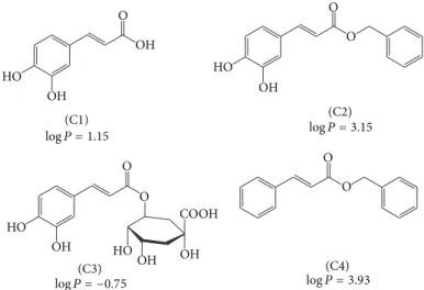
Figure 1: Molecular structures of cafeic acid, its derivatives, and hydrophobicity indexes (log�, partition coeicients n-octanol/water). Cafeic acid (C1), cafeic acid phenethyl ester (C2), chlorogenic acid (C3), and phenethyl cinnamate (C4).
pure MPO (50 nM), H2O2(50�M), and various concentra-tions of the tested compounds. he reacconcentra-tions were triggered by the addition of H2O2and were incubated at 37∘C. Ater 30 min, the reactions were stopped by the addition of catalase (20�g/mL) and the accumulated taurine chloramine was measured as described above. he chlorination inhibitory potency was calculated using the control, in which the MPO/H2O2 was incubated in the absence of the test com-pounds as a reference.
2.13. Cytokines Production by Activated Peripheral Blood Mononuclear Cells. Peripheral blood mononuclear cells (1× 10⁶ cells/mL per well) were cultured in RPMI-1640 (Gibco, Life Technologies, Foster City, CA, USA) medium added with fetal bovine serum (FBS) (Sigma, St. Louis, MO, USA) at 37∘C in an atmosphere of 5% CO2, overnight. Cells were stimulated withStaphylococcus aureus(10 to 1 microorganism per cell) ater 5 hours of incubation with the test compounds. Ater additional 18 hours of incubation the supernatants were stored at−80∘C. TNF-�and IL-10 were quantiied by enzyme-linked immunosorbent assay (ELISA) using BD OptEIA Human TNF ELISA Set (Cat. no. 555212) and BD OptEIA Human IL-10 ELISA Set (Cat. no. 555157), respectively, according to the manufacturer’s instructions.
2.14. Statistical Analysis. Comparisons were performed using one-way ANOVA multiple comparisons among means, with the Turkey’s post hoc test. Results were considered statisti-cally signiicant when� < 0.05. he results were expressed as mean and SEM.
3. Results and Discussion
3.1. Structures and Hydrophobicity Indexes. As already known, the capacity of a substance to cross a lipid barrier may be crucial for its pharmacological efect [30]. Considering that NADPH oxidases are membrane-bound multienzymatic complexes, we hypothesised that, besides the
redox properties, the hydrophobicity could also be relevant for the potency of NADPH oxidase inhibitors. his was our motivation for the study and comparison of cafeic acid and its derivatives as inhibitors of the production of oxidants by activated leukocytes.Figure 1shows the molecular structures and hydrophobicity indexes of cafeic acid (C1) and its derivatives, cafeic acid phenethyl ester (C2), chlorogenic acid (C3), and phenethyl cinnamate (C4). hese compounds were selected with the aim of increasing or decreasing the hydrophobicity compared to C1, without any alteration in the oxidisable catechol moiety, C2, and C3, respectively, or by keeping the hydrophobicity and altering the oxidisability as in C4. From the partitioning coeicient n-octanol/water values depicted inFigure 1, it can be concluded that this goal was reached.
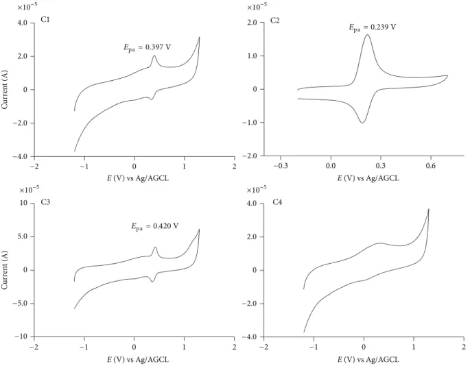
3.2. Oxidation Peak Potential. Most of the substances that have been proposed as inhibitors of NADPH oxidase are redox active compounds, for example, the potent, but non-selective, and toxic diphenyleneiodonium [31]. his com-pound has a mechanism of action based on the genera-tion of a transient radical and this inhibigenera-tion occurs ater direct phenylation of the redox active lavin prosthetic group of gp91phox, or of adjacent amino acid groups in the enzyme complex [31]. Another example is apocynin, which despite the same controversies about potency and selectivity, is the most commonly used NADPH oxidase inhibitor. For this molecule, the proposed mechanism of action is also dependent on its activation through MPO-mediated oxidation [6]. For this reason, prior to the cell-based studies, we measured and compared the oxidation potential of the tested compounds. Here, the oxidation peak potentials, which were measured as anodic peak potentials (�pa), were investigated by cyclic voltammetry using a glassy
C2
−0.3 0.0 0.3 0.6
2.0
1.0
0
−1.0
−2.0 ×10⁻⁵ C1
−2 −1 0 1 2
4.0
2.0
0
−2.0
−4.0 ×10⁻⁵
C
u
rr
en
t (A)
C3 C4
10
5.0
0 0
−5.0 ×10⁻⁵
C
u
rr
en
t (A)
−2 −1 0 1 2 −2 −1 0 1 2
4.0
2.0
−2.0
−4.0 ×10⁻⁵
E(V) vs Ag/AGCL
E(V) vs Ag/AGCL E(V) vs Ag/AGCL
E(V) vs Ag/AGCL Epa = 0.239 V
Epa= 0.397 V
Epa= 0.420 V
−10
Figure 2: Cyclic voltammograms for cafeic acid and its derivatives (0.1 mM) obtained in 0.2 M sodium phosphate bufer at pH 7.0. he anodic peak potential (�pa) is indicated. he scan rate was 5 Mv⋅s−1.
alteration in the oxidation peak potential, which was not unexpected, since esteriication of carboxylic moiety does not provoke signiicant alterations in the oxidisability of these compounds. For instance, a comparison can be made with other phenolic acids, such as gallic acid (�1/20.52 V) versus butyl gallate (�1/2 0.51 V) [32] or protocatechuic acid (�pa
0.222 V) versus heptyl protocatechuates (�pa 0.266 V) [18].
For the compounds studied here, the exception was C4, which did not show a deined anodic wave in the applied voltage range. Obviously, this is a consequence of the absence of a phenolic moiety, making it a redox inactive compound.
3.3. Antioxidant Activity (Cell-Free Assays). he cellular activation of the NADPH oxidase multienzymatic complex results in the generation of ROS; this phenomenon is usu-ally monitored by the use of oxidisable luminescent, chro-mogenic, and luorescent probes [33]. Hence, considering the importance of discrimination between the inhibition of the NADPH oxidase enzymatic activity and the simple and direct scavenging of ROS produced by the cells, we performed a complete panel of cell-free assays aiming to study the efects
provoked by hydrophobicity on the antioxidant capacity of the test compounds.
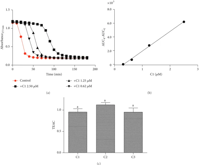
Firstly, the compounds were studied by their eicacy as a reducing agent of the stable free radical DPPH. his assay is based on the reduction of this free radical, which is monitored by a decrease in the absorbance; the efective concentration (EC50) value expresses the concentration that is necessary to decrease the absorbance of DPPH by 50%. he EC50 values obtained were15.7 ± 0.1,16.4 ± 0.3, and14.1 ± 0.1 �M, for C1, C2, and C3, respectively, demonstrating that an alteration in the hydrophobicity did not cause a signiicant alteration in the reducing capacity of the DPPH free radical. However, as expected due to the absence of the catechol moiety, C4 was totally unable to reduce the DPPH free radical.
0 50 100 150 200 0.0
0.5 1.0 1.5
Time (min)
Control
A
b
so
rba
n
ce27
3
nm
+C1 2.50 �M
+C1 1.25 �M
+C1 0.62 �M
(a)
0 1 2 3
0 2.0 4.0 8.0
×103
AU
Cs-A
UC
c
C1 (�M)
6.0
(b)
0.0 0.5
1.0 a
a
a
TEA
C
C1 C2 C3
(c)
Figure 3: Inhibitory efect of cafeic acid and its derivatives on triene degradation by ROO∙. (a) Bleaching of triene (eleostearic acid) by ROO∙ and the efect provoked by the addition of C1. (b) Linear relationship between AUC and concentrations of C1. he slopes were calculated from the liner regression curves. (c) Trolox equivalent antioxidant activity (TEAC = slope compound/slope trolox). he results are mean and SEM of triplicate experiments. Diferent letters denote signiicant diferences. One-way ANOVA and Tukey’s multiple comparison test,� < 0.05.
compounds, by scavenging ROO∙, delayed the bleaching and produced a concentration-dependent lag phase. he results depicted inFigure 3(a)show the kinetic proile of the luorescence bleaching and the efect of the addition of C1 at increasing concentrations.Figure 3(b)shows the relationship between the area under the curve and concentrations of C1 (similar results were obtained for C2 and C3, not shown). he slope of the linear regression (AUC/concentration) was used as an analytical parameter for assessment of the reac-tivity of the tested compounds with ROO∙. he antioxidant trolox, a water soluble derivative of vitamin E, was used for comparison of the antiradical eicacy of the tested compounds. he ratio slopesample/slopetrolox generated the trolox equivalent antioxidant capacity (TEAC) values, which we used to measure the relative antioxidant eicacy of the test compounds. From the results depicted inFigure 3, it can be observed that the tested compounds were as efective as
trolox, but no signiicant diference was obtained between them.
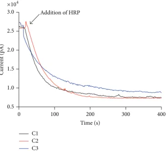
In the next step, cafeic acid and its derivatives were compared regarding their reactivity with H2O2. In these experiments, the reactions were monitored by amperometry using a H2O2 selective-electrode. he results displayed in
Figure 4 show that the tested compounds were completely
nonreactive with H2O2 in the absence of horseradish per-oxidase (HRP). However, the addition of a catalytic amount of HRP caused an instantaneous consumption of H2O2. No signiicant diference was obtained between the tested compounds.
0 100 200 300 400
C1 C2 C3
Addition of HRP
Time (s)
C
ur
ren
t (pA)
3.0
2.5
2.0
1.5
1.0
0.5 ×10⁴
Figure 4: Reactivity of the test compounds with hydrogen peroxide. he reaction mixtures were comprised of 0.25 mM of the test compounds, 0.25 mM H2O2, and 10 nM HRP in 0.01 M PBS, pH 7.4, at 25∘C. he results are representative of three experiments.
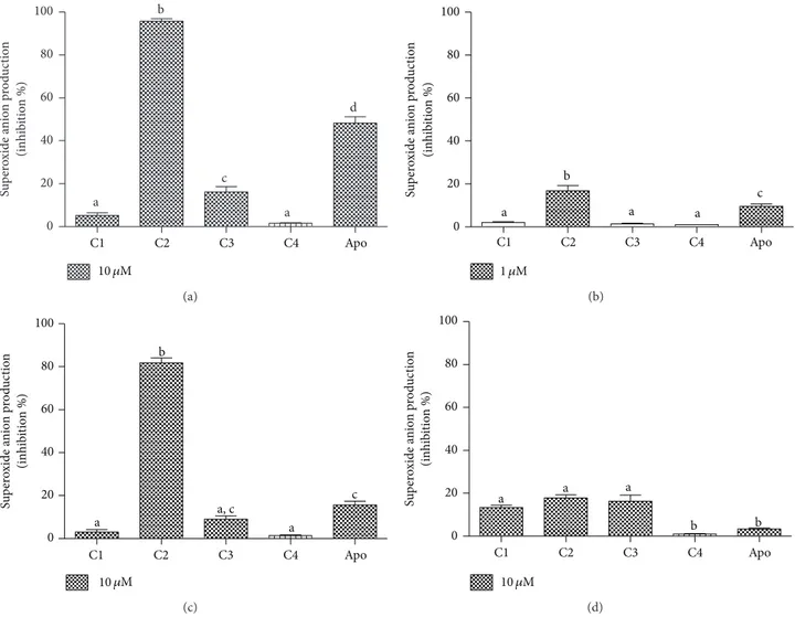
assays. For that, cafeic acid and its derivatives were initially studied as inhibitors of lucigenin-dependent chemilumi-nescence elicited by activated neutrophils. In this reaction, O2∙− produced by the activation of the NADPH oxidase multienzymatic complex reduces lucigenin to form its cation radical, which reacts with a second O2∙−to form the energy-rich dioxetane molecule that decomposes, emitting light [26]. In contrast to the previous antioxidant assays, C2 was found to be a signiicantly more potent inhibitor than the other compounds in this cell-based assay. As can be observed from the results depicted in Figures5(a)and 5(b), C1 and C4 were totally inefective, whereas C3 showed some activity, although this was much lower than that of C2.
he lucigenin-based assay is widely used for the detection of O2∙−; however, some controversies have appeared regard-ing its selectivity for this reactive species [34]. For this reason and considering the higher eicacy of C2 compared to the other compounds, we also studied the activation of NADPH oxidase in neutrophils using a speciic chromogenic probe to O2∙−. his probe is the sulphonated tetrazolium salt (WST-1), which is similar to nitroblue tetrazolium (NBT), which is a widely used compound for the cytochemical determination of NADPH oxidase in leukocytes by microscopy. Compared to NBT, the advantage of WST-1 is its water solubility and membrane-impermeability. Hence, the formazan salt produced by its speciic reaction with O2∙−can be detected in the extracellular medium using conventional absorbance measurements [27]. From the results depicted inFigure 5(c), we can conclude that the higher capacity of C2 was conirmed using the WST-1 assay. Indeed, the diference between C2 and the other compounds was still higher, which suggests its eicacy as an inhibitor of NADPH oxidase.
To conirm that C2 was indeed inhibiting the enzymatic activity of NADPH oxidase and not only acting as a scav-enger of O2∙−, an enzymatic and cell-free experiment was performed. For that, we used the xanthine/xanthine oxidase enzymatic system as a source of O2∙− (Figure 5(d)). As can be seen, using the same concentrations that were used in the cellular experimental model, the direct scavenger efects were minimal and, more importantly, the diferences between C1, C2, and C3 were not statistically signiicant. It is worth noting that the inhibitory efects were not the result of a cytotoxic efect on leukocytes, as conirmed by the trypan blue exclusion assay. At the higher concentration used for the leukocyte studies, the viability of the cells was>98% (results not shown).
From the results depicted in Figure 5, it can be seen that apocynin was used as a reference inhibitor [35]. he reason for this choice was, obviously, the large applicability of apocynin as an NADPH oxidase inhibitor. Hence, the higher eicacy of C2 compared to apocynin is an additional indicator of its potential application as an anti-inlammatory compound, as has been widely demonstrated for apocynin.
he inhibitory potency of C2 was also observed using PBMC instead of neutrophils and again the more lipophilic ester was the more efective one (Figure 6). Our motivation for the use of PBMC relied on the fact that monocytes have a lower content of MPO compared to neutrophils. his is relevant, since MPO may play a role in the mechanism of NADPH oxidase inhibition. his is the case for apocynin, which must be oxidised to perform its role through an MPO-dependent reaction and the consequence is its lower eicacy as an NADPH oxidase inhibitor in PBMC [36]. From the results in Figure 6, it can be seen that the diference between C2 and apocynin was still higher using PBMC than neutrophils. his is an indication that C2 is not dependent on MPO for its biological efect.
0 20 40 60 80 100 a b c a d Su p er o xide a nio n p ro d uc tio n (inhib it io n %)
10 �M
C1 C2 C3 C4 Apo
(a) 0 20 40 60 80 100 a b a a c
1 �M
C1 C2 C3 C4 Apo
Su p er o xide a nio n p ro d uc tio n (inhib it io n %) (b) 0 20 40 60 80 100 a b a, c a c Su p er o xide a nio n p ro d uc ti o n (inhib it io n %)
C1 C2 C3 C4 Apo
10 �M
(c) 0 20 40 60 80 100
a a a
b b
10 �M
C1 C2 C3 C4 Apo
Su p er o xide a nio n p ro d uc ti o n (inhib it io n %) (d)
Figure 5: Cafeic acid, its derivatives, and apocynin (apo) as inhibitors of the production of superoxide anion by stimulated PMN and xanthine/xanthine oxidase. (a, b) Lucigenin-dependent chemiluminescence elicited by opsonised zymosan-activated PMN. (c) WST-1 reduction elicited by PMA-activated PMN. (d) WST-1 reduction elicited by xanthine/xanthine oxidase. he results are mean and SEM of duplicates of three diferent experiments. Diferent letters denote signiicant diferences. One-way ANOVA and Tukey’s multiple comparison test,� < 0.05.
of C2 in this cell-based assay is one more conirmation of its efect on NADPH oxidase in leukocytes rather than this being a simple scavenging efect.
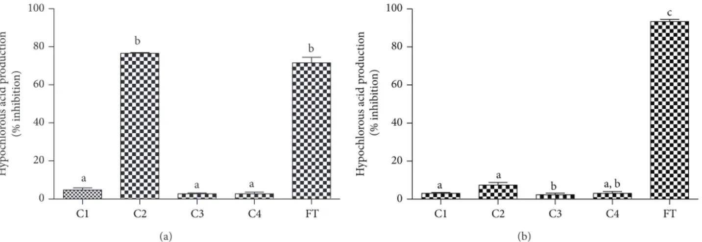
3.6. Inhibition of the Production of Hypochlorous Acid by Activated Leukocytes. he next step in the enzymatic cas-cade of reactions that characterises the oxidative burst of neutrophils is the production of HOCl through the MPO-mediated oxidation of chloride using H2O2as a cosubstrate. Hence, if C2 was indeed inhibiting the production of O2∙−, the production of HOCl should also be afected. To study this possibility, we used a technique based on trapping the HOCl produced with taurine, which is converted to taurine chloramine. his stable oxidant was detected by the iodide-catalysed oxidation of TMB [37]. From the results depicted
inFigure 8(a), it can be noted that, among the phenolic acid
derivatives, only C2 was able to inhibit the production of HOCl.
hree diferent reasons could explain the decreased pro-duction of HOCl by activated neutrophils in the presence of C2: (i) inhibition of the enzymatic activity of MPO, (ii) direct scavenging of HOCl, and (iii) inhibition of the production of O2∙−. To discriminate these efects, we measured the capacity of the tested compounds as inhibitors of the chlorinating activity of the puriied MPO. From the results depicted in Figure 8(b), we can note that all of the tested compounds, including C2, were unable to inhibit the formation of HOCl in the cell-free system. hese results also eliminated the direct scavenging efect upon HOCl. Hence, the inhibition of the production of HOCl without afecting the MPO enzymatic activity is an additional conirmation of the efect of C2 on NADPH oxidase in neutrophils.
0 20 40 60 80 100 a b
a a a
Su p er o xide a nio n p ro d uc ti o n (inhib it io n %)
1 �M
C1 C2 C3 C4 Apo
0 20 40 60 80 100 a b c, d c d Su p er o xide a nio n p ro d uc tio n (inhib it io n %)
10 �M
C1 C2 C3 C4 Apo
Figure 6: Cafeic acid, its derivatives, and apocynin (apo) as inhibitors of the production of superoxide anion by stimu-lated PBMC. Lucigenin-dependent chemiluminescence elicited by opsonised zymosan-activated PBMC. he results are mean and SEM of duplicates of three diferent experiments. Diferent letters denotes signiicant diference. One-way ANOVA and Tukey’s multiple com-parison test (� < 0.05).
production of HOCl in both cell and cell-free assays (Figures
8(a)and8(b)). However, C2 was only able to exert its efect
on neutrophils, which reinforces its speciic action upon the NADPH oxidase enzymatic system.
3.7. Inhibition of the Production of TNF-�and IL-10. Tumour necrosis factor-� (TNF-�) is a major proinlammatory cytokine involved in the inlammatory response. here is evidence of crosstalk between NADPH oxidase and TNF-� in many experimental models using several cell lineages. Some recent indings include the following: the inhibition of NADPH oxidase attenuated TNF-� impaired endothelium-dependent vasodilation [39], increased TNF-� expression associated with diabetes contributes to erectile dysfunction by promoting NAPDH oxidase-mediated ROS generation [40], and TNF-� produces oxidative stress in neuronal cells via the activation of NADPH oxidase through a ceramide-mediated mechanism [41] and so on. Moreover, TNF-�has been shown to prime the neutrophil respiratory
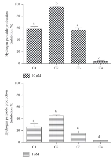
0 20 40 60 80 100 a b a c H ydr og en p er o xide p ro d uc tio n (inhib it io n %) 0 20 40 60 80 100 a b a d H ydr og en p er o xide p ro d uc tio n (inhib it io n %)
C1 C2 C3 C4
C1 C2 C3 C4
1 �M 10 �M
Figure 7: Cafeic acid and its derivatives as inhibitors of the production of hydrogen peroxide by stimulated PMN. he results are mean and SEM of duplicates of three diferent experiments. Diferent letters denote signiicant diferences. One-way ANOVA and Tukey’s multiple comparison test,� < 0.05.
burst through the partial phosphorylation of p47PHOX [42], which is a cytosolic component of the NADPH oxidase complex.
From the opposite point of view, oxidative stress, which can be initiated by the activation of NADPH oxidase, among other mechanisms, can promote the release of TNF-� through the activation of redox-sensitive transcription factor as nuclear factor-kappa B (NF-kappa B) [43, 44]. Hence, there are numerous demonstrations of the beneicial efect of natural compounds that, besides antioxidative efects, also decrease the production of TNF-�and other proinlamma-tory cytokines [10, 45, 46]. C2 is included in this class of natural antioxidants that are able to inhibit the release of TNF in many experimental models and using diferent cell/tissue models [2,47,48]. Indeed, C2 was described as a potent and a speciic inhibitor of NF-kappa B in the human U937 cell lineage [49].
0 20 40 60 80 100
a
b
a
b
a
H
yp
o
chlo
ro
u
s acid p
ro
d
uc
tio
n
(% inhib
it
io
n)
C1 C2 C3 C4 FT
(a)
0 20 40 60 80 100
a a b a, b
c
H
yp
o
chlo
ro
u
s acid p
ro
d
uc
ti
o
n
(% in
hib
it
io
n)
C1 C2 C3 C4 FT
(b)
Figure 8: Cafeic acid, its derivatives, and 5-luortryptamine (FT) as inhibitors of the production of hypochlorous acid by (a) stimulated PMN and (b) puriied MPO. he results are mean and SEM of duplicates of three diferent experiments. Diferent letters denote signiicant diferences. One-way ANOVA and Tukey’s multiple comparison test,� < 0.05.
0 1000 2000 3000
a a
b
c
d
0 15 30 45 400 800
1200 a a
b
c
d
IL
-10 (pg/mL)
C1 C1 C2 C2
Control
C1 C1 C2 C2
Control 70 �M 20 �M
70 �M 20 �M
TNF
-𝛼
(pg/mL)
Figure 9: Production of TNF-�and IL-10 by stimulated PBMC and the inhibitory efect of cafeic acid (C1) and cafeic acid phenethyl ester (C2). he results are mean and SEM of duplicates of six diferent experiments. Diferent letters denote signiicant diferences. One-way ANOVA and Tukey’s multiple comparison test,� < 0.05.
can be concluded that the higher hydrophobicity of C2 was also relevant regarding the inhibition of TNF-�. As discussed above, it would be expected that C2 showed this inhibitory efect, but the inding that it was signiicantly more efective than C1, which has similar antioxidant potency, is additional evidence that its hydrophobicity is an important factor in this biological efect, which is closely related to NADPH oxidase activation.
Similar results were obtained for IL-10, an anti-inlammatory cytokine whose production by stimulated PBMC is also inhibited by natural antioxidants [50]. Regarding the relationship between NADPH oxidase and IL-10, it has been proposed that this cytokine down regulates the polymorphonuclear neutrophil production of ROS [51]. Hence, similarly to TNF-�, our inding that C2 was also more efective than C1 as an inhibitor of the production of IL-10 could also be a consequence of the relationship between NADPH oxidase and this cytokine.
4. Conclusions
seems clear that these results are a consequence of the capacity of cafeic acid phenethyl ester to difuse into the cells and to reach the membrane-bound NADPH oxidase multienzymatic complexes.
As a potential NADPH oxidase inhibitor, this propolis component can be used for the treatment of chronic inlam-matory diseases; hence, a remaining question is what dosage should be used to reach a pharmacologically eicient plasma concentration. In this context, an analytical method to deter-mine cafeic acid phenethyl ester in rat plasma and urine was developed and the pharmacokinetic studies showed that it is rapidly absorbed and excreted in urine both as an unmodiied molecule and as a glucuronide conjugate [52]. In this study, 100 mg/kg was orally administered, but the recovered cafeic acid phenethyl ester was only 0.007 to 0.021%, which is an indication that it was promptly hydrolysed by plasmatic esterases [52], probably to its cafeic acid precursor. As we have demonstrated here, cafeic acid was signiicantly less eicient compared to the ester derivative, hence an efective dose of cafeic acid phenethyl ester forin vivostudies could be signiicantly higher than that obtained in cell-based studies. For this reason, we suggest that the efective concentrations of cafeic acid phenethyl ester found in cell-based studies should be only used as an initial parameter forin vivoapplications.
Conflict of Interests
he authors declare that there is no conlict of interests regarding the publication of this paper.
Acknowledgments
his work has been partially supported by the Brazil-ian agency, CAPES, the S˜ao Paulo Research Foundation (FAPESP), and the National Council for Scientiic and Tech-nological Development (CNPq).
References
[1] M. L. Khalil, “Biological activity of bee propolis in health and disease,”Asian Paciic Journal of Cancer Prevention, vol. 7, no. 1, pp. 22–31, 2006.
[2] C. Gebhard, B. E. St¨ahli, S. Largiad`er et al., “Cafeic acid phenethyl ester inhibits endothelial tissue factor expression,”
Biological and Pharmaceutical Bulletin, vol. 36, no. 6, pp. 1032– 1035, 2013.
[3] K. C. Pramanik, S. K. Kudugunti, N. M. Fofaria, M. Y. Moridani, and S. K. Srivastava, “Cafeic acid phenethyl ester suppresses melanoma tumor growth by inhibiting PI3K/AKT/XIAP path-way,”Carcinogenesis, vol. 34, no. 9, pp. 2061–2070, 2013. [4] J. S. dos Santos and A. Monte-Alto-Costa, “Cafeic acid
phenethyl ester improves burn healing in rats through anti-inlammatory and antioxidant efects,”Journal of Burn Care and Research, vol. 34, no. 6, pp. 682–688, 2013.
[5] K. Cui, W. Lu, L. Zhu, X. Shen, and J. Huang, “Cafeic acid phenethyl ester (CAPE), an active component of propolis, inhibits Helicobacter pylori peptide deformylase activity,” Bio-chemical and Biophysical Research Communications, vol. 435, no. 2, pp. 289–294, 2013.
[6] P. Kleniewska, A. Piechota, B. Skibska, and A. Gorąca, “he NADPH oxidase family and its inhibitors,”Archivum Immu-nologiae et herapiae Experimentalis, vol. 60, no. 4, pp. 277–294, 2012.
[7] D. C. Dale, L. Boxer, and W. Conrad Liles, “he phagocytes: neutrophils and monocytes,”Blood, vol. 112, no. 4, pp. 935–945, 2008.
[8] A. Simonyi, Y. He, W. Sheng et al., “Targeting NADPH oxidase and phospholipases A2 in alzheimer’s disease,”Molecular Neu-robiology, vol. 41, no. 2-3, pp. 73–86, 2010.
[9] H. Zhou, F. Zhang, S. Chen et al., “Rotenone activates phagocyte NADPH oxidase by binding to its membrane subunit gp91 phox,”Free Radical Biology and Medicine, vol. 52, no. 2, pp. 303– 313, 2012.
[10] M. T. Fischer, R. Sharma, J. L. Lim et al., “NADPH oxidase expression in active multiple sclerosis lesions in relation to oxidative tissue damage and mitochondrial injury,”Brain, vol. 135, no. 3, pp. 886–899, 2012.
[11] C. Lin and H. Wang, “NADPH oxidase is involved in H2O2-induced diferentiation of human promyelocytic leukaemia HL-60 cells,”Cell Biology International, vol. 36, no. 4, pp. 391–395, 2012.
[12] I. Takac, K. Schr¨oder, and R. P. Brandes, “he nox family of NADPH Oxidases: friend or foe of the vascular system?”
Current Hypertension Reports, vol. 14, no. 1, pp. 70–78, 2012. [13] G. R. Drummond, S. Selemidis, K. K. Griendling, and C. G.
Sobey, “Combating oxidative stress in vascular disease: NADPH oxidases as therapeutic targets,”Nature Reviews Drug Discovery, vol. 10, no. 6, pp. 453–471, 2011.
[14] J. Peˇcivov´a, T. Maˇciˇckov´a, K. Svitekov´a, and R. Nos´al’, “Quercetin inhibits degranulation and superoxide generation in PMA stimulated neutrophils,”Interdisciplinary Toxicology, vol. 5, no. 2, pp. 81–83, 2012.
[15] F. Chen, L. Qian, B. Deng, Z. Liu, Y. Zhao, and Y. Le, “Resveratrol protects vascular endothelial cells from high glucose-induced apoptosis through inhibition of nadph oxidase activation-driven oxidative stress,”CNS Neuroscience and herapeutics, vol. 19, no. 9, pp. 675–681, 2013.
[16] M. Ciz, P. Denev, M. Kratchanova, O. Vasicek, G. Ambrozova, and A. Lojek, “Flavonoids inhibit the respiratory burst of neutrophils in mammals,” Oxidative Medicine and Cellular Longevity, vol. 2012, Article ID 181295, 6 pages, 2012.
[17] I. H. Philippens, J. A. Wubben, B. Finsen, and B. A. ’T Hart, “Oral treatment with the NADPH oxidase antagonist apocynin mitigates clinical and pathological features of parkinsonism in the MPTP marmoset model,” Journal of Neuroimmune Pharmacology, vol. 8, no. 3, pp. 715–726, 2013.
[18] C. M. Q. G. de Faria, A. C. Nazar´e, M. S. Petrˆonio et al., “Pro-tocatechuic acid alkyl esters: hydrophobicity as a determinant factor for inhibition of NADPH oxidase,” Current Medicinal Chemistry, vol. 19, no. 28, pp. 4885–4893, 2012.
[19] A. Lojek, L. Kubala, H. ˇC´ıˇzov´a, and M. ˇC´ıˇz, “A comparison of whole blood neutrophil chemiluminescence measured with cuvette and microtitre plate luminometers,”Luminescence, vol. 17, no. 1, pp. 1–4, 2002.
[20] A. K. Ghose and G. M. Crippen, “Atomic physicochemical parameters for three-dimensional-structure-directed quantita-tive structure-activity relationships. 2. Modeling dispersive and hydrophobic interactions,”Journal of Chemical Information and Computer ScienceⓇ, vol. 27, pp. 21–35, 1987.
of myeloperoxidase,”Current Medicinal Chemistry, vol. 19, no. 31, pp. 5405–5413, 2012.
[22] W. Brand-Williams, M. E. Cuvelier, and C. Berset, “Use of a free radical method to evaluate antioxidant activity,”LWT-Food Science and Technology, vol. 28, no. 1, pp. 25–30, 1995. [23] M. Laguerre, L. J. L´opez-Giraldo, J. Lecomte et al., “Conjugated
autoxidizable triene (CAT) assay: a novel spectrophotometric method for determination of antioxidant capacity using triacyl-glycerol as ultraviolet probe,”Analytical Biochemistry, vol. 380, no. 2, pp. 282–290, 2008.
[24] M. S. Petrˆonio, M. L. Zeraik, L. M. da Fonseca, and V. F. Ximenes, “Apocynin: chemical and biophysical properties of a NADPH oxidase inhibitor,”Molecules, vol. 18, no. 3, pp. 2821– 2839, 2013.
[25] D. English and B. R. Andersen, “Single step separation of red blood cells, granulocytes and mononuclear leukocytes on discontinuous density gradients of Ficoll Hypaque,”Journal of Immunological Methods, vol. 5, no. 3, pp. 249–252, 1974. [26] A. C. de Almeida, O. C. Marques, C. Arslanian, A.
Condino-Neto, and V. F. Ximenes, “4-luoro-2-methoxyphenol, an apoc-ynin analog with enhanced inhibitory efect on leukocyte oxidant production and phagocytosis,” European Journal of Pharmacology, vol. 660, no. 2-3, pp. 445–453, 2011.
[27] A. S. Tan and M. V. Berridge, “Superoxide produced by activated neutrophils eiciently reduces the tetrazolium salt, WST-1 to produce a soluble formazan: a simple colorimetric assay for measuring respiratory burst activation and for screening anti-inlammatory agents,”Journal of Immunological Methods, vol. 238, no. 1-2, pp. 59–68, 2000.
[28] M. Zhou, Z. Diwu, N. Panchuk-Voloshina, and R. P. Haugland, “A stable nonluorescent derivative of resoruin for the luoro-metric determination of trace hydrogen peroxide: Applications in detecting the activity of phagocyte NADPH oxidase and other oxidases,”Analytical Biochemistry, vol. 253, no. 2, pp. 162– 168, 1997.
[29] V. F. Ximenes, I. M. M. Paino, O. M. M. de Faria-Oliveira, L. M. da Fonseca, and I. L. Brunetti, “Indole ring oxidation by activated leukocytes prevents the production of hypochlorous acid,”Brazilian Journal of Medical and Biological Research, vol. 38, no. 11, pp. 1575–1583, 2005.
[30] E. Rutkowska, K. PajIk, and K. J´o´zwiak, “Lipophilicity— methods of determination and its role in medicinal chemistry,”
Acta Poloniae Pharmaceutica, vol. 70, no. 1, pp. 3–18, 2013. [31] V. B. O’Donnell, D. G. Tew, O. T. G. Jones, and P. J. England,
“Studies on the inhibitory mechanism of iodonium compounds with special reference to neutrophil NADPH oxidase,” Biochem-ical Journal, vol. 290, no. 1, pp. 41–49, 1993.
[32] S. Gunckel, P. Santander, G. Cordano et al., “Antioxidant activity of gallates: an electrochemical study in aqueous media,”
Chemico-Biological Interactions, vol. 114, no. 1-2, pp. 45–59, 1998. [33] C. C. Winterbourn, “he challenges of using luorescent probes to detect and quantify speciic reactive oxygen species in living cells,”Biochimica et Biophysica Acta, vol. 1840, no. 2, pp. 730– 738, 2014.
[34] M. Barbacanne, J. Souchard, B. Darblade et al., “Detection of superoxide anion released extracellularly by endothelial cells using cytochrome c reduction, ESR, luorescence and lucigenin-enhanced chemiluminescence techniques,”Free Radical Biology and Medicine, vol. 29, no. 5, pp. 388–396, 2000.
[35] J. Stefanska and R. Pawliczak, “Apocynin: molecular aptitudes,”
Mediators of Inlammation, vol. 2008, Article ID 106507, 10 pages, 2008.
[36] A. C. de Almeida, M. M. dos Santos Vilela, A. Condino-Neto, and V. F. Ximenes, “he importance of myeloperoxidase in apocynin-mediated NADPH oxidase inhibition,”ISRN Inlam-mation, vol. 2012, Article ID 260453, 7 pages, 2012.
[37] A. J. Kettle, A. M. Albrett, A. L. Chapman et al., “Measuring chlorine bleach in biology and medicine,”Biochimica et Biophys-ica Acta, vol. 1840, no. 2, pp. 781–793, 2013.
[38] A. C. de Almeida, O. C. Marques, C. Arslanian, A. Condino-Neto, and V. F. Ximenes, “4-Fluoro-2-methoxyphenol, an apocynin analog with enhanced inhibitory efect on leukocyte oxidant production and phagocytosis,” European Journal of Pharmacology, vol. 660, no. 2-3, pp. 445–453, 2011.
[39] Y. Huang, L. Yan, S. Rong, H. Haller, and T. Kirch, “TNF-� induces endothelial dysfunction via PKC-�-dependent NADPH oxidase activation,”Journal of Huazhong University of Science and Technology. Medical Science, vol. 32, no. 5, pp. 642–647, 2012.
[40] T. Long, G. Liu, Y. Wang, Y. Chen, Y. Zhang, and D. Qin, “TNF-�, erectile dysfunction, and NADPH oxidase-mediated ROS generation in corpus cavernosum in high-fat diet/ streptozotocin-induced diabetic rats,”Journal of Sexual Medicine, vol. 9, no. 7, pp. 1801–1814, 2012.
[41] B. M. Barth, S. J. Gustafson, J. L. Hankins et al., “Ceramide kinase regulates TNF�-stimulated NADPH oxidase activity and eicosanoid biosynthesis in neuroblastoma cells,”Cellular Signalling, vol. 24, no. 6, pp. 1126–1133, 2012.
[42] C. Dewas, P. M. Dang, M. Gougerot-Pocidalo, and J. El-Benna, “TNF-�induces phosphorylation of p47phox in human neu-trophils: Partial phosphorylation of p47phox is a common event of priming of human neutrophils by TNF-�and granulocyte-macrophage colony-stimulating factor,”Journal of Immunology, vol. 171, no. 8, pp. 4392–4398, 2003.
[43] A. Csiszar, M. Wang, E. G. Lakatta, and Z. Ungvari, “Inlamma-tion and endothelial dysfunc“Inlamma-tion during aging: role of NF-�B,”
Journal of Applied Physiology, vol. 105, no. 4, pp. 1333–1341, 2008. [44] A. Mu˜noz and M. Costa, “Nutritionally mediated oxidative stress and inlammation,” Oxidative Medicine and Cellular Longevity, vol. 2013, Article ID 610950, 11 pages, 2013.
[45] M. S. Baliga, N. Joseph, M. V. Venkataranganna, A. Saxena, V. Ponemone, and R. Fayad, “Curcumin, an active component of turmeric in the prevention and treatment of ulcerative colitis: preclinical and clinical observations,”Food and Function, vol. 3, no. 11, pp. 1109–1117, 2012.
[46] C. Lin and H. Wang, “NADPH oxidase is involved in H2O2 -induced diferentiation of human promyelocytic leukaemia HL-60 cells,”Cell Biology International, vol. 36, no. 4, pp. 391–395, 2012.
[47] S. Juman, N. Yasui, K. Ikeda et al., “Cafeic acid phenethyl ester suppresses the production of pro-inlammatory cytokines in hypertrophic adipocytes through lipopolysaccharide-stimulated macrophages,” Biological and Pharmaceutical Bulletin, vol. 35, no. 11, pp. 1941–1946, 2012.
[48] T. C¸ akır, E. ¨Ozkan, E. Dulundu et al., “Cafeic acid phenethyl ester (CAPE) prevents methotrexate-induced hepatorenal oxidative injury in rats,” he Journal of Pharmacy and Pharmacology, vol. 63, no. 12, pp. 1566–1571, 2011.
[49] K. Natarajan, S. Singh, T. R. Burke Jr., D. Grunberger, and B. B. Aggarwal, “Cafeic acid phenethyl ester is a potent and speciic inhibitor of activation of nuclear transcription factor NF-�B,”
[50] P. M. Dang, C. Elbim, J. Marie, M. Chiandotto, M. Gougerot-Pocidalo, and J. El-Benna, “Anti-inlammatory efect of interleukin-10 on human neutrophil respiratory burst involves inhibition of GM-CSF-induced p47PHOX phosphorylation through a decrease in ERK1/2 activity,”he FASEB Journal, vol. 20, no. 9, pp. 1504–1506, 2006.
[51] M. Bergman, M. Djaldetti, H. Salman, and H. Bessler, “On the combined efect of statins and lycopene on cytokine production by human peripheral blood cells,”Heart and Vessels, vol. 25, no. 5, pp. 426–431, 2010.
Submit your manuscripts at
http://www.hindawi.com
Stem Cells
International
Hindawi Publishing Corporationhttp://www.hindawi.com Volume 2014
Hindawi Publishing Corporation
http://www.hindawi.com Volume 2014
INFLAMMATION
Hindawi Publishing Corporation
http://www.hindawi.com Volume 2014
Behavioural
Neurology
Endocrinology
International Journal of Hindawi Publishing Corporationhttp://www.hindawi.com Volume 2014 Hindawi Publishing Corporation
http://www.hindawi.com Volume 2014
Disease Markers
Hindawi Publishing Corporation
http://www.hindawi.com Volume 2014 BioMed
Research International
Oncology
Journal of Hindawi Publishing Corporationhttp://www.hindawi.com Volume 2014
Hindawi Publishing Corporation
http://www.hindawi.com Volume 2014
Oxidative Medicine and Cellular Longevity
Hindawi Publishing Corporation
http://www.hindawi.com Volume 2014
PPAR Research
The Scientiic
World Journal
Hindawi Publishing Corporation
http://www.hindawi.com Volume 2014
Immunology Research Hindawi Publishing Corporation
http://www.hindawi.com Volume 2014
Journal of
Obesity
Journal ofHindawi Publishing Corporation
http://www.hindawi.com Volume 2014
Hindawi Publishing Corporation
http://www.hindawi.com Volume 2014
Computational and Mathematical Methods in Medicine
Ophthalmology
Journal of Hindawi Publishing Corporationhttp://www.hindawi.com Volume 2014
Diabetes Research
Journal ofHindawi Publishing Corporation
http://www.hindawi.com Volume 2014
Hindawi Publishing Corporation
http://www.hindawi.com Volume 2014
Research and Treatment
AIDS
Hindawi Publishing Corporation
http://www.hindawi.com Volume 2014
Gastroenterology Research and Practice
Hindawi Publishing Corporation
http://www.hindawi.com Volume 2014
Parkinson’s
Disease
Evidence-Based Complementary and Alternative Medicine
Volume 2014