AbstrAct: Mango wilt, caused by Ceratocystis fimbriata, is one of the most important diseases affecting mango yields in Brazil.
Information regarding the biochemical mechanisms involved in mango
resistance against C. fimbriata is absent in the literature. Thus, the present study determined and quantified alkaloids and phenolics in
the stem tissue of mango plants from Palmer (susceptible) and Ubá
(resistant) cultivars. Furthermore, it was examined the effect of these
secondary metabolites against C. fimbriata growth in vitro. The high-performance liquid chromatography revealed that the concentration
of two alkaloids (theobromine and 7-methylxanthine) and six phenolic
compounds (caffeic acid, p-coumaric acid, gallic acid, protocatechuic acid, catechin and epicatechin) in the inoculated plants from cv. Ubá
was higher in comparison with inoculated plants from cv. Palmer.
PLANt PrOtEctION -
Article
Alkaloids and phenolics biosynthesis
increases mango resistance to infection by
Ceratocystis fimbriata
Leonardo Araujo1, Wilka Messner Silva Bispo1, Jonas Alberto Rios1, Sergio Antonio Fernandes2,
Fabrício de Ávila Rodrigues1*
1. Universidade Federal de Viçosa - Departamento de Fitopatologia - Viçosa (MG), Brasil. 2. Universidade Federal de Viçosa - Departamento de Química - Viçosa (MG), Brasil.
*Corresponding author: fabricio@ufv.br
Received: Jun. 19, 2015 – Accepted: Oct. 8, 2015
The concentration of the secondary metabolites was higher in the
non-inoculated plants from cv. Palmer than in the inoculated ones,
while the opposite was observed for plants of cv. Ubá. Peaks in the
concentrations of secondary metabolites in the inoculated plants
from both cultivars occurred at 7 and 14 days after inoculation. The
different concentrations (10 to 30 mg∙mL−1) of secondary metabolites added to the Petri dishes greatly inhibited C. fimbriata growth over time. These results suggest that secondary metabolites played an
important role in the resistance of mango plants against C. fimbriata
infection.
Key words:Ceratocystis fimbriata, Mangifera indica,high-performance liquid chromatography, host defense responses, secondary metabolism
in plants, vascular pathogen.
INtrODUctION
Mango (Mangifera indica L.) is one of the most important tropical fruit crops cultivated worldwide (FAO 2014). Among the factors that greatly improve mango quality and yield are the adaptability of the cultivars to different environmental conditions and their resistance to multiple diseases (Carvalho et al. 2004). Mango wilt caused by the fungus Ceratocystis fimbriata Ellis Halst.(Ferreira et al. 2010; Ribeiro 2005; Van Wyk et al. 2007; Viégas 1960) induces the death of the entire tree, either a few months after the fungus penetrates the roots or more slowly if it enters through wounded branches of the canopy, which is therefore a serious concern for growers (Ribeiro 2005; Van Wyk et al. 2007).
The acquisition of certified samplings free of C. fimbriata from suitable nursery and the eradication of mango trees with disease symptoms are some of the strategies employed by growers to reduce the yield losses caused by mango wilt (Ribeiro 2005; Van Wyk et al. 2007). In Brazil, the use of mango cultivars showing non-race specific resistance to C. fimbriata infection is the most effective strategy adopted by growers to control mango wilt, mainly because chemical control is not efficient (Carvalho et al. 2004; Ribeiro 2005; Rossetto et al. 1996).
metabolites are those produced by all plant cells that are directly involved in growth, development or reproduction, while the secondary metabolites are commonly involved in host defense response (Freeman and Beattie 2008; Mazid et al. 2011). Secondary metabolites are classified as alkaloids, terpenoids, compounds containing sulphur and phenolics according to the biosynthetic pathways (Freeman and Beattie 2008; Mazid et al. 2011).
Alkaloids are a large class of nitrogenous compounds found in many vascular plants; alkaloids include caffeine (1,3,7-trimethylxanthine), cocaine, morphine, nicotine and theobromine and can exhibit antimicrobial activity against pathogens (Freeman and Beattie 2008; Garba and Okeniyi 2012; Kim and Sano 2008; Mazid et al. 2011; Scarpari et al. 2005). Araujo et al. (2015) observed that preventive spray of mango plants with acibenzolar-S-methyl and potassium phosphite reduced mango wilt development, induced microscopic defense responses to infection by C. fimbriata and potentiated the synthesis of the phenolics compounds and alkaloids such as theobromine and 7-methylxanthine.
Terpenes constitute the largest class of secondary metabolites and are united by their common biosynthetic origin from acetyl-coA or glycolytic intermediates (Freeman and Beattie 2008; Mazid et al. 2011). A vast majority of the different terpenes structures produced by plants are presumed to be involved in defense (Freeman and Beattie 2008; Mazid et al. 2011). According to Hall et al. (2011), the accumulation of amorphous materials identified as terpenoids, i.e. hemigossypol, desoxyhemigossypol, desoxymethoxyhemigossypol and gossypol in the xylem vessels and adjacent parenchyma cells in the roots of a resistant cotton cultivar, restricted the colonization of Fusarium oxysporum f. sp. vasinfectum, a vascular pathogen.
Secondary metabolitescontaining sulphur (S) includes glucosinolates, glutathione, phytoalexins, thionins, defensins and allinin, which have been linked with the defense of plants against plant pathogens (Mazid et al. 2011). Williams et al. (2002) reported the presence of sulfate, glutathione, and cysteine in the cells of the stem tissue of a resistant variety of tomato in response to infection by Verticillium dahlia (vascular pathogen). X-ray microanalysis also revealed a higher accumulation of S in xylem parenchyma cells, vessel walls, vascular gels, and tyloses, structures in potential contact with hyphae and linked with defense to V. dahlia (Williams et al. 2002). Araujo et al. (2014b), using X-ray microanalysis, revealed that the tissues of Ubá cultivar (resistance mango
wilt) had higher levels of insoluble sulfur than those of Haden cultivar (susceptible).
Phenolics are produced primarily via shikimic acid and malonic acid pathways in plants (Freeman and Beattie 2008; Nicholson and Hammerschmidt 1992). Flavonoids (apigenin, catechin, epicatechin, kaempferol, luteolin, myricetin, naringin and quercetin), hydroxybenzoic acids (gallic acid, protocatechuic acid, salicylic acid, syringic acid and vanillic acid) and hydroxycinnamic acids (coumaric acid, caffeic acid, ferulic acid, p-coumaric acid and sinapic acid) are among the most important phenolics produced by plants to cope with infection by pathogens (Cushnie and Lamb 2005; Dixon and Paiva 1995; Nicholson and Hammerschmidt 1992). Phenolics that are constitutively present or that are produced during the infection process of a certain pathogen play a pivotal role in the host defense (Nicholson and Hammerschmidt 1992), primarily because of their effect on cell wall lignification (Benhamou and Bélanger 1998), antimicrobial activity (Cushnie and Lamb 2005; Rodrigues et al. 2003), modulation of plant hormones involved in defense signaling pathways and scavenging of reactive oxygen species (Dixon and Paiva 1995). Prusky and Keen (1993) reported the involvement of preformed antifungal compounds such as the resorcinol (phenolic lipids) in the resistance of avocado and mango fruits to fungal decay. Rodrigues et al. (2003) investigated the ultrastructural properties of the rice-Pyricularia oryzae interaction after Si treatment and observed an abundant occurrence of empty fungal hyphae surrounded by phenolic-like compounds that were further identified as momilactones A and B (Rodrigues et al. 2004). In leaves and fruits of many mango cultivars, several antimicrobial alkaloids and phenolic compounds have been identified (Ajila et al. 2010; Garba and Okeniyi 2012; Kaur et al. 2010; Prusky and Keen 1993; Ribeiro et al. 2008), but to the best of our knowledge, they were never associated with basal or constitutive resistance against mango wilt.
MAtErIALs AND MEtHODs
Plant materialMango plants from cultivars Palmer (susceptible) and Ubá (resistant) (scions) were obtained from a commercial nursery (Dona Euzébia city, Minas Gerais State, Brazil), and their rootstock was from the cv. Imbú. The 1.5-year-old plants were transplanted into plastic pots containing 8 kg of soil, sand and manure in a 2:1:1 proportion. Plants were kept in a greenhouse (temperature of 30 ± 2 °C during the day, 10 °C at night and a relative humidity of 70 ± 5%) for two months before starting the experiments. Plants were watered as needed.
Inoculation procedure
The isolate CEBS15 of C. fimbriata, used to inoculate the plants, was obtained from symptomatic mango plants collected in Brejo Santo, Ceará State, Brazil. The isolate was preserved using Castellani’s method (Dhingra and Sinclair 1995). Plugs of a malt extract agar medium containing fungal mycelia were transferred to Petri dishes containing potato dextrose agar (PDA). After three days, the PDA plugs containing fungal mycelia were transferred to Petri dishes containing the same culture medium and maintained in an incubator (temperature of 25 °C and 12-h photoperiod) for 14 days.
Inoculation was performed following the methods of Araujo et al. (2014a,b). Bark disks (10 mm diameter and 2 mm height) were removed from the stems of plants from both cultivars using a punch. The bark disks were removed approximately 5 cm above the graft scar. A plug (10 mm diameter), removed from the middle portion of each PDA plate obtained from 14-day-old colonies of C. fimbriata, was placed in the wound. Each wound containing the fungal mycelia PDA plug was carefully covered with a piece of moistened cotton and enclosed with parafilm to maintain adequate moisture allowing fungal infection. Wounds that only received plugs of PDA medium served as the controls.
Disease assessment
Disease progress in the stem tissue was evaluated at 7, 14 and 21 days after inoculation (dai). For each measured time point, five plants per treatment were eliminated or killed for the measurement of internal necrotic tissue in the scions from cultivars. The upward, downward and radial colonization of
the stem tissue by hyphae of C. fimbriata were accomplished by measuring the length (in cm) of internal necrotic tissue using an electronic digital caliper (Neiko 01407A, Stainless Steel, Mandaluyong, Philippines) according to Araujo et al. (2014a,b). Symptoms of internal necrotic tissue in both longitudinal and transverse stem sections from scions cultivars caused by C. fimbriata infection were photographed using a stereomicroscope (Stemi 2000-C,Carl Zeiss, Germany) coupled to a digital camera (PowerShot A640, Canon).
Processing of stem tissue for analysis of secondary metabolites
Longitudinal stem sections from scions (10 cm length) with wound point centered within the sample were obtained in five plants from Palmer and Ubá cultivars at 7, 14 and 21 dai. Sections taken from the stems from scions of non-inoculated plants served as the controls. The stem sections of all treatments were carefully covered with aluminum foil, placed into plastic bags, frozen in liquid nitrogen and stored at −80 °C (up to one month) until the extraction of the alkaloids andphenolic compounds, as described by Hukkanen et al. (2007), Petkovsek et al. (2009) and Wallis and Chen (2012).
Chemicals
Methanol and the standards caffeic acid, catechin, chlorogenic acid, epicatechin, epigallocatechin, gallic acid, kaempferol, myricetin, p-coumaric acid, p-hydroxybenzoic acid, phloridizin, protocatechuic acid, salicylhydroxamic acid, sinapinic acid, syringic acid, theobromine, 3-methylxanthine and 7-methylxanthine (Sigma-Aldrich, São Paulo, SP, Brazil) were used to quantify individual alkaloids and phenolics. Purified water was generated from a Millipore Direct-Q® ultrapure water system (Billerica, MA, USA). Standards were dissolved in methanol:water solution (80:20, v/v) at a concentration of 1.0 mg∙mL−1 and stored at −20 °C for 24 h until the injection in the equipment of ultrapure water.
Extraction of secondary metabolites
SP, Brazil) for 4 min. The extraction of soluble secondary metabolites from the stem sections was performed as described by Lisec et al. (2006) and Tohge and Fernie (2010), with some modifications. The fine powder (50 mg) of each lyophilized material was transferred to microcentrifuge tubes and extracted with 1.8 mL of a methanol:water solution (80:20, v/v). The suspension was incubated for 30 min at 30 °C under constant shaking (950 rpm, Thermomixer comfort, Eppendorf, Hamburg, Germany). The extracts from the stem tissue were centrifuged at 11,000 g for 10 min at 4 °C and the supernatant was transferred to new microcentrifuge tubes and stored at −20 °C (up to one month) until the determination of alkaloids and soluble phenolic compounds.
Determination and quantification of secondary metabolites
Soluble compounds present in the supernatant of each sample were analyzed using high-performance liquid chromatography (HPLC) with a Thermo Scientific Accela LC System (Accela PDA detector, Accela autosampler and Accela Pump) (Thermo Fisher Scientific, Austin, TX, USA) that was fitted with a C18 reverse-phase column (RP-18 LiChrospher, 5 μm, 150 × 4.6 mm, Supelco, Bellefonte, PA, USA).The elution solvents were: A (aqueous 0.01 M phosphoric acid) and B (100% methanol). The gradient operation was as follows: 85(A) − 15(B)% over 2 min, 75(A) − 25(B)% over 3 min, 70(A) − 30(B)% over 5 min, 65(A) − 35(B)% over 5 min, 50(A) − 50(B)% over 5 min, 40(A) − 60(B)% over 10 min, 20(A) − 80(B)% over 5 min, 00(A) − 100(B)% over 5 min, 95(A) − 5(B)% over 10 min, 85(A) − 15(B)% over 15 min and 100(A) − 00(B)% over 5 min. A total of 5 μL of each extract was injected into the column, which had a flow rate of 0.5 mL∙min−1. The compounds eluted were monitored at 280 nm and were identified by comparing the retention times and spectra as well as by Co-chromatography. The concentration of each alkaloid and soluble phenolic compound was calculated from the peak areas of the sample of the corresponding calibration standard. The threshold for quantification by peak areas was ≈ 10 AU∙min−1; therefore, compounds whose peak areas were below this value were not quantified. The concentration of each alkaloid and soluble phenolic compound was expressed as µg∙100 mg−1 of dry weight (DW) of stem tissue.
In vitro tests of alkaloids and phenolic
compounds against Ceratocystis fimbriata
Standards of alkaloids and phenolics previously identified and quantified in the stem tissue of mango plants were used in vitro against C. fimbriata growth according to the methodology of Rodrigues et al. (2004). The alkaloids (theobromine and 7-methylxanthine) and phenolic compounds (caffeic acid, p-coumaric acid, gallic acid, protocatechuic acid, catechin and epicatechin) were dissolved in methanol:water solution (80:20, v/v) at the concentrations of 10, 20 and 30 mg∙mL−1 according to Adrian et al. (1997), Funnell-Harris et al. (2014) and Mace et al. (1985).Petri dishes (9 cm diameter) were filled with 20 mL of PDA and a hole (7 mm diameter) was made in the center of each dish (Rodrigues et al. 2004). A volume of 150 µL of each secondary metabolite solution was transferred to each hole and allowed to dry for 30 min (Rodrigues et al. 2004). In separate Petri dishes, 150 µL of methanol was added to each hole and served as the control (Rodrigues et al. 2004). Two PDA plugs (7 mm diameter), obtained from the margin of an actively growing colony of C. fimbriata, were placed approximately 1.5 cm from the center of each hole (Rodrigues et al. 2004). Plates were transferred to an incubator (25 °C; 12 h light/12 h dark) and the fungal colony diameter was measured once at the same time of the day on multiple days (every 24 h) according to Blodgett and Stanosz (1997) and Funnell-Harris et al. (2014). Radial fungal growth rate (mm/day) was obtained by measuring fungal growth in each dish using an electronic digital caliper and was stopped when two fungal colonies reached confluence in the control treatment (Rodrigues et al. 2004). Representative plates of each treatment were digitally photographed (Coolpix L110; Nikon, Tokyo, Japan) at ten days of incubation to record the pattern of C. fimbriata radial growth.
Experimental design
the secondary metabolites (theobromine, 7-methylxanthine, caffeic acid, p-coumaric acid, gallic acid, protocatechuic acid, catechin and epicatechin) standards. Values were transformed to square root of × (value to be transformed) before statistical analysis. The data were analyzed by analysis of variance (ANOVA) and the treatment means were compared by Tukey’s test (p ≤ 0.05) using SAS (Release 8.02 Level 02M0 for Windows, SAS Institute, Inc. 1989, Cary, NC, USA).
rEsULts
Disease assessments
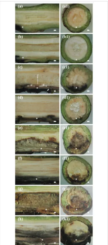
Small internal necrosis at the wound point without any progress in the subsequent evaluation times was observed in the stem tissue of non-inoculated plants from cultivars Palmer (Figure 1a, a1) and Ubá (Figure 1b, b1). The responses of plants from the Palmer and Ubá cultivars to C. fimbriata infection were classified as susceptible (Figure 1c, c1, e, e1, g, g1) or resistant (Figure 1d, d1, f, f1, h, h1) reactions, respectively.
Quantification of secondary metabolites
The factor cultivars, plant inoculation and evaluation times as well as their interactions were significant (p ≤ 0.05) for the concentrations of theobromine, 7-methylxanthine, caffeic acid, p-coumaric acid, gallic acid, protocatechuic acid, catechin and epicatechin (Figures 2, 3, 4, 5). Both non-inoculated and inoculated plants showed greater concentrations of p-coumaric acid in the stem tissue when compared with other secondary metabolites (Figures 2, 3, 4, 5).
Alkaloids
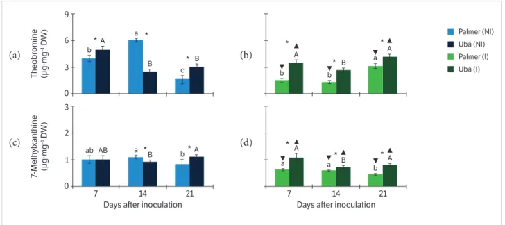
The concentration of theobromine in the stem tissue of non-inoculated plants from cv. Ubá showed a 0.25 – 0.96 fold increase in comparison with the non-inoculated plants from cv. Palmer at 7 and 21 dai (Figure 2a). In contrast, at 14 dai, the concentration of theobromine in the stem tissue of non-inoculated plants from cv. Palmer increased 1.48 fold in comparison with the non-inoculated plants from cv. Ubá (Figure 2a). The concentration of 7-methylxanthine in the stem tissue of non-inoculated plants from cv. Ubá was 0.35 higher than those in the non-inoculated plants from cv. Palmer at 21 dai (Figure 2c). In opposite, at 14 dai, stem tissue of non-inoculated plants from cv. Palmer contained0.18 more
Figure 1. Stem sections of non-inoculated plants of Palmer (a, a1) and Ubá (b, b1) cultivars. Internal necrotic tissue caused by Ceratocystis fimbriata infection in longitudinal (c, d, e, f, g, h) and transverse (c1, d1, e1, f1, g1, h1) stem sections of plants from the susceptible cultivar Palmer (a, a1, c, c1, e, e1, g, g1) and the resistant cultivar Ubá (b, b1, d, d1, f, f1, h, h1) at 7 (c, c1, d, d1), 14 (e, e1, f, f1) and 21 (g, g1, h, h1) days after inoculation.
7-methylxanthine in comparison with the non-inoculated plants from cv. Ubá (Figure 2c).
Stem tissue of inoculated plants from cv. Ubá contained 0.33 – 1.37 times more theobromine in comparison with the inoculated plants from cv. Palmer between 7 to 21 dai (Figure 2b). The concentration of 7-methylxanthine in the stem tissue of inoculated plants from cv. Ubá showed a 0.22 – 0.78 fold increase in comparison with the inoculated plants from cv. Palmer from 7 to 21 dai (Figure 2d).
The concentration of theobromine was higher in the stem tissue of non-inoculated plants from cv. Palmer than in the stem tissue of the inoculated ones (Figure 2a, b). In opposite, the concentration of theobrominewas higher in the stem tissue of inoculated plants from cv. Ubá than in their counterparts (Figure 2a, b). The concentration of 7-methylxanthine was higher in the stem tissue of non-inoculated plants from both cultivars than in the stem tissue of the inoculated ones (Figure 2c, d).
The concentrations of theobromine and 7-methylxanthine peaked at 14 dai in the stem tissue of non-inoculated plants from Palmer (Figure 2a, c). The concentrations of theobromine and 7-methylxanthine peaked at 7 and 21 dai, respectively, in the stem tissue of non-inoculated plants from cv. Ubá (Figure 2a, c). In the stem tissue of inoculated plants from both cultivars, peaks in the concentrations of
7-methylxanthine and theobromineoccurred at 7 and 21 dai, respectively (Figure 2b, d).
Hydroxycinnamic acids
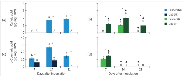
Caffeic acid was not detected in the stem tissue of non-inoculated plants from cv. Ubá, whereas in the tissue from cv. Palmer the compound was present at 14 and 21 dai (Figure 3a). The concentration of p-coumaric acid in the stem tissue of non-inoculated plants from cv. Palmer showed a 0.84 – 1.93 fold increase in comparison with the non-inoculated plants from cv. Ubá at 7 and 14 dai (Figure 3c). Stem tissue of inoculated plants from cv. Ubá contained 0.90 times more caffeic acid in comparison with the inoculated plants from cv. Palmer at 14 dai (Figure 3b). The concentration of p-coumaric acid in the stem tissue of inoculated plants from cv. Ubá showed a 0.40 fold increase in comparison with the inoculated plants from cv. Palmer at 7 dai (Figure 3d).
The concentrations of caffeic acid and p-coumaric acid were higher in the stem tissue of non-inoculated plants from cv. Palmer than in the stem tissue of the inoculated ones (Figure 3a, b, c, d). In contrast, the concentrations of caffeic acid and p-coumaric acid were higher in the stem tissue of inoculated plants from cv. Ubá than in their counterparts (Figure 3a, b, c, d).
Figure 2. Concentrations of the alkaloids — theobromine (a, b) and 7-methylxanthine (c, d) — in the stem tissue of mango plants from Palmer and Ubá cultivars that were not inoculated (NI; a, c) or inoculated (I; b, d) with Ceratocystis fimbriata at three different evaluation times.
9
6 A
b
a
B B
c
* *
*
3
0
Theobr
omine
(µg·mg
–1 D
W)
3
2
AB
ab a
B b*A
* 1
0
7 14 21
7-Meth
ylxanthine
(µg·mg
–1 D
W)
Palmer (NI) Ubá (NI) Palmer (I) Ubá (I) A
b b
B
A a
*
*
*
A
a a B b A
* *
*
7 14 21
Days aer inoculation Days aer inoculation
Means within a column followed by the same uppercase and lowercase letters are not significantly different (p ≤ 0.05) for Ubá NI or I and Palmer NI or I, respectively, as determined by Tukey’ test. *Significant difference between Palmer and Ubá NI or Palmer and Ubá I treatments for each evaluation time. ▼and ▲, when shown, indicate difference between Palmer (NI and I) and Ubá (NI and I), respectively,at each evaluation time. The error bars represent the standard error of the mean. n = 5; DW = Dry weight.
(a) (b)
In the stem tissue of non-inoculated plants from both cultivars, peaks in the concentrations of caffeic acid and p-coumaric acid occurred at 14 dai (Figure 3a, c). The concentrations of p-coumaric acid and caffeic acid peaked at 7 and 14 dai, respectively, in the stem tissue of inoculated plants from both cultivars (Figure 3b, d).
Hydroxybenzoic acids
The concentration of gallic acid in the stem tissue of non-inoculated plants from cv. Ubá showed a 0.67 – 0.70 fold increase in comparison with the non-inoculated plants from cv. Palmer at 7 and 21 dai (Figure 4a). In opposite, the concentration of protocatechuic acid in the stem tissue of non-inoculated plants from cv. Palmer showed a 0.49 – 0.88 fold increase in comparison with the non-inoculated plants from cv. Ubá at 7 and 14 dai (Figure 4c).
Stem tissue of inoculated plants from cv. Ubá contained 0.35 – 0.67 times more gallic acid in comparison with the inoculated plants from cv. Palmer at 7 and 14 dai (Figure 4b). The concentration of protocatechuic acid in the stem tissue of inoculated plants from cv. Ubá increased 0.20 – 0.60 fold in comparison with the inoculated plants from cv. Palmer from 7 to 21 dai (Figure 4d).
The concentration of gallic acid was higher in the stem tissue of non-inoculated plants from both cultivars than in
the stem tissue of inoculated plants (Figure 4a, b), whereas protocatechuic acid only showed greater accumulation in the stem tissue of non-inoculated plants from cv. Palmer (Figure 4c, d). The concentration of protocatechuic acid was higher in the stem tissue of inoculated plants from cv. Ubá than in their counterparts (Figure 4c, d).
In the stem tissue of non-inoculated and inoculated plants from both cultivars, peaks in the concentration of gallic acid occurred at 14 dai (Figure 4a, c). The concentration of protocatechuic acid peaked at 14 and 21 dai in the stem tissue of non-inoculated plants from cv. Palmer and Ubá, respectively (Figure 4c). In the stem tissue of inoculated plants from both cultivars, peaks in the concentration of protocatechuic acid occurred at 7 dai (Figure 4d).
Flavonoids
The concentration of catechin in the stem tissue of non-inoculated plants from cv. Ubá showed a 1.29 fold increase in comparison with the non-inoculated plants from cv. Palmer at 21 dai (Figure 5a). Epicatechin was not detected in the stem tissue of non-inoculated plants from cv. Ubá, whereas, in the tissue from cv. Palmer, the compound was present at all evaluation time (Figure 5c).
Stem tissue of inoculated plants from cv. Ubá contained 0.46 – 0.51 times more catechin in comparison with the Figure 3. Concentrations of the hydroxycinnamic acids — caffeic acid (a, b) and p-coumaric acid (c, d) — in the stem tissue of mango plants from Palmer and Ubá cultivars that were not inoculated (NI; a, c) or inoculated (I; b, d) with Ceratocystis fimbriata at three different evaluation times.
4
3
A A A
b
a * a *
2
1
0
C
aff
eic acid
(µg·mg
–1 D
W)
40
20 30
B b
a
A
C b *
*
*
10
0
7 14 21
p
-C
oumaric acid
(µg·mg
–1 D
W)
Palmer (NI) Ubá (NI) Palmer (I) Ubá (I) B
b b
a A
B
*
*
*
A a
b B b B
*
7 14 21
Days aer inoculation Days aer inoculation
Means within a column followed by the same uppercase and lowercase letters are not significantly different (p ≤ 0.05) for Ubá NI or I and Palmer NI or I, respectively, as determined by Tukey’ test. *Significant difference between the Palmer and Ubá NI or Palmer and Ubá I treatments for each evaluation time. ▼ and ▲, when shown, indicate difference between Palmer (NI and I) and Ubá (NI and I), respectively,at each evaluation time. The error bars represent the standard error of the mean. n = 5; DW = Dry weight.
(a) (b)
inoculated plants from cv. Palmer at 7 and 21 dai (Figure 5b). The concentration of epicatechin in the stem tissue of inoculated plants from cv. Ubá increased 0.27 fold in comparison with the inoculated plants from cv. Palmer at 14 dai (Figure 5d).
The concentration of catechin was higher in the stem tissue of inoculated plants from both cultivars than in the stem tissue of non-inoculated plants (Figure 5a, b). The concentrations of epicatechin were higher in the stem tissue of non-inoculated Figure 4. Concentrations of the hydroxybenzoic acids — gallic acid (a, b) and protocatechuic acid (c, d) — in the stem tissue of mango plants from Palmer and Ubá cultivars that were not inoculated (NI; a, c) or inoculated (I; b, d) with Ceratocystis fimbriata at three different evaluation times.
Figure 5. Concentrations of the flavonoids — catechin (a, b) and epicatechin (c, d) — in the stem tissue of mango plants from Palmer and Ubá cultivars that were not inoculated (NI; a, c) or inoculated (I; b, d) with Ceratocystis fimbriata at three different evaluation times.
4 3 B A A b a b * * 2 1 0 Gallic acid (µg·mg
–1 D
W) 4 2 3 B ab B a A b * * 1 0
7 14 21
P rot oc at echuic acid (µg·mg
–1 D
W) Palmer (NI) Ubá (NI) Palmer (I) Ubá (I) A b a a A A * * A
a a A B
b
*
* *
7 14 21
Days aer inoculation Days aer inoculation
9 6 C A B b b a * * 3 0 C at echin (µg·mg
–1 D
W) 6 2 4 A a A a A a * * * 0
7 14 21
Epic
at
echin
(µg·mg
–1 D
W) Palmer (NI) Ubá (NI) Palmer (I) Ubá (I) B b a a B A * * A a a A B b * *
7 14 21
Days aer inoculation Days aer inoculation
Means within a column followed by the same uppercase and lowercase letters are not significantly different (p ≤ 0.05) for Ubá NI or I and Palmer NI or I, respectively, as determined by Tukey’ test. *Significant difference between the Palmer and Ubá NI or Palmer and Ubá I treatments for each evaluation time.
▼ and ▲, when shown, indicate difference between Palmer (NI and I) and Ubá (NI and I), respectively,at each evaluation time. The error bars represent the standard error of the mean. n = 5; DW = Dry weight.
plants from cv. Palmer than in the stem tissue of the inoculated ones (Figure 5c, d). In opposite, the concentration of epicatechin was higher in the stem tissue of inoculated plants from cv. Ubá than in their counterparts (Figure 5c, d).
The concentration of catechin peaked at 21 dai in the stem tissue of non-inoculated plants from both cultivars (Figure 5a). Peaks in the concentration of epicatechin were not detected in the stem tissue of non-inoculated plants from both cultivars (Figure 5c). In the stem tissue of inoculated plants from both cultivars, peaks in the concentrations of epicatechin and catechin occurred at 7 and 21 dai, repectively (Figure 5b, d).
In vitro tests
The confluence of C. fimbriata colonies for the control treatment occurred at approximately ten days of incubation (Figure 6a,b,c). At the same evaluation time, C. fimbriata colonies confluence was not observed on the Petri dishes containing the different concentrations of two alkaloids (theobromine and 7-methylxanthine) (Figure 6a1, b1, c1, a2,b2,c2), two hydroxycinnamic acids (caffeic acid and p-coumaric acid) (Figure 6a3, b3, c3, a4, b4, c4), two hydroxybenzoic acids (gallic acid and protocatechuic acid) (Figure 6a5, b5, c5, a6, b6, c6) and two flavonoids (catechin and epicatechin) (Figure 6a7, b7, c7, a8, b8, c8). All secondary metabolites greatly affected fungal growth over time compared to the control treatment (Figure 6). The C. fimbriata growth was lower at the highest concentrations (10 to 30 mg∙mL−1) mainly for caffeic acid (Figure 6a3, b3, c3), p-coumaric acid (Figure 6a4, b4, c4), gallic acid (Figure 6a5, b5, c5)and protocatechuic acid (Figure 6a6, b6, c6) in comparison with the control treatment. The greatest zone of fungal growth inhibition and lowest C. fimbriata growth rate was observed on the Petri dishes containing caffeic acid (Figure 6a3, b3, c3) and p-coumaric acid (Figure 6a4, b4, c4) treatments.
DIscUssION
The results from the present study support previous findings that phenolic-like compounds and possibly other antimicrobial compounds belonging to other biochemical classes are involved in mango resistance against C. fimbriata infection (Araujo et al. 2014a,b; 2015). To the best of the author’s knowledge, however, the present study is the first to describe
Figure 6. In vitro tests with alkaloids and phenolic compounds against Ceratocystis fimbriata. Confluence of C. fimbriata colonies for the control treatment (PDA medium amended with methanol) at ten days of incubation (a, b, c).
Effect of the concentrations of 10 (a1 to a8), 20 (b1 to b8) and 30 (c1 to c8) mg∙mL−1 of alkaloids (a1, b1, c1 = theobromine; a2, b2, c2 = 7-methylxanthine), hydroxycinnamic acids (a3, b3, c3 = caffeic acid; a4, b4, c4 = p-coumaric acid), hydroxybenzoic acids (a5, b5, c5 = gallic acid; a6, b6, c6 = protocatechuic acid) and flavonoids (a7, b7, c7 = catechin; a8, b8, c8 = epicatechin) on C. fimbriata growth in Petri dishes.
The concentration of two alkaloids (theobromine and 7-methylxanthine) and six phenolic compounds (caffeic acid, p-coumaric acid, gallic acid, protocatechuic acid, catechin and epicatechin) in the stem tissue of inoculated plants from cv. Ubá was higher in comparison with inoculated plants from cv. Palmer. It has been proven that the concentration of secondary metabolites present in tissue infected by pathogens is cultivar-dependent (Hall et al. 2011; Nicholson and Hammerschmidt 1992; Petkovsek et al. 2009; Prusky and Keen 1993; Treutter 2005; Veberic et al. 2005; Williams et al. 2002). According to Petkovsek et al. (2009) and Veberic e t a l . ( 2 0 0 5 ) , t h e d i f fe re n c e b e t we e n re s i s t ant and susceptible apple cultivars to scab was due to higher concentrations of caffeic acid, catechin, epicatechin, p-coumaric acid and protocatechuic acid in the resistant cultivar. High concentrations of methylxanthines (caffeine) and theobromine in the tissue of tobacco and cocoa plants were linked to the induction of plant defense mechanisms against infection by Moniliophthora perniciosa, Pseudomonas syringae and Tobacco mosaic virus (Aneja and Gianfagna 2001; Kim and Sano 2008; Scarpari et al. 2005). It is known that the fungitoxic effect of most plant secondary metabolites on fungal cells is attributed to their interaction with lipids or an increase in phospholipid concentration and therefore membrane permeability, leakage of cellular contents and cytoplasm aggregation (Weete 1980). Araujo et al. (2014a,b) observed an abundance of dead C. fimbriata hyphae surrounded by phenolic-like compounds in the cells of stem tissue of plants from cultivars resistant to mango wilt. These findings agree with the data from the present study, which indicated that the concentrations of secondary metabolites were higher in the stem tissue of plants from the resistant cv. Ubá, therefore contributing to a reduction in the symptoms caused by C. fimbriata infection. In contrast, in the stem tissue of plants from the susceptible cv. Palmer, the concentration was not sufficient to counteract fungal colonization.
Interestingly, the concentration of most secondary metabolites was higher in the stem tissue of non-inoculated plants from cv. Palmer than in the stem tissue of inoculated ones, while the inverse was observed for plants of cv. Ubá. Plants respond to physical injury, pathogen infection or different types of abiotic stress by increasing the concentration of pre-existing alkaloids and
phenolics or by producing new ones through different metabolic pathways (Nicholson and Hammerschmidt 1992; Treutter 2005). Thus, high concentrations of secondary metabolites are often found in the tissue of plants infected by pathogens in comparison with non-infected plants (Aneja and Gianfagna 2001; Hukkanen et al. 2007; Petkovsek et al. 2009; Rodrigues et al. 2004; Scarpari et al. 2005). However, grapevine plants exhibiting symptoms of Pierce’s disease caused by Xylella fastidiosa (vascular pathogen) showed a significant reduction in phenolic concentrations in the vascular vessels in comparison with non-infected plants (Wallis and Chen 2012). Pierce’s disease causes grapevines to enter into a state of unreturned decline; therefore, the plants no longer have the resources to support the production of secondary metabolites with antimicrobial activity (Wallis and Chen 2012). It is known that, in a state of decline, the physiology of the diseased plants cannot be returned to adequate levels, therefore causing them to enter into a survival mode (Park et al. 2013; Wallis and Chen 2012). Thus, higher concentrations of phenolics could be found in tissues of non-infected plants than in the infected ones (Wallis and Chen 2012). In the present study, the intense internal necrotic tissue on the stem of plants from cv. Palmer (due to C. fimbriata infection) may have caused the plants to enter into a state of decline that negatively affected the production of secondary metabolites. Alternatively, plants from the resistant cv. Ubá that showed reduced symptoms of mango wilt most likely maintained active defense responses during infection by C. fimbriata. Araujo et al. (2015) also reported that plants with induced resistance to mango wilt showed higher concentrations of alkaloids and phenolics during the infection process of C. fimbrita in the stem tissue.
2011; Scarpari et al. 2005; Wallis and Chen 2012; Araujo et al. 2015). As a rule, the most resistant plants from a certain cultivar rapidly initiate the production of secondary metabolites in response to pathogens infection (Nicholson and Hammerschmidt 1992; Treutter 2005), explaining, at least in part, the final amount of disease (Wallis and Chen 2012). Araujo et al. (2014a, b) observed that the variation in the susceptibility of mango cultivars to C. fimbriata infection was attributed to differences in their ability to reduce fungal spread in the stem tissue through rapid deposition of phenolic-like compounds. In the present study, the concentrations of secondary metabolites in the stem tissue of mango plants were high in the early stages of fungal infection; however, the concentrations in the stem tissue were cultivar-dependent. This could explain why plants from the Palmer cultivar exhibited more rapid and greater disease symptoms than plants from cv. Ubá. The different concentrations of secondary metabolites added to the Petri dishes inhibited C. fimbriata growth over time. According to Funnell-Harris et al. (2014), one possible effect of changes in phenolics composition in the plant is due to their direct effect on inhibiting pathogens growth. The authors provided in vitro evidence that ferulic acid inhibited several fungi, which could explain the increase of sorghum lines resistance to some diseases (Funnell-Harris et al. 2014). Blodgett and Stanosz (1997) observed that
different concentrations of phenolic compounds and monoterpenes of red pine inhibited in vitro the spore germination and mycelial growth of Sphaeropsis sapinea. Blodgett and Stanosz (1997) reported that the changes in the concentration of secondary metabolites may prevent colonization of S. sapinea on red pine, explaining, in part, the differential responses of resistant plants to isolates of the fungus. In the present study, it was observed reduced C. fimbriata growth at higher concentrations of secondary metabolites during the in vitro tests, especially using the caffeic acid and p-coumaric acid (hydroxycinnamic acids). Inoculated plants from Ubá cultivar showed the highest concentration of hydroxycinnamic acids in stem tissue, explaining, therefore, why these plants were more resistant to C. fimbriata infection than plants from Palmer cultivar.
cONcLUsION
The data from the present study seem to support the concept that secondary metabolites, such as theobromine, 7-methylxanthine, caffeic acid, p-coumaric acid, gallic acid, protocatechuic acid, catechin and epicatechin, played an important role in the resistance of mango plants against C. f imbr iata infec tion, esp ecially in plants from cv. Ubá.
rEFErENcEs
Adrian, M., Jeandet, P., Veneau, J., Weston, L. A. and Bessis, R.
(1997). Biological activity of resveratrol, a stilbenic compound from
grapevines, against Botrytis cinerea, the causal agent for gray mold.
Journal of Chemical Ecology, 23, 1689-1702. http://dx.doi.org/
10.1023/B:JOEC.0000006444.79951.75.
Ajila, C. M., Rao, L. J. and Rao, U. J. S. P. (2010). Characterization
of bioactive compounds from raw and ripe Mangifera indica L.
peel extracts. Food and Chemical Toxicology, 48, 3406-3411.
http://dx.doi.org/10.1016/j.fct.2010.09.012.
Aneja, M. and Gianfagna, T. (2001). Induction and accumulation
of caffeine in young, actively growing leaves of cocoa
(Theobromacacao L.) by wounding or infection with Crinipellis
perniciosa. Physiological and Molecular Plant Pathology, 59,
13-16. http://dx.doi.org/10.1006/pmpp.2001.0337.
Araujo, L., Bispo, W. M. S., Cacique, I. S., Cruz, M. F. A. and
Rodrigues, F. A. (2014a). Histopathological aspects of mango
resistance to the infection process of Ceratocystis fimbriata. Plant
Pathology, 63, 1282-1295. http://dx.doi.org/10.1111/ppa.12208.
Araujo, L., Bispo, W. M. S., Cacique, I. S., Moreira, W. R. and
Rodrigues, F. A. (2014b). Resistance in mango against infection
by Ceratocystis fimbriata. Phytopathology, 104, 820-833. http://dx.doi.org/10.1094/PHYTO-11-13-0316-R.
Araujo, L., Bispo, W. M. S., Rios, V. S., Fernandes, S. A. and
Rodrigues, F. A. (2015). Induction of the phenylpropanoid
pathway by acibenzolar-S-methyl and potassium phosphite
increases mango resistance to Ceratocystis fimbriata infection.
Plant Disease, 99, 447-459. http://dx.doi.org/10.1094/
Benhamou, N. and Bélanger, R. R. (1998).
Benzothiadiazole-mediated induced resistance to Fusarium oxysporum f.sp.
radicis-lycopersici in tomato. Plant Physiology, 118, 1203-1212.
http://dx.doi.org/10.1104/pp.118. 4.1203.
Blodgett, J. T. and Stanosz, G. R. (1997). Differential inhibition
of Sphaeropsis sapinea morphotypes by a phenolic compound
and several monoterpenes of red pine. Phytopathology, 87,
606-609. http://dx.doi.org/10.1094/PHYTO.1997.87.6.606.
Carvalho, C. R. L., Rossetto, C. J., Mantovani, D. M. B., Morgano,
M. A., Castro, J. V. and Bortoletto, N. (2004). Avaliação de
cultivares de mangueira selecionadas pelo Instituto Agronômico
de Campinas comparadas a outras de importância comercial.
Revista Brasileira de Fruticultura, 26, 264-271. http://dx.doi.
org/10.1590/S0100-29452004000200021.
Cushnie, T. P. T. and Lamb, A. J. (2005). Antimicrobial activity
of flavonoids. International Journal of Antimicrobial Agents, 26,
343-356. http://dx.doi.org/10.1016/j.ijantimicag.2005.09.002.
Dhingra, O. D. and Sinclair, J. B. (1995). Basic plant pathology
methods. Boca Raton: Lewis Publisher.
D i xo n , R . A . a n d Pa i va , N . L . (19 9 5 ) . St re ss i n d u ce d
phenylpropanoid metabolism. Plant Cell, 7, 1085-1097.
http://dx.doi.org/10.1105/tpc.7.7.1085.
Ferreira, E. M., Harrington, T. C., Thorpe, D. J. and Alfenas,
A. C. (2010). Genetic diversity and interfertility among
highly differentiated populations of Ceratocystisfimbriata
in Brazil. Plant Pathology, 59, 721-735. http://dx.doi.org/
10.1111/j.1365-3059.2010.02275.x.
Food and Agriculture Organization of the United Nations (2014).
Medium-term prospects for agricultural commodities; [accessed
2014 Nov 1]. www.fao.org/docrep/006/y5143e/y5143e1a.htm.
Freeman, B. C. and Beattie, G. A. (2008). An overview of plant
defenses against pathogens and herbivores. The Plant Health
Instructor. http://dx.doi.org/10.1094/PHI-I-2008-0226-01.
Funnell-Harris, D. L., Sattler, S. E. and Pedersen, J. F. (2014).
Response of Fusarium thapsinum to sorghum brown midrib
lines and to phenolic metabolites. Plant Disease, 98,
1300-1308. http://dx.doi.org/10.1094/PDIS-09-13-0980-RE.
Garba, S. and Okeniyi, S. O. (2012). Antimicrobial activities
of total alkaloids extracted from some Nigerian medicinal
plants. Journal of Microbiology and Antimicrobials, 4, 60-63.
http://dx.doi.org/10.5897/JMA11.081.
Hall, C., Heath, R. and Guest, D. I. (2011). Rapid and intense
accumulation of terpenoid phytoalexins in infected xylem tissues
of cotton (Gossypium hirsutum) resistant to Fusarium oxysporum
f.sp. vasinfectum. Physiological and Molecular Plant Pathology,
76, 182-188. http://dx.doi.org/10.1016/j.pmpp.2011.09.002.
Hukkanen, A. T., Kokko, H. I., Buchala, A. J., Mcdougall, G. J., Stewart,
D., Karenlampi, S. O. and Karjalainen, R. O. (2007). Benzothiadiazole
induces the accumulation of phenolics and improves resistance to
powdery mildew in strawberries. Journal of Agricultural and Food
Chemistry, 55, 1862-1870. http://dx.doi.org/ 10.1021/jf063452p.
Kaur, J., Rathinam, X., Kasi, M., Leng, L. M., Ayyalu, R., Kathiresan,
S. and Subramaniam, S. (2010). Preliminary investigation on the
antibacterial activity of mango (Mangifera indica L: Anacardiaceae)
seed kernel. Asian Pacific Journal of Tropical Medicine, 3,
707-710. http://dx.doi.org/10.1016/S1995-7645(10)60170-8.
Kim, Y. S. and Sano, H. (2008). Pathogen resistance of transgenic
tobacco plants producing caffeine. Phytochemistry, 69,
882-888. http://dx.doi.org/10.1016/j.phytochem.2007.10.021.
Lisec, J., Schauer, N., Kopka, J., Willmitzer, L. and Fernie, A.
R. (2006). Gas chromatography mass spectrometry-based
metabolite profiling in plants. Nature Protocols, 1, 387-396.
http://dx.doi.org/10.1038/nprot.2006.59.
Mace, M. E., Stipanovic, R. D. and Bell, A. A. (1985). Toxicity
and role of terpenoid phytoalexins in verticillium wilt resistance
in cotton. Physiological Plant Pathology, 26, 209-218. http://
dx.doi.org/10.1016/0048-4059(85)90021-9.
Mazid, M., Khan, T. A. and Mohammad, F. (2011). Role of
secondary metabolites in defense mechanisms of plants.
Biology and Medicine, 3, 232-249.
Nicholson, R. L. and Hammerschmidt, R. (1992). Phenolic
compounds and their role in disease resistance. Annual Review
of Phytopathology, 30, 369-389. http://dx.doi.org/ 10.1146/
annurev.py.30.090192.002101.
Park, J. H., Juzwik, J. and Cavender-Bares, J. (2013). Multiple
Ceratocystis smalleyi infections associated with reduced stem
water transport in bitternut hickory. Phytopathology, 103,
565-574. http://dx.doi.org/ 10.1094/PHYTO-10-12-0272-R.
Petkovsek, M. M., Stampar, F. and Veberic, R. (2009).
Accumulation of phenolic compounds in apple in response
to infection by the scab pathogen, Venturia inaequalis.
Physiological and Molecular Plant Pathology, 74, 60-67.
Prusky, D. and Keen, N. T. (1993). Involvement of preformed antifungal
compounds in the resistance of subtropical fruits to fungal decay.
Plant Disease, 77, 114-119. http://dx.doi.org/10.1094/PD-77-0114.
Ribeiro, I. J. A. (2005). Doenças da mangueira (Mangifera indica L.).
In H. Kimati, L. Amorim, A. Bergamin Filho, L. E. A. Camargo and J. A.
M. Rezende (Eds.), Manual de Fitopatologia: doenças das plantas
cultivadas (p. 457-465). São Paulo: Editora Agronômica Ceres.
Ribeiro, S. M. R., Barbosa, L. C. A., Queiroz, J. H., Knodler, M. and
Schieber, A. (2008). Phenolic compounds and antioxidant capacity
of Brazilian mango (Mangifera indica L.) varieties. Food Chemistry,
110, 620-626. http://dx.doi.org/10.1016/j.foodchem.2008.02.067.
Rodrigues, F. A., Benhamou, N., Datnoff, L. E., Jones, J. B. and
Bélanger, R. R. (2003). Ultrastructural and cytochemical aspects
of silicon-mediated rice blast resistance. Phytopathology, 93,
535-546. http://dx.doi.org/10.1094/PHYTO.2003.93.5.535.
Rodrigues, F. A., McNally, D. J., Datnoff, L. E., Jones, J. B., Labbé,
C., Benhamou, N., Menzies, J. G. and Bélanger, R. R. (2004).
Silicon enhances the accumulation of diterpenoid phytoalexins in
rice: a potential mechanism for blast resistance. Phytopathology,
94, 177-183. http://dx.doi.org/10.1094/PHYTO.2004.94.2.177.
Rossetto, C. J., Ribeiro, I. J. A., Igue, T. and Gallo, P. B. (1996).
Seca-da-mangueira: XV. Resistência varietal a dois isolados
de Ceratocystis fimbriata. Bragantia, 55, 117-121. http://dx.doi.
org/10.1590/S0006-87051996000100013.
Scarpari, L. M., Meinhardt, L. W., Mazzafera, P., Pomella, A. W.
V., Schiavinato, M. A., Cascardo, J. C. M. and Pereira, G. A. G.
(2005). Biochemical changes during the development of witches’
broom: the most important disease of cocoa in Brazil caused by
Crinipellisperniciosa. Journal of Experimental Botany, 56, 865-877.
http://dx.doi.org/10.1093/jxb/eri079.
Tohge, T. and Fernie, A. R. (2010). Combining genetic diversity,
informatics and metabolomics to facilitate annotation of plant
gene function. Nature Protocols, 5, 1210-1227. http://dx.doi.
org/10.1038/nprot.2010.82.
Treutter, D. (2005). Significance of flavonoids in plant resistance
and enhancement of their biosynthesis. Plant Biology, 7,
581-591. http://dx.doi.org/10.1055/s-2005-873009.
Van Wyk, M., Al Adawi, A. O., Khan, I. A., Deadman, M. L., Al
Jahwari, A. A., Wingfield, B. D., Ploetz, R. and Wingfield, M. J.
(2007). Ceratocystis manginecans sp. nov., causal agent of a
destructive mango wilt disease in Oman and Pakistan. Fungal
Diversity, 27, 213-230.
Veberic, R., Trobec, M., Herbinger, K., Hofer, M., Grill, D. and
Stampar, F. (2005). Phenolic compounds in some apple
(Malus domestica Borkh) cultivars of organic and integrated
production. Journal of the Science of Food and Agriculture,
85, 1687-1694. http://dx.doi.org/10.1002/jsfa.2113.
Viégas, A. P. (1960). Seca da mangueira. Bragantia, 19, 163-182.
Wallis, C. M. and Chen, J. (2012). Grapevine phenolic compounds
in xylem sap and tissues are significantly altered during
infection by Xylella fastidiosa. Phytopathology, 102, 816-826.
http://dx.doi.org/10.1094/PHYTO-04-12-0074-R.
Weete, E. R. (1980). Lipid biochemistry of fungi and other
organisms. New York: Plenum Press.
Williams, J. S., Hall, S. A., Hawkesford, M. J., Beale, M. H. and
Cooper, R. M. (2002). Elemental sulfur and thiol accumulation in
tomato and defense against a fungal vascular pathogen. Plant