Journal of the Renin- Angiotensin-Aldosterone System
(Including other peptidergic systems)
March 2001 Volume 2 Supplement 1
S77
Prevention of atherosclerosis by specific AT1-receptor
blockade with candesartan cilexetil
Vasilios Papademetriou, Philippe Mammillot, Robert Redman, Aldo Notargiacomo, Puneet Narayan, Raj Lakshman
Keywords: telmisartan, lisinopril, antihypertensive, hypertension, diabetes, renoprotection, angiotensin II receptor antagonist The Va Medical Center and Georgetown University and the George Washington University Washington D.C. USA
Correspondence to: Dr Vasilios Papademetriou VA Medical Center, 151E,
50 Irving St., N.W. Washington, D.C. 20422, USA
Tel: +1 202 745 8333 Fax: +1 202 745 8636 E-mail: papavip@ aol.com
JRAAS2001;2 (suppl 1):S77-S80
Abstract
Several studies indicate that blockade of the renin-angiotensin-aldosterone system (RAAS) can prevent atherosclerosis and vascular events, but the precise mechanisms involved are still unclear. In this study, we investigated the effect of the AT1-receptor blocker,
candesartan, in the prevention of atherosclerosis in Watanabe heritable hyperlipidaemic (WHHL) rabbits and also the effect of AT1-receptor blockade in the
uptake of oxidised LDL by macrophage cell cultures. In the first set of experiments, 12 WHHL rabbits were randomly assigned to three groups: placebo, atenolol 5 mg/kg daily or candesartan 2 mg/kg daily for six months. Compared with controls and atenolol-treated rabbits, candesartan treatment resulted in a significant 50–60% reduction of atherosclerotic plaque formation and a 66% reduction in cholesterol accumulation in the thoracic aorta.
Studies in macrophage cultures indicated that candesartan prevented uptake of oxidised LDL-(oxLDL)-cholesterol by cultured macrophages. Candesartan inhibited the uptake of oxLDL in a dose-dependent manner, reaching a maximum inhibition of 70% at concentrations of 5.6 µg/ml. Further studies in other animal models and well-designed trials in humans are warranted to further explore the role of AT1-receptor
blockade in the prevention of atherosclerosis.
Introduction
Activation of the renin-angiotensin-aldosterone system (RAAS) is a major contributor to the devel-opment and progression of atherosclerosis. Observational data in humans indicate that plasma renin activity can predict the risk of myocardial infarction in patients with hypertension, irrespective of cholesterol levels.1 Furthermore, a deletion
poly-morphism in the gene for ACE has been shown to be a potent predictor for myocardial infarction (MI).2
Further support for the role of the RAAS as a pro-motor of atherosclerotic vascular events is provided from large interventional trials.3-5At molecular and
cellular levels, angiotensin II (Ang II) has been shown to exert a number of complex interactions that may contribute to the development and pro-gression of atherosclerosis. Ang II has been shown to be a promotor of endothelial dysfunction,6,7
smooth muscle cell proliferation and migration8and
a facilitator of LDL oxidation.8 Ang II has also been
shown to play a role in the recruitment of mono-cytes/macrophages in the arterial wall9,10 and to
promote the uptake of oxidised LDL.11 The role of
AT1-receptor activation or blockade in the uptake
of oxidised LDL by macrophages has not been clarified.
Studies that investigated the effect of RAAS blockade on atherosclerosis in animal models, pro-vided conflicting results. Over a decade ago, Chobanian and co-workers demonstrated that early treatment of Watanabe heritable hyperlipi-daemic (WHHL) rabbits with angiotensin-convert-ing enzyme inhibitors (ACE-I)12,13can diminish the
development of atherosclerosis. Antiatherogenic effects of ACE-I have been reported in several other animal models,14-16but the role of blood
pres-sure-lowering of these agents has not been clari-fied.17 The use of AT
1-receptor blockers (ARB)
demonstrated prevention of atherosclerosis in some studies,18 but not in others.14 In a recent
report, Hope et al.19indicated that low-dose
irbe-sartan, that did not lower blood pressure (BP), had no effect on atherosclerosis, whereas a higher dose, that also caused reduction in BP, significant-ly reduced aortic atherosclerosis. Because ARBs differ in the way they bind to the AT1-receptor, in
the present study we investigated the effect of candesartan, an ARB with insurmountable binding properties, on the prevention of atherosclerosis in WHHL rabbits. In an attempt to further clarify the mechanism of prevention of atherosclerosis by ARBs, we also investigated the role of AT1-receptor
blockade with candesartan on the uptake of oxi-dised LDL in a macrophage cell-culture model.
Materials and methods
Animal experiments
Twelve WHHL rabbits were acquired from Covance Research Products, Inc. at the age of four months. All rabbits were acclimatised for two weeks in our facil-ity prior to experiments. Animals were randomly-assigned to three groups: control, candesartan 2 mg/kg or atenolol 10 mg/kg. The dose of can-desartan was selected based on prior experiments (personal communication, Peter Morsing, AstraZeneca). The dose of atenolol was selected based on prior experiments in our laboratory. Medications were added to the Agwa Prolab high-fibre rabbit chow (Agway, Syracuse, NY), which also contained 4% Karo syrup to improve palatability and adherence of the drug to the food, as described by Chobanian et al.17
Journal of the Renin- Angiotensin-Aldosterone System
(Including other peptidergic systems)
March 2001 Volume 2 Supplement 1
sure Monitor (A.J. Buck & Sons). Each animal was handled by the same researcher throughout the study. The BP was measured in a quiet room. The animal was allowed to settle for 5 minutes and lie flat on a padded table. The femoral artery was pal-pated and a #3 pediatric cuff placed around the same hind leg. BP was measured in triplicate and averaged for the session. All animals were sacri-ficed using pentobarbital (100 mg/kg) at the end of the six month treatment period. The tissues were fixed by whole body perfusion with 10% acetate-buffered formalin.12 Segments of rabbit
aorta were removed for histological analysis from the arch, thorax and abdomen. A portion of each segment was cut open longitudinally and immersed in 4.0% formaldehyde, prepared fresh from paraformaldehyde, and 2.5% dimethylsulfox-ide (DMSO) in 0.1 M NaPO4 buffer for 4–8 hours,
washed in the buffer, and cut into 3–4 pieces. All pieces of aorta oriented with the plaque and aortal wall on opposite sides were analysed. Areas occupied by plaque alone and plaque-plus-aortic wall were measured and tallied using Jandel’s soft-ware Sigmanscan, by tracing the outline of the camera lucida images on a digitising pad. The per-centage plaque for each specimen was computed as the sum of the areas of plaque divided by sum of the areas of plaque plus aorta x 100.
Cholesterol content of the thoracic and abdominal aortic regions was assayed using previ-ously-described methods20 and expressed as
mg/gram tissue.
Oxidised-LDL uptake experiments
We used a mouse macrophage cell line (J774A) routinely used in our laboratory. Development and maintenance of this cell line has been previ-ously described.21
Macrophage cultures at a density of 1 x 10⁶
cells per well were plated for 24–48 hours prior to actual uptake experiments. Cells were cultured in 4 ml DMEM medium, supplemented with 10% heat inactivated foetal bovine serum. Cells were incubated at 37°C in a humidified incubator, in an atmosphere of 5% CO2. Uptake assay was started when cells were about 90% confluent, by
remov-ing the spent medium and addremov-ing fresh medium, without FBS, containing indicated amounts of labelled oxidised LDL plus or minus candesartan. Incubations were continued for up to 24 hours. At the end of the incubation period, medium was col-lected and kept for further analysis. Cells were washed three times with 1 ml FBS, then lysed with 2 ml of 0.1 N NaOH, followed by two washes with FBS (H 7.4). Lysate and final washes were combined and analysed for radioactivity and protein content. For radioactivity counting, an equal volume of glacial acetic acid was added to the cell lysate in order to prevent any quenching due to NaOH.
LDL oxidation and lipoprotein isolation were prepared using previously-described methods.22
Oxidised LDL was labelled by incubating HNE-LDL, in a mixture of H3-cholesteryl-oleate in DMSO and tris-HCL for two hours at 37°C, followed by overnight incubation at 4°C. After this procedure, the sample was dialysed extensively against the same buffer without EDTA and stored on ice until use.
Initially, experiments were performed to iden-tify optimal concentrations of oxidised LDL needed for optimal uptake by macrophages. Subsequent experiments determined the optimal concentration of candesartan required to prevent uptake of oxidised LDL. Lastly, experiments were performed with and without induction of uptake by Ang II, with and without AT1-receptor blockade.
Results
Animal experiments
Candesartan and atenolol had no effect on systolic and diastolic BP, compared with control (Figure 1). Body weight increased with time, but in a similar way in all three groups.
S78
PAPER
Figure 1 Shows blood pressure values during the six month treatment period.There were no statistically significant differences between groups.
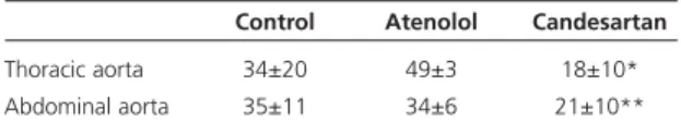
Table 1 Average plaque formation in rabbit aortas (% surface covered).
Control Atenolol Candesartan
Thoracic aorta 34±20 49±3 18±10*
Abdominal aorta 35±11 34±6 21±10**
Mean±SD; *p<.05 for both atenol and control; **p=.07 for both
Figure 2 Representative aortic specimens from the three treatment groups. Note that the candesartan-treated rabbit had very little atherosclerosis compared to the other two groups.
100
Weight (kg)
DBP SBP Control
Candesartan Atenolol
80
60
0 2 4 6
40
20
Blood pr
essur
e (mmHg)
Journal of the Renin- Angiotensin-Aldosterone System
(Including other peptidergic systems)
March 2001 Volume 2 Supplement 1
S79 Aortic atherosclerosis
Atherosclerotic involvement was similar between control and atenolol-treated animals. Candesartan treatment, however, significantly reduced athero-sclerotic plaque formation in the thoracic aorta and marginally in the abdominal aorta (Table 1). Figure 2 shows a sample of thoracic aorta of an animal in each group. The candesartan-treated rabbit showed almost total elimination of the ath-erosclerotic plaque.
Cholesterol content was significantly decreased by 66% (p<0.02) in the thoracic aortae of candesar-tan-treated animals, compared with controls or atenolol-treated rabbits (Table 2, Figure 3). There were no significant differences in abdominal aortic cholesterol content between the three groups.
Macrophage cultures: oxidised LDL uptake From prior experiments in our lab, it was found that oxidised LDL, at a concentration of 10–100 µ/ml, would yield adequate uptake by macrophage cul-tures within 3–24 hours of incubation. For this reason, concentrations of 10, 20 and 50 µg/ml were used in these experiments. As shown in Figure 4, there was an incremental uptake of oxi-dised LDL in the three concentrations used (control panel Figure 4).When Ang II was added to cell cultures, uptake of oxidised LDL was signifi-cantly enhanced, particularly in the cells containing 50 µg/ml oxidised LDL (mid panel).This effect was prevented when candesartan 0.56 µg/ml was added with Ang II. In a second series of experi-ments, oxidised LDL, 50 µg/ml, was added to macrophage cultures, either alone or in combina-tion with log concentracombina-tions of candesartan (from 0.0056–5.6 µg/ml). A dose-response curve of
pre-vention of uptake of oxidised LDL cholesterol by macrophages was noted (Figure 5). Concentrations of candesartan higher than 0.56 µg/ml prevented uptake of oxidised LDL by 70% or more. Viability studies did not show any cellular toxicity, even with the highest concentrations of candesartan.
Discussion
Results from this study indicate that treatment of WHHL rabbits with candesartan, 2 mg/kg daily, can substantially decrease the development of athero-sclerotic plaques. This effect was independent of BP changes, since systolic and diastolic pressure remained unchanged for the duration of the study and were similar to control and atenolol-treated animals. This protective effect of candesartan seems to be specific for AT1-receptor blockers, since it was not noted with atenolol. In previous studies, other antihypertensive agents such as nifedipine,23verapamil24and propranolol25failed to
prevent atherosclerosis in the WHHL rabbit model. The protective effect of candesartan noted in the present study differs from previous obser-vations, in which only hypotensive doses of irbe-sartan were able to inhibit atherosclerosis.19 This
PAPER
Figure 4 Demonstrates an uptake of oxidised-LDL - cholesterol (6 hours) by macrophage cell cultures. Left panel (control) shows an incremental uptake of oxidised LDL from 10–50 µg/ml.The mid-panel (AII) demonstrates an enhanced uptake of the cell cultures containing 50 µg/ml oxidised LDL, when Angiotensin II was added to the cultures. Right panel (AII+CAN) demonstrates reversal of the angiotensin effect when candesartan 50 ng/ml was added.
Figure 3 Demonstrates the cholesterol content of thoracic aortas, in mg/gram tissue, in the three treatment groups. There was significantly less cholesterol in candesartan-treated animals.
Table 2 Average cholesterol content in rabbit aortas (mg/gram tissue).
Control Atenolol Candesartan
Thoracic Aorta 15.2±8 13.2±6 5.1±3*
Abdominal Aorta 13.8±12 8.9±7 10.3±5
Mean±SD; *p<.02 compared to control and p<.01 compared to atenolol
Figure 5 Demonstrates a dose-related reduction of uptake of oxidised LDL cholesterol with log-concentrations of candesartan. Concentrations of candesartan more than 0.56 mg/ml prevented uptake of oxidised LDL by more than 70% *p<.05.
16
14
12
10
8
6
4
2
0
Control
8000
7000
6000
5000
4000
3000
2000
1000
Control Ang II
10 µg OXLDL
20 µg OXLDL 50 µg OXLDL
Ang II + CAN
Candesartan Atenolol
0
Candesartan µg/ml
Uptake (nmol CE/mg cell pr
otein) 50 µg OXLDL
0 4%
Placebo 0.0056 0.056 0.56 5.6 8%
Journal of the Renin- Angiotensin-Aldosterone System
(Including other peptidergic systems)
March 2001 Volume 2 Supplement 1
S80
difference is potentially attributable to the differ-ent receptor binding properties of the two ARBs. In contrast, these data are in agreement with the recently-published findings of Strawn et al.26
These investigators demonstrated that losartan inhibited the development of fatty streaks, and reduced the media area of coronary arteries, in non-human primates by a mechanism indepen-dent of BP and plasma lipids.The reduction of fatty streak formation was accompanied by a decrease in carotid artery cholesterol content, a finding which is also consistent with our own results.
The second important finding of our study is the observation that candesartan inhibited the uptake of oxidised LDL-cholesterol by murine macrophages in a dose-dependent manner. It is well known that in the early stages of atherosclerosis, fatty streaks are defined by the presence of large numbers of macrophages in the vascular intima and by the for-mation of foam cells.27 Ang II has been shown
to facilitate the recruitment of monocytes/ macrophages by stimulating the production of monocyte chemoattractant protein9and vascular cell
adhesion molecule-1.10Furthermore, Ang II may
con-tribute to enhanced oxidation of LDL and degrada-tion of nitric oxide.11 Recent evidence suggests that
Ang II may regulate the newly-characterised oxi-dised LDL (oxLDL) receptor, which is biochemically distinct from the scavenger receptor.28 Our data
indi-cate that blockade of AT1-receptors with candesartan
can prevent the uptake of oxidised LDL.
Although the precise mechanism by which this is achieved is not known, an interaction or cross-talk between the AT1-receptor and the scavenger
and/or the oxLDL receptor is probable.
In summary, data from this study indicate that treatment of WHHL rabbits with the specific AT1 -receptor blocker candesartan can retard athero-sclerosis independently of BP reduction. Data from macrophage cultures indicate that AT1
-recep-tor blockade can prevent uptake of oxidised LDL in a dose-dependent manner.
Acknowledgements
We acknowledge the technical support of Bijal Desaikalaiki and the superb secretarial support of Mrs Patricia Mohan.
References
1. Aldeman MH, Madhaven S, Ooi WL, Cohen H, Sealy JE, Laragh JH. Association of the renin-sodium profile with the risk ofmyocardial infarction in patients with hypertension. N Engl J Med 1991;324:1098-104.
2. Gombien F, Poirier O, Lecerf L et al. Deletion polymor-phism in the gene for angiotensin converting enzyme is a potent risk factor for myocardial infarction. Nature
1992;359:641-44.
3. Rutherford JD, Pfeffer MA, Moye LA et al. Effects of capto-pril on ischemic events after myocardial infarction: results of the Survival and Ventricular Enlargement Trial: SAVE Investigators,Circulation1994;90:1731-8.
4. Yusuf S, Pepine CJ, Garces C et al. Effect of enalapril on myocardial infarction and unstable angina in patients with low ejection fractions.Lancet1992;340:1173-8.
5. The Heart Outcomes Prevention Evaluation Study (HOPE) Investigators. Effects of an angiotensin-converting-enzyme inhibitor, ramipril, on cardiovascular events in high-risk patients.N Engl J Med2000;342:145-3.
6. Ferrario CM, Deitch JS, Dean RH, Strawn WB. Hypertension and atherosclerosis: a mechanistic understanding of disease progression.Cardiovasc Risk Factors1996;6:299-310. 7. Clozel M, Kuhn H, Hefti F, Baumgartner HR. Endothelial dysfunction and subendothelial monocyte macrophages in hypertension.Hypertension 1991;18:132-41.
8. Yanagitani Y, Rakugi A, Okamura A et al. Angiotensin II type 1 receptor-mediated peroxide production in human macrophages.Hypertension1999;33(pt II):II-335-9.
9. Chen XL, Tummala PE, Olbrych MT, Alexander RW, Medford RM. Angiotensin II induces monocyte chemoattrac-tant protein-1 gene expresson in rat vascular smooth muscle cells.Circ Res1998;83:952-9.
10. Tummala PE, Chen XL, Sundell CL et al. Ang II induces vas-cular cell adhesion molecule-1 expression in rat vasculature: a potential link between the renin-angiotensin system and ather-osclerosis.Circulation1999;100:1223-9.
11. Rajagopalan S, Kurz S, Munzel T et al. Ang II-mediated hyper-tension in the rat increases vascular superoxide production via membrane NADH/NADPH oxidase activation: contribution to alterations of vasomotor tone.J Clin Invest1996;97:1916-23. 12. Chobanian AV, Haudenschild CC, Nickerson C, Drago R. Anti-atherogenic effect of captopril in the Watanabe heritable hyperlipidemic rabbit.Hypertension1990;1:327-31.
13. Chobanian AV, Haudenschild CC, Nickerson C, Hope S. Trandolapril inhibits atherosclerosis in the Watanabe Heritable Hyperlipidemic Rabbit.Hypertension1992;20:473-7. 14. Schuh JR, Blehmn DJ, Friedrich GE et al. Differential effects of renin-angiotensin blockade on atherogenesis in cho-lesterol-fed rabbits.J Clin Invest1993;91:1453-8.
15. Charpiot P, Rolland Ph, Friggi A et al.ACE-I with perindopril and atherogenesis-induced structural and functional changes in minipig arteries.Arterioscler Thromb1993;13:1125-38. 16. Kowala MC, Grove RI, Aberg G. Inhibitors of angiotensin-converting enzyme decrease early atherosclerosis in hyperlipi-demic hamsters. Fosinopril reduces plasma cholesterol and captopril inhibits macrophase-foam cell accumulation inde-pendently of BP and plasma lipids. Atherosclerosis
1994;108:61-72.
17. Chobanian AV, Hope S, Brecher P. Dissociation between antiatherosclerotic effect of trandolapril and suppression of serum and aortic angiotensin-converting enzyme activity in the Watanabe Heritable Hyperlipidemic rabbit. Hypertension
1995;25:1306-10.
18. Sugano M, Makino N, Yanaga T. The effects of renin-angiotensin system inhibition on aortic cholesterol content in cholesterol-fed rabbits.Atherosclerosis1996;127:123-9. 19. Hope S, Brecher P, Chobanian AV. Comparison of the effects of AT1receptor blockade and angiotensin converting enzyme inhibi-tion on atherosclerosis.Am J Hypertens1999;12:28-34. 20. Allain CA, Poon LS, Chan CSG, Richmond W, Fu PC. Enzymatic determination of total serum cholesterol.Clin Chem
1974;20:470-3.
21. Basu SK, Brown MS, Ho YK, Havel RJ, Goldstein JL. Mouse macrophages synthesize and secrete a protein resembling apolipoprotein E.Proc Nat Acad Sci1981;78:7545-9. 22. Esterbauer H, Striegl G, Puhl H, Rothhender M. Continuous monitoring of in vitro oxidation of human low density lipoprotein.Free Rad Res Comms1989;6:67-75.
23. Van Niekerk JLM, Hendriks T, Deboer HHM, Van’t Laar A. Does nifedipine suppress atherogenesis in WHHL rabbits:
Atherosclerosis1984:53:91-8.
24. Tilton GD, Buja LM, Bilheimer DW et al. Failure of a slow channel calcium antagonist, verapamil, to retard atherosclerosis in the Watanbe heritable hyperlipidemic rabbit: an animal model of familial hypercholsterolemia.J Am Coll Cardiol1985;6:141-4. 25. Lichtenstein AH, Drago R, Nickerson C et al.The effect of propranolol on atherogenesis in the Watanabe heritable hyper-lipidemic rabbit.J Vasc Med Biol1989;1:248-54.
26. Strawn WB, Chappell MC, Dean RH, Kivlighn S, Ferrario CM. Inhibition of early atherogenesis by losartan in monkeys with diet-induced hypercholesterolaemia. Circulation
2000;101:1586-93.
27. Stary HC, Chandler AB, Glagov S et al. A definition of initial, fatty streak, and intermediate lesions of atherosclerosis: a report from the Committee on Vascular Lesions of the Council on Arteriosclerosis. American Heart Association.
Arterioscler Thromb1994;14:840-56.
28. Li DY, Zhang YC, Philips MI, Sawamura T, Mehta JL. Upregulation of endothelial receptor for oxidised low-density liproprotein (LOX-1) in cultured human coronary artery endothelial cells by Ang II type 1 receptor activation.Cir Res