Methoxyflavon (4
9
-
O
-Methylfisetin), Promotes
Melanogenesis in B16F10 Melanoma Cells
Ayako Kumagai1,2, Nanao Horike1,2¤, Yudai Satoh1, Tatsuya Uebi2, Tsutomu Sasaki3, Yumi Itoh2, Yoshiyuki Hirata1, Kozue Uchio-Yamada4, Kazuo Kitagawa3, Shinichi Uesato1, Hidehisa Kawahara1, Hiroshi Takemori2*, Yasuo Nagaoka1
1Department of Life Science and Biotechnology, Faculty of Chemistry, Materials and Bioengineering, Kansai University, Osaka, Japan,2Laboratory of Cell Signaling and Metabolic Disease, National Institute of Biomedical Innovation, Osaka, Japan,3Department of Neurology, Osaka University Graduate School of Medicine, Osaka, Japan, 4Laboratory of Animal Models for Human Diseases, National Institute of Biomedical Innovation, Osaka, Japan
Abstract
Flavonoids, which are plant polyphenols, are now widely used in supplements and cosmetics. Here, we report that 49 -methylflavonoids are potent inducers of melanogenesis in B16F10 melanoma cells and in mice. We recently identified salt inducible kinase 2 (SIK2) as an inhibitor of melanogenesis via the suppression of the cAMP-response element binding protein (CREB)-specific coactivator 1 (TORC1). Using an in vitro kinase assay targeting SIK2, we identified fisetin as a candidate inhibitor, possibly being capable of promoting melanogenesis. However, fisetin neither inhibited the CREB-inhibitory activity of SIK2 nor promoted melanogenesis in B16F10 melanoma cells. Conversely, mono-methyl-flavonoids, such as diosmetin (49-O-metlylluteolin), efficiently inhibited SIK2 and promoted melanogenesis in this cell line. The cAMP-CREB system is impaired inAy/amice and these mice have yellow hair as a result of pheomelanogenesis, whileSik2+/2;Ay
/a mice also have yellow hair, but activate eumelanogenesis when they are exposed to CREB stimulators. FeedingSik2+/2;Ay
/a mice with diets supplemented with fisetin resulted in their hair color changing to brown, and metabolite analysis suggested the presence of mono-methylfisetin in their feces. Thus, we decided to synthesize 49-O-methylfisetin (49MF) and found that 49MF strongly induced melanogenesis in B16F10 melanoma cells, which was accompanied by the nuclear translocation of TORC1, and the 49-O-methylfisetin-induced melanogenic programs were inhibited by the overexpression of dominant negative TORC1. In conclusion, compounds that modulate SIK2 cascades are helpful to regulate melanogenesis via TORC1 without affecting cAMP levels, and the combined analysis ofSik2+/2mice and metabolites from these mice is an effective strategy to identify beneficial compounds to regulate CREB activityin vivo.
Citation:Kumagai A, Horike N, Satoh Y, Uebi T, Sasaki T, et al. (2011) A Potent Inhibitor of SIK2, 3, 39, 7-Trihydroxy-49-Methoxyflavon (49-O-Methylfisetin), Promotes Melanogenesis in B16F10 Melanoma Cells. PLoS ONE 6(10): e26148. doi:10.1371/journal.pone.0026148
Editor:Mikhail V. Blagosklonny, Roswell Park Cancer Institute, United States of America
ReceivedAugust 1, 2011;AcceptedSeptember 20, 2011;PublishedOctober 13, 2011
Copyright:ß2011 Kumagai et al. This is an open-access article distributed under the terms of the Creative Commons Attribution License, which permits unrestricted use, distribution, and reproduction in any medium, provided the original author and source are credited.
Funding:This study was supported by Grants-in-aid for Scientific Research from the Ministry of Education, Culture, Sports, Science, and Technology, Japan; Natural Scientists and the Strategic Project to Support the Formation of Research Bases at Private Universities; and a grant from the Sumitomo Foundation. The funders had no role in study design, data collection and analysis, decision to publish, or preparation of the manuscript.
Competing Interests:The authors have declared that no competing interests exist.
* E-mail: [email protected]
¤ Current address: Laboratory of Animal Models for Human Diseases, National Institute of Biomedical Innovation, Osaka, Japan
Introduction
Melanin plays an important role in animals by preventing the cellular damage induced by ultraviolet (UV) light. When keratino-cytes in the skin are exposed to UV irradiation, alpha-melanocyte stimulating hormone (alpha-MSH), a peptide hormone, is processed from the precursor peptide proopiomelanocortin and is secreted as a paracrine factor [1,2,3,4]. Secreted alpha-MSH subsequently binds to its receptor, the melanocortin 1 receptor, on the membrane of melanocytes and activates adenylyl cyclase, resulting in increased levels of intracellular cAMP. cAMP then activates protein kinase A (PKA), which phosphorylates the transcription factor cAMP response element (CRE)-binding protein (CREB) at Ser133, initiating the transcriptional cascades of the melanogenic program, e.g., the induction of microphthalmia-associated transcription factor (Mitf) expression [5,6]. Finally, MITF induces the expression of
tyrosinase, which initiates the catalysis of melanin from tyrosine by the sequential hydroxylation [7].
Flavonoids are polyphenolic compounds that are widely distributed in vegetables and fruits and protect organisms from damage caused by UV exposure and reactive oxygen species [8,9]. Flavonoids consist of two parts: one is a basic skeleton having three rings (A, B, and C) with one or two oxygen molecules (e.g., flavan or flavone, respectively), while the other part consists of modified side chains,e.g., hydroxy, methoxy, andO-glycosyl groups [10].
was shown to stimulate melanogenesis by upregulating the extracellular signal-regulated kinase (ERK) pathway, which induces the expression of tyrosinase via the activation of CREB. In addition to this pathway, nobiletin inhibits phosphodiesterase leading to an elevation of intracellular cAMP levels [14], which bypasses the alpha-MSH pathways.
We previously found that the CREB-specific coactivator TORC1 (transducer of CREB activity, also called CRTC1) and its repressor, salt-inducible-kinase 2 (SIK2) [15,16,17], are fundamental determinants of the melanogenic program in mice [18]. Exposure of B16F10 melanoma cells to UV light results in the immediate nuclear translocation of TORC1, which is inhibited by SIK2. Overexpression of dominant negative TORC1 also inhibits UV-induced Mitf gene expression and melanogenesis. alpha-MSH signaling regulates hair pigmentation, and a decrease in alpha-MSH activity in hair follicle melanocytes switches the synthesis of melanin from eumelanin (black) to pheomelanin (yellow). Mice with the lethal yellow allele of agouti (Ay/a) have yellow hair due to the impaired activation of the alpha-MSH receptor.Ay/amice withSik22/2have brown hair, indicating that SIK2 represses eumelanogenesis in mice.
Here we report that flavonoids with anO-methyl group at their 49
position efficiently inhibit SIK2 action in cultured melanoma cells and promote the melanogenic program in a TORC1-dependent
manner. Diosmetin (49-O-methylluteolin) and fisetin (after its conversion into 49-O-methylfisetinin vivo) enhance eumelanogenesis inAy/a mice whose CREB-cascades were sensitized by theSik2 heterozygous (Sik2+/2) background.
Results
SIK2 inhibitory activity of the flavonoids
To identify SIK2 inhibitory substances, we employed an enzyme-linked immunosorbent assay (ELISA) system and screened the compounds using a kinase inhibitor library (BioMol). Most candidates,e.g., staurosporine, hypericin, etc. [19], were nonspe-cific kinase inhibitors and were considered difficult to utilize in structure-activity-related studies. However, quercetin, a flavonoid, has a number of derivatives despite its weak inhibitory activity (IC50= 500 nM); therefore, we decided to examine the
SIK2-inhibitory activity of quercetin derivatives.
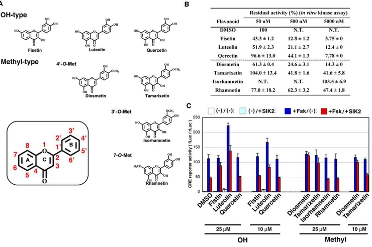
The structures of flavonoids (Figure 1A) and their SIK2-inhibitory activity (Figure 1B) are shown. Fisetin was found to inhibit SIK2 even at a low concentration (50 nM). Some of the O-methylated derivatives, such as diosmetin, inhibited SIK2 at medium concentrations (50–500 nM).
To monitor the SIK2-inhibitory activity in cultured cells (HEK293), we employed the CRE-reporter assay. As shown in
Figure 1. Inhibition of SIK2 by flavonoids.(A) Structure of the flavonoids used in this study. (B)In vitrokinase assay of SIK2. GST-SIK2 expressed in COS-7 cells was used as the enzyme, while GST-TORC2 peptide [42], expressed inEscherichia coli, was used as the substrate for the ELISA. The optical density (OD) value in the absence of a flavonoid was set as 100%. n = 2, means and differences are shown. (C) HEK293 cells transformed with the CRE-Luc firefly luciferase plasmid (200 ng) and pRL-Tk Int- (internalRenillaluciferase: 30 ng) in the presence or absence of pTarget-SIK2 (50 ng) were treated with forskolin (Fsk: 20mM) in the presence of the indicated dose of flavonoids. The ratio of firefly luciferase toRenillaluciferase is shown.
Figure 1C, 25mM fisetin inhibited the SIK2-mediated suppression
of CRE activity that had been upregulated by the cAMP-agonist forskolin. However, a low dose of fisetin (10mM) failed to inhibit SIK2 activity in cultured cells, while diosmetin was able to inhibit SIK2 even at a low dose (10mM), suggesting that other parameters, such as cell permeability, may affect their SIK2-inhibitory activity in cultured cells. On the other hand, it is also important that theO-methyl group at the 49-position of the B-ring more increased their SIK2-inhibitory activity in cultured cells than theO-methyl group at the 39-position or at the 7- position.
O-methyl-flavonoids promote melanogenesis in B16F10 cells
Because one of the representative phenomena of SIK2 inhibition is the promotion of melanogenesis, we employed a melanogenesis assay using B16F10 melanoma cells to evaluate SIK2-inhibitory flavonoids. As shown in Figure 2, non-methylated flavonoids did not induce melanogenesis. In contrast, the 49 -O-methyl flavonoids diosmetin and tamarixetin efficiently induced melanogenesis, while the 39-O-methyl flavonoid isorhamnetin had a modest effect. A small induction of melanogenesis was observed when the 7-O-methyl flavonoid rhamnetin was added into the cultured medium.
The requirement of the methyl group at the 49-position of the B-ring for melanogenesis in B16F10 melanoma cells was similar to that for the inhibition of the SIK2-mediated suppression of CREB activity in HEK293 cells, suggesting that 49-O-methyl flavonoids may induce melanogenesis mainly due to the inhibition of SIK2. The effect of fisetin on melanogenesis was not affected by other factors, such as cell-permeability and stability, which are different between cell types, because fisetin did not affect CREB activity in B16F10 melanoma cells (shown later).
Flavonoids promote eumelanogenesisin vivo
CREB activity determines the ratio of eumelanogenesis to pheomelanogenesis in hair follicle melanocytes in vivo, and inhibition of SIK2 facilitates eumelanogenesis due to the constitutive activation of CREB. Mice with thelethal yellowallele ofagouti(Ay) have yellow hair due to the impaired activation of the alpha-MSH receptor followed by the inactivation of the cAMP-CREB cascade. The Sik22/2genetic background reactivates the CREB cascade inAy/amice, which restores the yellow hair color to wild-type mice (brown).
The Sik2 heterozygous (Sik2+/2) background partially
re-stored hair color, butSik2+/2;Ay
/amice were highly sensitive to CREB agonists, such as UV irradiation, which appeared as a hair color change (Figure 3A). Therefore, we decided to use Sik2+/2; Ay
/a mice to evaluate the effect of flavonoids on melanogenesis in vivo. We assessed the activity of fisetin, quercetin, and diosmetin because their low cost would facilitate their use as dietary supplements. As shown in Figure 3B, fisetin and diosmetin changed the hair color of Sik2+/2; Ay/a
mice, while quercetin had a modest effect. This hair color change was reversible. The difference between fisetin and quercetin could be explained by their inhibitory efficiency toward SIK2 in HEK293 cells (Figure 1C); however, the fact that fisetin promoted eumelanogenesis at the same level as diosmetin disagreed with the results observed in B6F10 melanoma cells (Figure 2). Therefore, we surmised that some of the metabolites, probably O-methylfisetin, might promote eumelanogenesis in mice consuming fisetin. To confirm this hypothesis, we analyzed fisetin metabolites in feces and identified mono-methylfisetin in the metabolites (Figure 3C).
49-O-methylfisetin strongly promotes melanogenesis in B16F10 melanoma cells
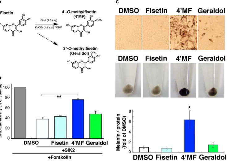
The LC system is not able to separate 49-O-methyl flavonoids from their 39-O-methyl isomers, and 49-O-methylfisetin is not commercially available, while 39-O-methyl fisetin is available as geraldol. Therefore, we decided to synthesize 49-O-methylfisetin using CH3I to confirm its potential as a promoter of melanogenesis
(Figure 4A). The 49-OH group of fisetin might more actively
Figure 2. Induction of melanogenesis by flavonoids in B16F10 melanoma cells.(A) B16F10 melanoma cells were treated with 10mM
flavonoids for 3 d with a medium change at day 2. (B) The cells were recovered in test tubes. (C) Melanin was extracted with an alkaline method. After normalization of the melanin content to the protein amount in each sample, the melanin level was expressed as fold of control (DMSO-treated cells). n = 3, means and standard deviations (S.D.) are shown.
Figure 3. Induction of melanogenesis by flavonoidsin vivo.(A) 1.5-week-oldAy/a
male mice with differentSik2backgrounds (Sik2+/+
orSik2+/2)
accept the methyl group than the 39-OH group did because the yield of 49-O-methylfisetin (5.3%, 5.9 mg) was higher than the 39 -O-isomer (,1.0%). The identity of 49-O-methylfisetin was confirmed by1H NMR,13C NMR, and ESI-MS [20] (Figure S1). When 49-O-methylfisetin was added into the culture medium of B16F10 melanoma cells, the SIK2-mediated suppression of CREB activity was weakened (Figure 4B) and melanogenesis was strongly promoted (Figure 4C), suggesting that eumelanogenesis in fisetin-treated mice might be induced by 49-O-methylfisetin.
49-O-methylfisetin promotes melanogenesis dependent on TORC1 and independent of cAMP
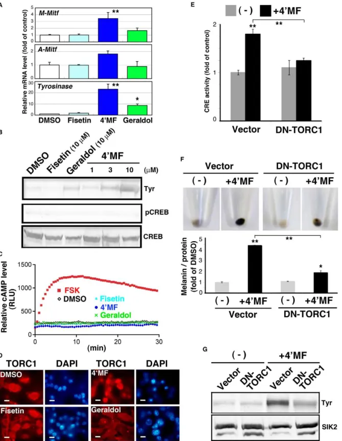
To examine the molecular mechanisms underlying 49 -O-methylfisetin-induced melanogenesis, we monitored the mRNA expression of the melanogenic genes, M-type Mitf, A-type Mitf, andTyrosinase. As shown in Figure 5A, 49-O-methylfisetin induced these mRNAs in B16F10 melanoma cells. The Tyrosinase protein level (Tyr) was also elevated in 49-O-methylfisetin-treated cells
(Figure 5B), which was observed from 3mM. Geraldol was also
able to induce Tyrosinase expression, but its efficiency was less than one-third of 49-O-methylfisetin.
When we examined CREB phosphorylation levels (Figure 5B), we noticed that 49-O-methylfisetin induced melanogenesis without elevating the phosphorylation of CREB at Ser133. This was confirmed by an assay indicating that these flavonoids had little effect on intracellular cAMP levels in B16F10 melanoma cells (Figure 5C). In addition to cAMP/PKA cascade, 49 -O-methylfi-setin did not alter the phosphorylation levels of Erk and GSK-3beta, while fisetin and geraldol enhanced pGSK-3 beta signals (Figure S2).
Since the loss of SIK2 induces melanogenesis by activating TORC1, we monitored the activation of TORC1 by its intracellular distribution. As shown in Figure 5D, 49 -O-methylfi-setin induced the nuclear accumulation of TORC1, but other flavonoids did not. We then examined whether 49-O-methylfisetin was able to activate CREB, and if so, whether this activation was ethyl acetate and detected by LC-MS with a scan range of ms 285–299, as described in the Materials and Methods. The positions of authentic flavonoids are also shown in left panels.
doi:10.1371/journal.pone.0026148.g003
Figure 4. 49-O-methylfisetin (49MF) inhibits SIK2-mediated CRE suppression and induces melanogenesis in B16F10 melanoma cells.
(A) Synthesis of 49MF. (B) B16F10 melanoma cells transformed with CRE-Luc firefly luciferase plasmid (200 ng) with pRL-Tk Int- (internalRenilla
luciferase: 30 ng) in the presence or absence of pTarget-SIK2 (50 ng) were treated with forskolin (Fsk: 20mM) in the presence of the indicated dose of
flavonoids. The relative units of firefly luciferase were normalized toRenillaluciferase, and expressed as % of control (without flavonoid or SIK2). n = 3, means and S.D. are shown. **,p,0.01. (C) B16F10 melanoma cells were treated with 10mM flavonoids for 3 d, with a medium change at day 2, and
Figure 5. 49MF induces melanogenesis by activating TORC1 without enhancing the cAMP level in B16F10 melanoma cells.(A) Quantitative PCR analyses were performed with total RNA prepared from flavonoid-treated B16F10 melanoma cells (10mM for 3 d, with a medium
change at day 2). The mRNA levels are shown as fold of control. n = 3, means and S.D. are shown. 49MF: 49-O-methylfisetin. * and **,p,0.05 and
,0.01, respectively. (B) Tyrosinase (Tyr) protein was detected by western blot analysis. 49MF was added at the indicated concentration. pCREB (pSer133) and total CREB were also examined using the same cell lysate. The panels represent the findings from one of the duplicated experiments. (C) B16F10 cells transformed with the cAMP-indicator plasmid pGloSensor-22F were treated with 10mM flavonoid or forskolin (Fsk: 20mM) in the
presence of luciferin. The relative light units are shown as relative cAMP levels. (D) B16F10 cells were treated with 10mM flavonoids for 72 h and then
dependent on TORC1. Expectedly, 49-O-methylfisetin upregu-lated CRE-reporter activity, which was inhibited by the overex-pression of DN-TORC1.
Finally, we tested whether DN-TORC1 was able to inhibit 49 -O-methylfisetin-induced melanogenesis. As shown in Figure 5E, DN-TORC1 inhibited melanin synthesis (Figure 5F), which was accompanied by the suppression of Tyrosinase expression (Figure 5G). These results suggest that 49-O-flavonoids, especially 49-O-methylfisetin, are potent inhibitors of SIK2 and capable of activating TORC1 followed by the induction of the melanogenic program in mice.
Discussion
We have shown that 49-O-methyl flavones can inhibit SIK2 activity and promote melanogenesis via the activation of TORC1 in B16F10 melanoma cells [18]. However, first, we have to discuss about the discrepancy found between thein vitroand cultured cell assays for structure activity correlation. The in vitro kinase assay using the TORC peptide suggested that non-methylated flavones more potently inhibited SIK2 than their methylated derivatives. However, in HEK293 cells and B16F10 melanoma cells, 49 -O-methylflavone inhibited SIK2 more efficiently, suggesting several mechanisms exist by which flavones can inhibit SIK2 in cultured cells. This hypothesis is also supported by the observation that 39 -O-methylflavones, such as isorhamnetin and geraldol, do not inhibit SIK2 in vitro, while they weakly induce melanogenesis in B16F10 melanoma cells. These results suggested that methylated flavonoids induce the melanogenic program by several mecha-nisms, such as enhanced cell permeability and SIK2-independent signaling pathways.
Meanwhile, it was also true that the efficiency of SIK2 inhibition and the potency of melanogenic promotion by 49 -O-methylflavones in cultured cells (49-O-methylfisetin.diosmetin.
tamarixetin) correlated well with the efficiency of SIK2-kinase inhibition in vitro by their non-methylated cognates (fisetin .
luteolin .quercetin). Moreover, fisetin promoted eumelanogen-esis inAy/a;Sik2+/2mice more potently than quercetin, suggesting
that a synergistic effect between the direct inhibition of SIK2 by a structural dependence of flavones and an indirect effect via a mechanism depending on their 49-O-methoxy groups may efficiently promote melanogenesis in mice.
A number of factors and related compounds have been reported to intricately modulate the melanogenic program. For example, tyrosine kinases and glycogen synthase kinase 3 beta (GSK-3 beta) play opposing roles in the regulation of melano-genesis in melanocytes [21]. The transcriptional activity of the MITF protein is modulated by protein kinase cascades that are induced by the stem cell factor and its receptor kinase c-KIT. The activation of c-KIT invokes two opposing pathways: the MEK and PI3K-AKT pathways. The RAS-RAF-MEK pathway activates ERK-p90RSK, which phosphorylates CREB at Ser133 and MITF at Ser73 and Ser409 [22] and promotes melanogenesis, whereas AKT inhibits the MITF-activating kinase GSK-3 beta and downregulates melanogenesis [23]. The plant steroid diosgenin also inhibits melanogenesis by activating PI3K signaling [24].
However, the action of GSK-3 beta in the regulation of melanogenesis is complicated and paradoxical. The promoter activity of theMitfgene is upregulated by the beta-catenin-TCF/ LEF complex [25], and the phosphorylation of beta-catenin by GSK-3 beta [26] destabilizes beta-catenin and leads to the suppression of MITF-induced melanogenesis [27]. The observa-tion that indirubin derivatives, potent inhibitors of GSK-3 beta [27,28], stabilize the beta-catenin-TCF/LEF complex and promote melanogenesis in B16F10 melanoma cells suggests that Mitf expression, rather than the phosphorylation-dependent activation of MITF, is the rate-limiting step of the melanogenic program [29].
The GSK-3 beta-mediated regulation of melanogenesis is often accompanied by the activation of the cAMP-PKA-CREB pathway. The plant steroid glycyrrhizin inhibits GSK-3 beta activity, while stimulating CREB-mediated transcription by activating PKA, which results in the promotion of melanogenesis [30]. Meanwhile, we reported that the GSK-3 beta inhibitor indirubin induces the degradation of SIK1 and SIK2 proteins in COS-7 cells [31] and in differentiating C2C12 myocytes [32]. GSK-3 beta is capable of phosphorylating (activating) sites in the activation loop of SIK1/2, and the activated SIK1/2 proteins are stable [31], suggesting that B16F10 melanoma cells that have been treated with GSK-3 beta inhibitors have low levels of SIK2, which would promote melanogenesis. Meanwhile, 49-O-methylfisetin did not modulate the AKT-GSK-3 beta and MEK cascades, suggesting that the melanogenic programs induced by 49 -O-methylflavones may be different from those induced by plants compounds modulating the AKT-GSK-3 beta and MEK cascades.
Some methylated flavonoids, such as nobiletin [14] and ayanin [33], inhibit phosphodiesterase, which increases the intracellular cAMP levels [34]. In contrast to these polymethylated flavonoids, 49-O-methylfisetin elevates neither CREB phosphorylation levels nor cAMP-indicator luciferase activity, irrespective of the length of treatment, suggesting that 49-O-methylfisetin upregulates CREB activity independently of cAMP. The mechanism of 49 -O-methylfisetin-induced CREB activity may depend on the activity of TORC1 induced by SIK2 inhibition.
TORC1, or its other isoforms, plays important roles in neuronal activity, such as memory in the hippocampus [35,36], behavior (food intake) in the arcuate and ventromedial nuclei [37], and corticotrophin-releasing hormone synthesis in the hypothalamus [38]. In addition to these roles, we also found that TORC1 is essential for neuronal survival after brain ischemia [19], which is evident in Sik22/2 mice. Interestingly, fisetin was found to enhance memory function in the brain and long term potentiation in cultured PC12 cells via MEK-ERK-mediated CREB activation [39]. Because 49-O-methylfisetin did not activate ERK in B16F10 melanoma cells, the upregulation of TORC activity by SIK2 inhibition has been suggested be a beneficial strategy for the treatment of neuronal diseases, and fisetin or 49-O-methylfisetin may be helpful to perform this strategy.
On the other hand, the present study also revealed that heterozygous insufficiency of theSik2allele increases the sensitivity of CREB-mediated gene expressionin vivo, such as switching to eumelanogenesis in hair melanocytes. This phenomenon may be luciferase plasmid (200 ng) and pRL-Tk Int- (internalRenillaluciferase: 30 ng). After 24 h, the cells were treated with 10mM 49MF for an additional
24 h. CRE activity was expressed as fold of control (the cells were transfected with the empty adenovirus and not treated with 49MF). n = 3, means and S.D. are shown. Bars indicate 10mm. (F) B16F10 melanoma cells transfected with the adenoviruses as in (E) were treated with 10mM 49MF for 3 d with
a medium change at day 2, and the melanin content was measured. n = 3, means and S.D. are shown. (G) Tyrosinase protein levels in B16F10 melanoma cells (the same sample as in F) were examined by western blot analyses. SIK2 was detected as a loading control.
helpful to screen CREB activatorsin vivo. Given that the daily food intake ofAy/amice is,4 g on average, the present dose of fisetin,
400 mg/kg, is not extremely high. Unfortunately, fisetin intake elevates the blood glucose levels ofAy/amice, while diosmetin did slightly (data not shown). As there was no significant difference in blood glucose levels between wild-type andSik22/2mice [18,40], fisetin may affect blood glucose homeostasis in a SIK2-indepen-dent manner.
In conclusion, by modulating SIK2 signaling, we were able to identify a biologically active substance, 49-O-methylfisetin, which initiated CREB-mediated transcription via TORC1 activation. In this study, we also found that the hair color ofSik2+/2mice and
the analysis of metabolites in their feces and blood may act as beneficial indicators to develop compounds that modulate CREB activity.
Materials and Methods
Flavonoids
Luteolin, diosmetin, quercetin, tamarixetin, isorhamnetin, rhamnetin, and geraldol were obtained from Extrasynthese (Genay Cedex, France). Fisetin and forskolin were purchased from Wako Pure Chemicals Co. Ltd., (Osaka, Japan) and Sigma-Aldrich (St. Louis, MO, USA), respectively. These compounds were dissolved in dimethyl sulfoxide (DMSO) as 61000 stock solutions.
Cell culture, flavonoid treatment, and melanin measurement
B16F10 murine melanoma cells and HEK293 cells were obtained from the American Type Culture Collection (Manassas, VA, USA). B16F10 cells were growth at 37uC under 5% CO2in
Dulbecco’s modified Eagle’s medium (DMEM; high glucose) (Wako) supplemented with 10% fetal bovine serum (FBS), penicillin (100 U/mL), and streptomycin (50mg/mL). HEK293 cells were growth at 37uC under 5% CO2in DMEM (low glucose)
(Wako) supplemented with 10% FBS and penicillin/streptomycin. B16F10 were seeded in 6-well plates at a density of 3.46105 cells/well. After 24 h, the culture medium was replaced with fresh medium supplemented with flavonoids, and, after 48 h, the medium was changed again with fresh medium containing the same flavonoids. After an additional 24 h, the cells were harvested for the melanin or mRNA/protein assays.
To measure melanin, the cells were washed twice with phosphate-buffered saline (PBS), suspended in PBS, and recovered by centrifugation at 8,000 rpm for 1.5 min. The cell pellet was suspended in 300mL of 1 N NaOH and incubated at 45uC for
2 h, and, then, melanin was extracted with a chloroform-methanol mixture (2:1). Melanin was detected with a spectrophotometer (BIO-RAD Model 680 MICRO PLATE READER; Bio-Rad, Hercules, CA, USA) at 405 nm. The standard curve was obtained by using purified melanin (0–1000mg/mL). The protein concen-tration of the cell pellets was determined using the Bradford reagent (Bio-Rad) and used for normalization of the melanin content.
Animal experiments and liquid chromatography-mass spectrometry (LC-MS) analysis of flavonoids
The experimental protocols for mice were approved by the committee at the National Institute of Biomedical Innovation (approval ID: DS20-55).Sik2+/2;Ay
mice (4-week-old male mice; the mice were gifts from ProteinExpress Co. Ltd., Chiba, Japan) were housed under standard light (08:00–20:00) and temperature (23uC/60% humidity) conditions.
Mice feces (3.0 g dry weight) were soaked in water : ethyl acetate (1:1), and the flavonoids were recovered from the organic phase. Ethyl acetate was evaporated under N2gas, and the dried
residues were dissolved in 25% acetonitrile/water and subjected to LC-MS analysis (API 3000 mass spectrometer; Applied Biosys-tems, Foster City, CA, USA). To separate the flavonoids, a C18
column (2.0650 mm i.d., particle size 5mm) (Nacalai tesque, Kyoto, Japan) was used. A linear gradient was prepared with 0.1% formic acid in water (solvent A) and acetonitrile (solvent B): from 20% solvent B to 100% solvent B in 25 min at 30uC. The flavonoids were monitored by a UV detector at 255 nm.
Synthesis of 49-O-methylfisetin
The methods for the synthesis of methylflavonids were described in [41]. To a solution of fisetin (122.4 mg, 4.361024mol) in N,N-dimethylformamide (10 ml) was added CH3I (26.4mL,
4.361024mol), and K2CO3(71.7 mg, 5.16102 4
mol). After being stirred for 14 h at room temperature, the reaction mixture was concentrated in vacuo, dissolved in ethyl acetate, washed with sat NaCl, dried over Na2SO4 and evaporated. The resulting residue
was separated with preparative SiO2 thin layer chromatography
(eluent: chloroform/methanol (9/1)) followed by HPLC using a gel filtration colum, JAIGEL GS-320 (Japan Analytical Industry Co., Ltd, Tokyo, Japan) with an eluent methanol, and finally 49 -O-methylfisetin was recovered as yellow crystals (5.9 mg, 5.3% yield). The identity and structure of 49-O-methylfisetin was confirmed with electrospray ionization mass spectroscopy (ESI-MS) and1H and
13
C nuclear magnetic resonance (NMR) [20], respectively. 49 -O-methylfisetin (C16H13O6): 1H NMR (399.65 MHz, CD3OD) d:
3.94 (1 H, d,J= 3.6 Hz), 6.91 (2 H, m), 7.06 (1 H, d,J= 8.4 Hz), 7.77 (1 H, m), 7.98 (1 H, d,J= 9.6 Hz).13C NMR (399.65 MHz, CD3OD): 56.35 (OCH3) 102.98, 112.24, 115.45, 115.65, 116.08,
121.45, 127.55, 147.42, 150.61, 158.57. The spectral data of 49 -O-methylfisetin and its derivatives is shown in Figure S1.
ESI-MS spectra were measured on AB SCIEX API-3000 mass spectrometer. NMR siganl was recorded on a JEOL JNM-JSX400 spectrometer using CD3OD as a solvent and tetramethylsilane
(TMS) as the internal standard.
Quantitative real-time PCR
Total RNA was isolated from B16F10 cells by using the EZ1 RNA Universal Tissue Kit (Qiagen, Venlo Park, the Netherlands), according to the manufacturer’s protocol. cDNA was synthesized using the Transcriptor cDNA First Strand Synthesis Kit (Roche Diagnostics Corp., Indianapolis, IN, USA). PCR amplification was performed using Platinum Quantitative PCR SuperMix (Invitro-gen). The resulting cDNA was amplified using the specific primers: GAPDH-F, 59-ACTCACGGCAAATTCAACGG and GAPDH-R, 59-GACTCCACGACATACTGAGC; Tyrosinase-F, 59 -TGG-GGATGAGAACTTCACTG and Tyrosinase-R, 59 -ACGTAA-TAGTGGTCCCTCAGGT; A-Mitf-F, 59 -GGAAATGCTAGAA-TACAGTCACTA and Pan-Mitf-R, 59 -GTCGCCAGGCTG-GTTTGGACA; and M-Mitf-F 59 -GGAAATGCTAGAATA-CAGTCACTA and Pan-Mitf-R. The reactions were performed for 42 cycles at 95uC for 20 s, 58uC for 20 s, 72uC for 20 s, and 31 cycles at 75uC for 10 s.
Western blot analysis
membranes were blocked with Blocking-One (Nacalai tesque, Kyoto, Japan) and then incubated with the following primary antibodies: anti-Tyrosinase goat polyclonal antibody (Santa Cruz Biotechnology, Santa Cruz, CA, USA), anti-SIK2 rabbit poly-clonal antibody [15], and anti-CREB and anti-phospho CREB rabbit polyclonal antibodies (Cell Signaling Technology, MA, USA) at 4uC overnight. After washing, the membranes were incubated with peroxidase-conjugated secondary antibody at room temperature for 4 h. Detection was performed using the KONICA MINOLTA immunostaining HRP-1000 Kit (KONICA MIN-OLTA, Tokyo, Japan).
Immunocytochemistry
To perform immunocytochemistry, B16F10 cells were seeded on glass cover-slips. The medium was changed with fresh medium supplemented with 10mM flavonoid for 72 h. The cells were fixed
with 4% formaldehyde and stained with the anti-TORC1/3 rabbit polyclonal antibody. To detect the TORC1-antibody complex, anti-rabbit IgG conjugated with Alexa Fluor-594 (Eugene, OR, USA) was used. Nuclei were stained with 49, 6-diamino-2-phenylindole (DAPI).
Expression vector, adenoviruses, transfection, and luciferase/cAMP assay
The reporter plasmids and adenoviruses were previously described [42,43]. Briefly, B16F10 cells in a 24-well plate were co-transfected with the pTAL-CRE vector (200 ng/well) with the internal reporter pRL-TK (30 ng) in the presence or absence of the SIK2 expression vector (pTarget-SIK2 50 ng) using Lipofec-tamine2000 (Invitrogen, Carlsbad, CA, USA). After 24 h, the cells were treated with forskolin (20mM) and cultured for an additional
6 h. Reporter activity was monitored using the Dual Luciferase Reporter Assay Kit (Promega, Madison, WI, USA).
The dominant negative TORC1 (DN-TORC1) adenovirus was previously described [19]. B16F10 cells plated in 6-well dishes were infected with adenoviruses (DN-TORC1 or lacZ at a multiplicity of infection of 10). After a 3 h incubation, the medium was changed with new medium that did not contain adenoviruses, and the cells were cultured for 72 h with a medium change after 48 h.
Fluctuation of the intracellular cAMP level was monitored by the PKA regulatory subunit-liked luciferase reporter system, the GloSensorTM cAMP Assay kit (Promega). Briefly, B16F10 cells
were seeded in 96-well plate at a density of 56103cells/well and incubated for 24 h and transfected with the pGloSensorTM-22F cAMP-reporter plasmid (1 ng/well) using LipofectAMIN2000. After 18 h, cells were incubated with GloSensorTMcAMP reagent for 2 h, and, then, forskolin (20mM) or flavonoids (10mM) was added into the culture medium.
Statistical analysis
Student’s t-test was used to assess all experimental data in Microsoft Excel. The mean and standard deviation (S.D.) are shown.
Supporting Information
Figure S1 NMR analysis of 49-O-methylfisetin and its
derivatives (authentic). The structure of 49-O-methylfisetin was confirmed by comparison of its13C-NMR chemical shifts in B-ring positions with those of other similar flavonoids owing 49 -OH or 49-OMe with 39-OMe or 39-OH groups. 13C-NMR chemical shifts of 49-O-methylfisetin for the B-ring positions, from C-19 to 69, are similar to those of (49-OMe, 39-OH)-type tamarixetin [20] and different from those of (49-OH, 39 -OMe)-type isorhamnetin and geraldor.
(TIF)
Figure S2 49-O-methylfisetin (49MF) does not affect the
MEK or GSK-3 beta pathways.B16F10 cells cultured in FCS-free medium overnight were treated with fisetin, 49MF, or geraldol (10 mM) for 30 min. MEK/pMEK and GSK-3 beta/pGSK-3 beta were examined. The photographs indicate a representative set from the duplicate experiments.
(TIF)
Acknowledgments
We are grateful to Mrs. Junko Morita (NIBIO), Ms. Yuko Shimono, Ms. Tomoko Onishi, Mr. Yuki Shiota, and Mr. Akihiro Hojyo (Kansai University) for their technical assistance. We thank ProteinExpress Co. Ltd. (Chiba, Japan) for their providing usSik22/2mice.
Author Contributions
Conceived and designed the experiments: HT. Performed the experiments: AK. Analyzed the data: KK SU HK YN. Contributed reagents/materials/ analysis tools: NH YS TU TS YI YH KU. Wrote the paper: HT.
References
1. Lin JY, Fisher DE (2007) Melanocyte biology and skin pigmentation. Nature 445: 843–850.
2. Hocker TL, Singh MK, Tsao H (2008) Melanoma genetics and therapeutic approaches in the 21st century: moving from the benchside to the bedside. J Invest Dermatol 128: 2575–2595.
3. Yamaguchi Y, Hearing VJ (2009) Physiological factors that regulate skin pigmentation. Biofactors 35: 193–199.
4. Vachtenheim J, Borovansky J (2010) ‘‘Transcription physiology’’ of pigment formation in melanocytes: central role of MITF. Exp Dermatol 19: 617–627. 5. Busca R, Ballotti R (2000) Cyclic AMP a key messenger in the regulation of skin
pigmentation. Pigment Cell Res 13: 60–69.
6. D’Orazio JA, Nobuhisa T, Cui R, Arya M, Spry M, et al. (2006) Topical drug rescue strategy and skin protection based on the role of Mc1r in UV-induced tanning. Nature 443: 340–344.
7. Jimbow K, Alena F, Dixon W, Hara H (1992) Regulatory factors of pheo- and eumelanogenesis in melanogenic compartments. Pigment Cell Res Suppl 2: 36–42.
8. Winkel-Shirley B (2002) Biosynthesis of flavonoids and effects of stress. Curr Opin Plant Biol 5: 218–223.
9. Kovacic P, Somanathan R (2011) Cell signaling and receptors with resorcinols and flavonoids: redox, reactive oxygen species, and physiological effects. J Recept Signal Transduct Res 31: 265–270.
10. Nakayama T (1994) Suppression of hydroperoxide-induced cytotoxicity by polyphenols. Cancer Res 54: 1991s–1993s.
11. Shoji T, Masumoto S, Moriichi N, Kobori M, Kanda T, et al. (2005) Procyanidin trimers to pentamers fractionated from apple inhibit melanogenesis in B16 mouse melanoma cells. J Agric Food Chem 53: 6105–6111. 12. Fujii T, Saito M (2009) Inhibitory effect of quercetin isolated from rose hip (Rosa
canina L.) against melanogenesis by mouse melanoma cells. Biosci Biotechnol Biochem 73: 1989–1993.
13. Yoon HS, Lee SR, Ko HC, Choi SY, Park JG, et al. (2007) Involvement of extracellular signal-regulated kinase in nobiletin-induced melanogenesis in murine B16/F10 melanoma cells. Biosci Biotechnol Biochem 71: 1781–1784.
14. Nagase H, Omae N, Omori A, Nakagawasai O, Tadano T, et al. (2005) Nobiletin and its related flavonoids with CRE-dependent transcription-stimulating and neuritegenic activities. Biochem Biophys Res Commun 337: 1330–1336.
15. Horike N, Takemori H, Katoh Y, Doi J, Min L, et al. (2003) Adipose-specific expression, phosphorylation of Ser794 in insulin receptor substrate-1, and activation in diabetic animals of salt-inducible kinase-2. J Biol Chem 278: 18440–18447.
16. Katoh Y, Takemori H, Min L, Muraoka M, Doi J, et al. (2004) Salt-inducible kinase-1 represses cAMP response element-binding protein activity both in the nucleus and in the cytoplasm. Eur J Biochem 271: 4307–4319.
18. Horike N, Kumagai A, Shimono Y, Onishi T, Itoh Y, et al. (2010) Downregulation of SIK2 expression promotes the melanogenic program in mice. Pigment Cell Melanoma Res 23: 809–819.
19. Sasaki T, Takemori H, Yagita Y, Terasaki Y, Uebi T, et al. (2011) SIK2 is a key regulator for neuronal survival after ischemia via TORC1-CREB. Neuron 69: 106–119.
20. Blasko G, Shieh HL, Pezzuto JM, Cordell GA (1989) 13C-nmr spectral assignment and evaluation of the cytotoxic potential of rotenone. J Nat Prod 52: 1363–1366.
21. Blume-Jensen P, Jiang G, Hyman R, Lee KF, O’Gorman S, Hunter T (2000) Kit/stem cell factor receptor-induced activation of phosphatidylinositol 39-kinase is essential for male fertility. Nat Genet 24: 157–162.
22. Wu M, Hemesath TJ, Takemoto CM, Horstmann MA, Wells AG, et al. (2000) c-Kit triggers dual phosphorylations, which couple activation and degradation of the essential melanocyte factor Mi. Genes Dev 14: 301–312.
23. Oka M, Nagai H, Ando H, Fukunaga M, Matsumura M, et al. (2000) Regulation of melanogenesis through phosphatidylinositol 3-kinase-Akt pathway in human G361 melanoma cells. J Invest Dermatol 115: 699–703.
24. Lee J, Jung K, Kim YS, Park D (2007) Diosgenin inhibits melanogenesis through the activation of phosphatidylinositol-3-kinase pathway (PI3K) signaling. Life Sci 81: 249–254.
25. Takeda K, Yasumoto K, Takada R, Takada S, Watanabe K, et al. (2000) Induction of melanocyte-specific microphthalmia-associated transcription factor by Wnt-3a. J Biol Chem 275: 14013–14016.
26. Meijer L, Skaltsounis AL, Magiatis P, Polychronopoulos P, Knockaert M, et al. (2003) GSK-3-selective inhibitors derived from Tyrian purple indirubins. Chem Biol 10: 1255–1266.
27. Bellei B, Flori E, Izzo E, Maresca V, Picardo M (2008) GSK3beta inhibition promotes melanogenesis in mouse B16 melanoma cells and normal human melanocytes. Cell Signal 20: 1750–1761.
28. Cho M, Ryu M, Jeong Y, Chung YH, Kim DE, et al. (2009) Cardamonin suppresses melanogenesis by inhibition of Wnt/beta-catenin signaling. Biochem Biophys Res Commun 390: 500–505.
29. Khaled M, Larribere L, Bille K, Aberdam E, Ortonne JP, et al. (2002) Glycogen synthase kinase 3beta is activated by cAMP and plays an active role in the regulation of melanogenesis. J Biol Chem 277: 33690–33697.
30. Lee J, Jung E, Park J, Jung K, Park E, et al. (2005) Glycyrrhizin induces melanogenesis by elevating a cAMP level in b16 melanoma cells. J Invest Dermatol 124: 405–411.
31. Hashimoto YK, Satoh T, Okamoto M, T H (2008) Importance of autophosphorylation at Ser186 in the A-loop of salt inducible kinase 1 for its sustained kinase activity. J Cell Biochem 104: 1724–1739.
32. Takemori H, Katoh Hashimoto Y, Nakae J, Olson EN, Okamoto M (2009) Inactivation of HDAC5 by SIK1 in AICAR-treated C2C12 myoblasts. Endocr J 56: 121–130.
33. Chan AL, Huang HL, Chien HC, Chen CM, Lin CN, et al. (2008) Inhibitory effects of quercetin derivatives on phosphodiesterase isozymes and high-affinity [(3) H]-rolipram binding in guinea pig tissues. Invest New Drugs 26: 417–424. 34. Peluso MR (2006) Flavonoids attenuate cardiovascular disease, inhibit phosphodiesterase, and modulate lipid homeostasis in adipose tissue and liver. Exp Biol Med (Maywood) 231: 1287–1299.
35. Zhou Y, Wu H, Li S, Chen Q, Cheng XW, et al. (2006) Requirement of TORC1 for late-phase long-term potentiation in the hippocampus. PLoS One 1: e16.
36. Li S, Zhang C, Takemori H, Zhou Y, Xiong ZQ (2009) TORC1 regulates activity-dependent CREB-target gene transcription and dendritic growth of developing cortical neurons. J Neurosci 29: 2334–2343.
37. Altarejos JY, Goebel N, Conkright MD, Inoue H, Xie J, et al. (2008) The Creb1 coactivator Crtc1 is required for energy balance and fertility. Nat Med 14: 1112–1117.
38. Liu Y, Coello AG, Grinevich V, Aguilera G (2010) Involvement of transducer of regulated cAMP response element-binding protein activity on corticotropin releasing hormone transcription. Endocrinology 151: 1109–1118.
39. Maher P, Akaishi T, Abe K (2006) Flavonoid fisetin promotes ERK-dependent long-term potentiation and enhances memory. Proc Natl Acad Sci U S A 103: 16568–16573.
40. Muraoka M, Fukushima A, Viengchareun S, Lombes M, Kishi F, et al. (2009) Involvement of SIK2/TORC2 signaling cascade in the regulation of insulin-induced PGC-1alpha and UCP-1 gene expression in brown adipocytes. Am J Physiol Endocrinol Metab 296: E1430–1439.
41. Bouktaib M, Lebrun S, Atmani A, Rolando C (2002) Hemisynthesis os all the O-monomethylated analogues of quercetin including the major metabolites, through selective protection of phenolic functions. Tetrahedron 58: 10001–10009.
42. Katoh Y, Takemori H, Lin XZ, Tamura M, Muraoka M, et al. (2006) Silencing the constitutive active transcription factor CREB by the LKB1-SIK signaling cascade. Febs J 273: 2730–2748.