Autores
André Antunes Poitevin1 Christian Viezzer1
Denise Cantarelli Machado1 Bartira Ercilia Pinheiro da Costa1
Ana Elizabeth Figueiredo1
Domingos d'Avila1 Carlos Eduardo Poli-de-Figueiredo1
1 Pontifical Catholic University
of Rio Grande do Sul.
Data de submissão: 29/03/2012. Data de aprovação: 10/12/2013.
Correspondência para:
Bartira Ercilia Pinheiro da Costa. Pontifícia Universidade Católica do Rio Grande do Sul. Av. Ipiranga, nº 6690, Hospital São Lucas da PUCRS, 2º andar Laboratório de Nefrologia, Instituto de Pesquisas Biomédicas. Porto Alegre, RS, Brazil. CEP: 90610-000. E-mail: [email protected] Tel: (51) 3367-700. Fax: (51) 3367-700.
Peritosteril® and Balance® bags
were provided by Fresenius Medical Care.
Effect of standard and neutral-pH peritoneal dialysis solutions
upon fibroblasts proliferation
Efeito da solução de diálise peritoneal com pH neutro e padrão
sobre a proliferação de fibroblastos
Introdução: A exposição contínua da membrana peritoneal a soluções conven-cionais de diálise é um importante fator de risco para induzir alterações estruturais e funcionais. Objetivo: Comparar a viabili-dade in vitro dos fibroblastos NIH-3T3 de camundongo após exposição à solução de diálise com pH neutro com células expos-tas à solução padrão. Métodos: Estudo ex-perimental; ambas as soluções foram tes-tadas em todas as concentrações de glicose comercialmente disponíveis. A viabilidade celular foi avaliada por ensaio colori-métrico de sal tetrazólio. Resultados: A viabilidade de fibroblastos foi melhor na solução de pH neutro em relação ao con-trole nas três concentrações de glicose (densidade óptica em nm-médias ± DP: 1,5% 0,295 ± 0,047 vs. 0,372 ± 0,042, p < 0,001; 2,3% 0,270 ± 0,036 vs. 0,337 ± 0,051, p < 0,001; 4,25% 0,284 ± 0,037 vs. 0,332 ± 0,032, p < 0,001; controle vs. pH neutro respectivamente, teste t de Student). Não houve diferença significa-tiva na viabilidade celular entre as três concentrações de glicose quando solução padrão foi utilizada (ANOVA p = 0,218), embora a viabilidade celular tenha sido superior após exposição aos fluidos de diálise peritoneal neutros, pH 1,5% em comparação com 2,3 e 4,25% de concen-trações de glicose (ANOVA p = 0,008: Bonferroni 1,5% vs. 2,3% p = 0,033, 1,5% vs. 4,25% p = 0,014, 2,3% vs. 4,25% p = 1,0). Conclusão: A viabilidade celular foi melhor em solução neutra de pH de diálise, especialmente nas menores concentrações de glicose. O pH fisiológico e com menos produtos de degradação de glicose podem ser responsáveis por estes resultados.
RESUMO
Palavras-chave: diálise peritoneal; fibro-blastos; teste de materiais; técnicas de cultura de células.
Introduction: Continuous exposition of the peritoneal membrane to conventional dialysis solutions is an important risk fac-tor for inducing structural and functional alterations. Objective: To compare in vitro
mouse fibroblast NIH-3T3 cell viability after exposition to a neutral pH dialysis solution in comparison to cells exposed to a standard solution. Methods: Experi-mental study to compare the effects of a conventional standard or a neutral-pH, low-glucose degradation products peri-toneal dialysis solution on the viability of exposed fibroblasts in cell culture. Both so-lutions were tested in all the commercially available glucose concentrations. Cell vi-ability was evaluated with tetrazolium salt colorimetric assay. Results: Fibroblast via-bility was significantly superior in the neu-tral pH solution in comparison to control, in all three glucose concentrations (Optical density in nm-means ± SD: 1.5% 0.295 ± 0.047 vs. 0.372 ± 0.042, p < 0.001; 2.3% 0.270 ± 0.036 vs. 0.337 ± 0.051, p < 0.001; 4.25% 0.284 ± 0.037 vs. 0.332 ± 0.032, p < 0.001; control vs. neutral pH respectively,
Student t Test). There was no significant difference in cell viability between the three concentrations of glucose when standard solution was used (ANOVA p = 0.218), although cell viability was higher after ex-position to neutral pH peritoneal dialysis fluid at 1.5% in comparison to 2.3 and 4.25% glucose concentrations (ANOVA
p = 0.008: Bonferroni 1.5% vs. 2.3% p = 0.033, 1.5% vs. 4.25% p = 0.014, 2.3%
vs. 4.25% p = 1.00). Conclusion: Cell vi-ability was better in neutral pH dialysis so-lution, especially in the lower glucose con-centration. A more physiological pH and lower glucose degradation products may be responsible for such results.
A
BSTRACTKeywords: cell culture techniques; fi-broblasts; materials testing; peritoneal dialysis.
INTRODUCTION
Long-term peritoneal dialysis (PD) as therapy for end-stage renal disease (ESRD) patients is only fea-sible if adequate peritoneal membrane structure and function are preserved. Standard PD solutions are acid (pH = 5.5), lactate-buffered, with high glucose con-centrations (75-220 mmol/L), hyperosmolar (334-486 mOsm/L) and contain high concentration of glucose degradation products (GDP) - known to affect peri-toneal function.1 Lasting PD therapy with an intact
peritoneal membrane has been, as yet, an unreached goal and the main drive to develop biocompatible PD solutions. Peritoneal membrane continuous exposi-tion to acid soluexposi-tions seems to be an important risk factor for structural and functional changes. Loss of mesothelial cells, small vessels changes, thickened and fibrotic sub-mesothelial compact zone have been observed at peritoneal biopsies.2 Functional
chan-ges have included ultrafiltration loss, small solutes clearance decline and, eventually, technique failure.3
Using biocompatible dialysis solutions is paramount in accomplishing long-term membrane survival.4 A
neutral-pH, poor GDP dialysis solution, presented in a double-chambered bag has been developed in antici-pation of improved biocompatibility.3 Previous clinical
and experimental studies have suggested its beneficial effects.3,5,6 The aim of the current study was to
compa-re mouse fibroblast NIH-3T3 - a well characterized cell lineage - viability after exposition to either the standard acid or the neutral-pH dialysis solution.
METHODS
Mouse fibroblast NIH-3T3 cell cultures were placed in media containing conventional (Control) PD solu-tion or a neutral-pH PD fluid. All the commercially available glucose (1.5, 2.3 and 4.25%) concentrations were tested.
DIALYSISSOLUTIONS
Both solutions used glucose as osmotic agent. The control solution was Peritosteril®(Fresenius Medical
Care, Jaguariúna, Brazil) and the neutral-pH solution was Balance®(Fresenius Medical Care, Bad Homburg,
Germany) provided in a dual-chambered bag. The composition of both dialysis solutions is presented in Table 1. The 1.5% glucose Balance® bag contains
an alkaline chamber with sodium lactate (sodium: 75 mmol/L; lactate: 70 mmol/L) and a second acid
fluid chamber with electrolytes and glucose (sodi-um: 193 mmol/L; calci(sodi-um: 3.5 mmol/L; magne-sium: 1 mmol/L; chloride: 203 mmol/L; glucose: 166.5 mmol/L). The Balance® solution is ready for
use by opening the seal between the two chambers and mixing their contents, resulting in a fluid with pH in the range of 6.8 to 7.4. Differently, Peritosteril®
has a pH of 5.5. Both Peritosteril® and Balance® bags
were provided by Fresenius Medical Care.
Components in mmol/L Control Neutral pH
Sodium 134 134
Calcium 1.75 1.75
Magnesium 0.5 0.5
Chloride 103.5 101.5
Lactate 35 35
pH 5.5 7.0
Osmolarity (mOsm/L) 358 358
TABLE 1 COMPOSITIONOFTHEPERITONEALDIALYSIS
SOLUTIONSBEFOREMIXINGWITHCULTURE MEDIUM
CELLCULTURES
Mouse fibroblast cell line NIH-3T3 (American Type Culture Collection CRL-1658, Manassas, VA, USA) grown in 25 ml bottles, containing Dulbecco’s mo-dified eagle’s medium (DMEM) cell culture media, gentamycin at 0.025 g/L, streptomycin/penicillin at 0.1 g/L and supplemented with 10% fetal bovine serum (FBS) (Gibco, Grand Island, NY, USA). Incubations we-re performed in a humid atmosphewe-re at 37 ºC and 5% CO2. All the procedures were performed on a laminar flow hood, observing strict aseptic techniques to mi-nimize the risk of microbial contamination. Fibroblast growth in culture occurred as an adherent monolayer. Cells were washed with phosphate-buffered saline (PBS) (Gibco, Grand Island, NY, USA) and detached by trypsin-EDTA 0.05% (Gibco, Grand Island, NY, USA). FBS was used to inactivate trypsin action. Cells were counted on a Neubauer chamber and seeded in DMEM on 96-well tissue culture plates at a density of 0.5 x 10⁴ cells/well and allowed to grow for 24
reached after a series of pilot tests that evaluated cell proliferation with different proportions of test so-lution and culture media (25, 50, 75 and 100% of peritoneal dialysis fluid), or different incubation times (24, 48 and 72-hour). Fibroblast growth was poor at any incubation time in the presence of 75 or 100% PD solutions. Control wells (DMEM 100%) best cell viability was obtained at 48-hour incubation time.
MTT CELLVIABILITYASSESSMENT
Cell viability was evaluated with 3-[4.5-dimethylthiazol-2-yl]-2.5-diphenyl tetrazolium bromide (MTT)(Sigma, Saint Louis, MO, USA). After a 48-hour incubation, added PD solution/DMEM was removed from each well and cells washed with PBS (100 µL). DMEM with 10% MTT 5 mg/ml solution (50 µL/well) was added for a further 4-hour incubation time. Next, MTT was taken out, dimethylsulphoxide [(DMSO); 100 µL; Sigma Co.) was added. Conversion of the MTT product - an estimate of cell viability - was measured by optical density (OD) at 570 nm, using a spectrophotometer fitted with a Microplate Reader (Bio-Rad, Hercules, CA, USA). The OD for the negative control (simply DMEM) represented 100% cell viability. All the experiments were thrice repeated and sample analyses were performed in triplicate.
STATISTICALANALYSIS
Variables are presented as mean and standard deviation (SD). Student’st test was used to compare two continuous variables; normality was tested by Kolmogorov-Smirnov test. ANOVA, with ad hoc Bonferroni test, was employed for multiple comparisons. Differences were considered significant at a p value of 0.05 or less. A Statistical Package for Social Sciences s100 µL software (SPSS, version 17 for Windows, SPSS Inc, Chicago, IL, USA) was used in all statistical analyses.
RESULTS
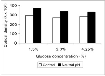
Cells viability after exposition to standard (Control) and neutral-pH solutions with different glucose concentrations is depicted in Figure 1. Cells viability was significantly greater on the neutral pH than on the acid solution, at all tested glucose concentrations, measured by optical density (in nm): 0.295 ± 0.047
vs. 0.372 ± 0.042, p < 0.001 for 1.5% glucose control and neutral pH, respectively; 0.270 ± 0.036
vs. 0.337 ± 0.051, p < 0.001 for 2.3% control and
neutral pH, respectively; 0.284 ± 0.037 vs. 0.332 ± 0.032, p < 0.001 for 4.25% control and neutral pH solution, respectively. No significant differences in cell viability among the three control solution glu-cose concentrations were observed (repeated-mea-sures ANOVA: p = 0.218). However, cells viability on neutral-pH solution was greater after exposition to 1.5 than to 2.3 or 4.25% glucose concentrations [ANOVA with post hoc Bonferroni: p = 0.033 (1.5%
vs. 2.3%); p = 0.014 (1.5% vs. 4.25%); p = 1.00 (2.3%
vs. 4.25%)].
Figure 1. Fibroblast viability after cells were exposed to acid and
neutral pH solutions at different glucose concentrations.
DISCUSSION
The current experiments demonstrated that cells viability is greater when fibroblasts are exposed,
in vitro, to a neutral pH than to an standard PD solution. Besides, greater viability of cells exposed to 1.5%, in comparison to 2.3 and 4.25% glucose on the neutral pH solution was evidenced. The significance of such findings is not yet clear, but it is conceivable that a neutral pH and less glucose degradation products are better for cell viability and that such effect may be amplified at lower glucose concentrations. Improved cell viability with the neutral pH solution does not mean that it is nontoxic or does not induce fibrosis. Fibroblasts were used as an experimental model7-9 and
evaluating the effects of such exposition in other cell models, or in experimental animals, would be desired.10,11 Use of the neutral pH dialysis
solution, in vivo, resulted in improved peritoneal ultrafiltration and peritoneal membrane integrity markers.3,12 It also improved mesothelial cells
A 3-[4.5-dimethylthiazol-2-yl]-2.5-diphenyl tetrazolium bromide-based (MTT) colorimetric assay was used to evaluate cell viability. Its use allows measuring citotoxicity, cell proliferation and cell activation. The assay is based on viable cell mitochondrial dehydrogenases to transform MTT into a water-insoluble blue formazan. The assay method is not time consuming, cheap and allows for using multiwall scanning spectrophotometry.14
Fibroblasts have also been used in a previous study, which found no difference in cell proliferation with incubation in either icodextrin-containing or standard glucose dialysis solution.15 The study
was carried out on pH-adjusted solutions mixed with culture medium, a major distinction from the present work, where test solutions pH difference was a major characteristic being evaluated. Interestingly, heat sterilized 4.25% glucose solution caused a significant reduction in in vitro cell growth, compared with filter-sterilized solution, supposedly associated with increased GDP.15 Occurrence of GDP and low pH seem to
be important factors related with biocompatibility of PD solutions. GDP have been implicated in the formation of advanced glycation end products (AGEs) occurring during the heat sterilization process. The presence of AGEs has been associated with reduce mesothelial cell growth in vitro.16 It
has been previously shown that in vitro cells exposed to lower glucose concentrations (1.5 or 2.27%) grow better than at higher (3.86%).17
Such results are in agreement with findings of this study, suggesting that GDP are highly inhibitory of in vitro cell growth (17) and could be related with clinical consequences.18,19 The neutral pH
peritoneal dialysis solution was designed as a two-chamber bag, one containing the alkaline component with lactate and the other with electrolytes and glucose. Such approach allows for a higher pH and sterilization with reduced formation of GDP. The two-compartments fluids are mixed immediately before infusion, by rupturing a seal between the chambers.
In conclusion, fibroblasts viability was higher on neutral-pH dialysis solution, especially with lower glucose concentrations. It is possible that a more physiological pH and lower GDP are associated with such results.
ACKNOWLEDGMENTS
Both Peritosteril® and Balance® bags were
pro-vided by Fresenius Medical Care. The Nephrology Laboratory received support from Fundação de Amparo à Pesquisa no Rio Grande do Sul (FAPERGS), Conselho Nacional de Pesquisa (CNPq), Coordenadoria de Aperfeiçoamento de Pessoal de Ensino Superior (CAPES), and Pontifícia Universidade Católica do Rio Grande do Sul (PUCRS). Poli-de-Figueiredo is a CNPq researcher.
CONFLICT OF INTERESTS/DISCLOSURE
Both Peritosteril® and Balance® bags were provided
by Fresenius Medical Care. André A. Poitevin, Ana E. Figueiredo, Domingos O. d’Avila and Carlos E. Poli-de-Figueiredo have received travel awards by both companies with commercially available perito-neal dialysis products in Brazil (Fresenius Medical Care and Baxter) in the past.
REFERENCES
1. Topley N. Membrane longevity in peritoneal dialysis: impact of infection and bio-incompatible solutions. Adv Ren Replace Ther 1998;5:179-84.
2. Williams JD, Craig KJ, von Ruhland C, Topley N, Williams GT; Biopsy Registry Study Group. The natural course of peritoneal membrane biology during peritoneal dialysis. Kidney Int Suppl 2003:S43-9. DOI: http://dx.doi.org/10.104 6/j.1523-1755.2003.08805.x
3. Williams JD, Topley N, Craig KJ, Mackenzie RK, Pischetsrieder M, Lage C, et al.; Euro Balance Trial Group. The Euro-Balance Trial: the effect of a new biocompatible pe-ritoneal dialysis fluid (balance) on the pepe-ritoneal membrane. Kidney Int 2004;66:408-18. DOI: http://dx.doi.org/10.1111/ j.1523-1755.2004.00747.x
4. Mackenzie R, Holmes CJ, Jones S, Williams JD, Topley N. Clinical indices of in vivo biocompatibility: the role of ex vivo cell function studies and effluent markers in peritoneal dialysis patients. Kidney Int Suppl 2003:S84-93. PMID: 14870881 DOI: http://dx.doi.org/10.1046/j.1523-1755.2003.08809.x 5. Lage C, Pischetsrieder M, Aufricht C, Jörres A, Schilling H,
Passlick-Deetjen J. First in vitro and in vivo experiences with Stay-Safe Balance, a pH-neutral solution in a dual-chambered bag. Perit Dial Int 2000;20:S28-32
6. Lee HY, Park HC, Seo BJ, Do JY, Yun SR, Song HY, et al. Superior patient survival for continuous ambulatory peritoneal dialysis patients treated with a peritoneal dialysis fluid with neutral pH and low glucose degradation product concentration (Balance). Perit Dial Int 2005;25:248-55.
7. Wieslander AP, Nordin MK, Kjellstrand PT, Boberg UC. Toxicity of peritoneal dialysis fluids on cultured fibroblasts, L-929. Kidney Int 1991;40:779. DOI: http://dx.doi. org/10.1038/ki.1991.182
9. Ogata S, Naito T, Yorioka N, Kiribayashi K, Kuratsune M, Koh-no M. Effect of lactate and bicarbonate on human peritoneal me-sothelial cells, fibroblasts and vascular endothelial cells, and the role of basic fibroblast growth factor. Nephrol Dial Transplant 2004;19:2831-7. DOI: http://dx.doi.org/10.1093/ndt/gfh478 10. Fernández-Perpén A, Pérez-Lozano ML, Bajo MA,
Albar-Vizcaino P, Sandoval Correa P, del Peso G, at al. In-fluence of bicarbonate/low-GDP peritoneal dialysis fluid (BicaVera) on in vitro and ex vivo epithelial-to-mesenchymal transition of mesothelial cells. Perit Dial Int 2012;32:292-304. DOI: http://dx.doi.org/10.3747/pdi.2010.00315 11. Leung JC, Chan LY, Tam KY, Tang SC, Lam MF, Cheng AS,
et al. Regulation of CCN2/CTGF and related cytokines in cul-tured peritoneal cells under conditions simulating peritoneal dialysis. Nephrol Dial Transplant 2009;24:458-69. DOI: http:// dx.doi.org/10.1093/ndt/gfn524
12. Choi HY, Kim DK, Lee TH, Moon SJ, Han SH, Lee JE, et al. The clinical usefulness of peritoneal dialysis fluids with neutral pH and low glucose degradation product concentration: an open randomized prospective trial. Perit Dial Int 2008;28:174-82. 13. Witowski J, Korybalska K, Ksiazek K, Wisniewska-Elnur J, Jörres
A, Lage C, et al. Peritoneal dialysis with solutions low in glucose degradation products is associated with improved biocompatibility profile towards peritoneal mesothelial cells. Nephrol Dial Trans-plant 2004;19:917-24. DOI: http://dx.doi.org/10.1093/ndt/gfh013
14. Gerlier D, Thomasset N. Use of MTT colorimetric assay to measure cell activation. J Immunol Methods 1986;94:57-63. DOI: http://dx.doi.org/10.1016/0022-1759(86)90215-2 15. Cooker LA, Choo CG, Luneburg P, Lamela J, Holmes CJ.
Effect of icodextrin peritoneal dialysis solution on cell proliferation in vitro. Adv Perit Dial 1999;15:17-20. 16. Breborowicz A, Witowski J, Polubinska A, Pyda M, Oreopoulos D.
L-2-oxothiazolidine-4-carboxylic acid reduces in vitro cyto-toxicity of glucose degradation products. Nephrol Dial Trans-plant 2004;19:3005-11. DOI: http://dx.doi.org/10.1093/ ndt/gfh539
17. Cooker LA, Luneburg P, Faict D, Choo C, Holmes CJ. Reduced glucose degradation products in bicarbonate/lactate-buffered peritoneal dialysis solutions produced in two-chambered bags. Perit Dial Int 1997;17:373-8.
18. Yokoi H, Kasahara M, Mori K, Ogawa Y, Kuwabara T, Imamaki H, et al. Pleiotrophin triggers inflammation and increased peritoneal permeability leading to peritoneal fibrosis. Kidney Int 2012;81:160-9. PMID: 21881556 DOI: http://dx.doi.org/10.1038/ki.2011.305