J of Evolution of Med and Dent Sci/ eISSN- 2278-4802, pISSN- 2278-4748/ Vol. 3/ Issue 24/June 16, 2014 Page 6781
MANAGEMENT OF WRIST GANGLIA BY TRANSFIXATION
TECHNIQUE: OUR EXPERIENCE IN A RURAL TEACHING INSTITUTION
Sharma Man Mohan1, Batra Kasturi Mohan2, Kakria Hira Lal3, Sharma Shubham Mohan4HOW TO CITE THIS ARTICLE:
Sharma Man Mohan, Batra Kasturi Mohan, Kakria Hira Lal, Sharma Shubham Mohan. Management of Wrist Ganglia by Transfixation Technique: Our Experience in a Rural Teaching Institution. Journal of Evolution of Medical and Dental Sciences 2014; Vol. 3, Issue 24, June 16; Page: 6781-6789,
DOI: 10.14260/jemds/2014/2819
ABSTRACT: BACKGROUND: Ganglion is one of the commonly seen lesions in minor surgical practice.
Treatment of wrist ganglia includes simple reassurance, watchful waiting, non-operative aspiration, injection, surgical excision and even sometimes advanced endoscopic excision. Although treatment is not often necessary, many patients seeking consultation ask for some form of definitive treatment. High incidence of recurrence is seen with cyst aspiration/injection or after surgical excision. Recurrence is the common problem, irrespective of the treatment modality. MATERIALS AND
METHODS: This study evaluates results of a modified minimally invasive surgical technique used for
the management of ganglia of the wrist. Thirty six wrist ganglia have been treated by using the transfixation technique. RESULTS: The successful results for the procedure were seen in 94.44% patients. Consequent to the procedure, an average of 2days off work were lost. Recurrence in the 2 year follow-up was seen in two patients. DISCUSSION: On comparing with other modalities this method is minimally invasive, cost-effective, less time consuming, low rate of recurrence and at the same time can be carried out under local anesthesia as a day case procedure and even not requiring any specific instrument and further this technique can be easily learned.
KEYWORDS: Ganglion, minimally invasive, transfixation.
INTRODUCTION: Although ganglion is one of the commonest but troublesome lesions met in minor
surgical practice. About their cause or treatment, there is little agreement. These are the benign soft tissue tumors occurring around the wrist, consisting of mucin-filled cyst connected to a tendon, tendon sheath, or joint capsule.[1] They may also present as intraosseous or intra tendinous.[2] Most
ganglions are seen in females in their second, third or fourth decades of life.[3] Spontaneous resolution
is commonly seen with 50% of untreated patients when assessed at six years.[4] The cause of
ganglions remains uncertain but only small minority gives a history of previous trauma.[5] The
capsule of the ganglion consists of compressed stroma without a cellular lining. This may be linked to the joint by a narrow channel which can act as a one way valve.[6]
The majority of this kind of tumor (60-70%) is found on the dorsum of the wrist over the scapholunate interval.[7] Though most ganglions are painless yet pain may be the chief presenting
complaint. Other patients present for consultation because they are worried about how the ganglion looks or worried about malignancy.[4] The diagnosis can be made clearly by history and physical
examination but the treatment options have many options and each treatment option yields different success results.[8]
J of Evolution of Med and Dent Sci/ eISSN- 2278-4802, pISSN- 2278-4748/ Vol. 3/ Issue 24/June 16, 2014 Page 6782 manually or puncturing by a percutaneous needle or by percutaneously incising with a tenotome and radically excising the cyst either by open or endoscopic methods.
The best results in terms of recurrence have been reported with surgical excision. However, the treatment can only be offered successfully in a specialist hand department and is associated with complications including wound healing problems such as infections, neuroma, keloid and the scar formation. Other common complications include scapho-lunate dissociation, joint stiffness and sometimes damage to the posterior interosseous nerve`s terminal branch resulting in decreased grip strength along with risks of general anesthesia and upper limb tourniquet application.[9]
Thus the treatment is complicated by the recurrence as the major problem, no matter what treatment modality is used. It is a fact that not all patients need or demand treatment nor all patients can afford the most advanced treatment. No matter which treatment modality is used, what patients want is the successful result.
In this study we present the results of treatment of wrist ganglia by using the transfixation technique-a minimally invasive day case procedure with successful results in terms of cure of patient`s complaints but without the complications mentioned above.
MATERIALS AND METHODS: A prospective study was conducted on patients presenting to the
orthopedic out-patient department at the institute hospital for wrist ganglions (dorsal and volar) from January 2012 to January 2014. Full medical and surgical history was taken before starting the procedure along with proper hand examination. Patients were given detailed explanation of the adopted technique and proper consent was taken.
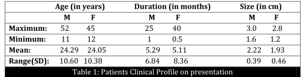
This study comprised 36 patients with ganglia who were followed up for a minimum period of 6 months (range 6-24 months). The male/female ratio was 1:1.57 with ages varying from 11 to 52 years and a median age of 24.14 years. There was no significant difference in the mean age of males (24.29) and females (24.05) (Table 1). The highest incidence was in the 15-<25 age group with 21 cases (58.33%) followed by the 25-<35 age group with 6 cases (16.67%) (Fig.1). The dominant hand was affected in 29 (80.56%) cases. Dominant hand ganglia presented earlier after the onset of the first symptoms (ganglia in the dominant hand presented in the first 2 months in 62% as compared to 28.5% of the ganglia in the non-dominant hand).
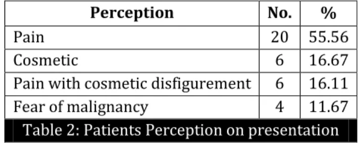
Most common presenting complaint was pain (55.56%) followed by cosmetic (19.44%) irrespective of position of ganglion, sex or dominance (Table 2). Pain overall was present in 69.44% of the ganglia. The mean time of presentation was 5.18 months (range from 15 days to 40 months). Fifty six percent of the patients presented within the first 2months. It was noticed that 50% of the patients presented within 2 months when the chief complaint was pain compared to only 33% if it was cosmetic. The mean size at the time of the presentation was 1.86cms (varying from 0.7cm to 3cms).
J of Evolution of Med and Dent Sci/ eISSN- 2278-4802, pISSN- 2278-4748/ Vol. 3/ Issue 24/June 16, 2014 Page 6783 Patients having complicated palmer ganglion, ganglion near the radial artery, infected ganglion, ganglion of other sites, ganglion less than 5mms in size, previous treatment taken in any form, ganglion associated with arthritic disorder and patients with diabetes were excluded from the study.
MATERIALS: 0.5% bupivacaine, Disposable 16G needle, Linen thread no. 2-0.
Surgical Technique: The procedure was done on out-patient day care basis. The patients were given
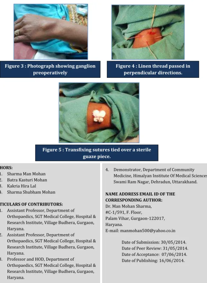
a single dose of Erythromycin -500 mg, on the day of the procedure 1 hour before the procedure. Preoperatively, the skin over the ganglion was painted with chlorhexidine. To confirm that the swelling under consideration was a ganglion, an aspiration was performed. A clear jelly like fluid aspiration was considered to be confirmatory. Following this, area surrounding the lesion was infiltrated with 0.5% bupivacaine. A sterile linen suture thread no (2-0) was passed through the cyst and taken out from the opposite side [Fig. 4].
The thread shows a glistening appearance on coming out because of the mucin content thus confirming its passage through the cyst. Another thread was passed at right angle to the former thread in the same way. Continued firm pressure and gentle massage at the centre of the ganglion, resulted expulsion of the mucinous contents of the ganglion out on to the skin surface with the complete disappearance of swelling. Over a sterile gauze piece the thread was then tied over the cyst firm enough to hold it in place.
Care was taken to avoid puckering of the skin [Fig. 5]. Sterile compression dressing was given. The patient was sent home with analgesics and an antibiotic. No restriction of activity was advised. On 4th day, the thread was removed at the time of the 1st dressing. Patients were advised follow-up at 6 weeks, 6 months, end of the 1st and 2nd year after removal of thread. The size of the swelling along with any other complications was assessed during each of these visits.
RESULTS: Thirty four cases out of the total 36 included in this study, were completely symptoms free, thus giving a success rate of 94.44%. The mean age in the study group was 24.14 years (range − . There were % females and % males. Of the total 36 patients, 20patients (55.56%) had come to seek consultation for the treatment because of pain in the swelling, 6 patients (16.67%) presented for the non-cosmetic appearance, 6 patients (16.67%) for pain along-with cosmetic disfigurement and only 4 patients (11.11%) presented with fear of malignancy. Ganglions on the dorsal aspect of wrist (72.22%) were more common than on the volar aspect (27.78%).
Ganglions were more common in midline (55.56%) than on radial side (44.44%). The dominant hands were involved more (80.56%) commonly than the non-dominant one (19.44%). Sizes of ganglions were ranged from 1cm to 2.5cms in diameter (table 2). The mean size of the ganglia was 2.04±0.45 cm. The females presented with lesser size of the swelling (1.93 cm) compared to males (2.22 cm). None of the patients was treated before by any previous aspiration or surgery. The mean time of presentation for the treatment after the development of swelling was 5.18 months (range 0.5-40months).
J of Evolution of Med and Dent Sci/ eISSN- 2278-4802, pISSN- 2278-4748/ Vol. 3/ Issue 24/June 16, 2014 Page 6784 completely relieved of pain and 12 patients who were having apprehension of cosmetic disfigurement, were completely satisfied with their resolution of swellings. There was no complaint of the restriction of movement at wrist joint in any patient. Two patients developed a mild localized rash and another one developed some mild restriction of hand movement, all of which resolved with time.
At the site of entry point of threads, there was a superficial infection at ten days of follow-up in one case, which was treated with antibiotics satisfactorily. The final result was equally good. Recurrence was the only complication seen in 2 patients, within 6 months following the procedure for which patients were advised re-procedure or surgical excision of the cyst for which patients refused.
DISCUSSION: Ganglion (Greek ganglia: node tissue) is defined as a cystic swelling which is connected to the joint capsule or tendon sheaths and filled with a thick gel like material characterized as a clear, highly viscous, sticky jelly mucine made up of albumin, globulin, glucosamine and high concentrations of hyaluronic acid.
There are many treatment modalities available for treating it, which in itself indicates that no single effective treatment modality is available without complications. So regarding the treatment of ganglion, a large number of methods have been reported in the literature, bursting of the ganglion manually thumping with a heavy book (traditionally the Bible being the most ancient one.[10]
It was noted that ganglia taking no treatment occasionally give history of spontaneous disappearance.[11] The methods in use include simply reassurance to the patient, simple aspiration,
aspirations and injection of corticosteroid with or without hyaluronidase[10] injection of sclerosing
agent[9], manual rupture, cyst wall puncture with a needle, trans-fixation with silk suture [5, 6], radical
surgical excision,[10] arthroscopic excision[7] and x-ray therapy.[8]
Varying rates of success of these methods have been reported which shows considerable variations. (igh recurrence rates ranging from − % [10, 12] have been reported following various
methods of treatment of the ganglion in the literature.
The recurrence rate of ganglions is quite high after surgery. In most reviews, the recurrence rate was around 40%.[13]
In a series of 347 patients Zachariae et al reported a recurrence rate of 34% who were operated upon even in a well-established hand clinic.[14] A recurrence rate of 15% to 20% was
reported by De Orsay et al[13] and Posch[15] in their series of surgically treated patients. McEvedy reported a % failure rate following simple excision.[16] Other modes of therapy such as crushing,
aspiration and injection also reported comparable recurrence rates.[13]
Following simple excision the high recurrence rate of ganglia was improved in the following years by the introduction of radical excision, in which the ganglia were excised with an underlying portion of the joint capsule. During excision the ganglia were traced to their origin from the scapho-lunate joint. Angelides and Wallace (1976)[17] and Clay and Clement (1988)[11] reported recurrence
rates of 1-5%.
This low recurrence rate was also attributed to the fact that these patients were operated by highly experienced hand surgeons, at other centers which is not always possible. This procedure however had its own complications. Angelides and Wallace[17] reported a loss of volar flexion from 0º
J of Evolution of Med and Dent Sci/ eISSN- 2278-4802, pISSN- 2278-4748/ Vol. 3/ Issue 24/June 16, 2014 Page 6785 reported in other series include joint stiffness, persistent pain, scapholunate dissociation, and decreased grip strength. There were also the risks of general anesthesia and upper limb tourniquets application.[9,11, and 18]
Cure rates varying from − % with a recurrence rate of − %were reported by surgical excision of ganglia.[10,13and 19] The low recurrence rate observed by them was because of proper
procedure of excision as they have followed and removed the ganglion from the base.[19] The analysis
of 62 operative cases revealed the pedicles of ganglia arising from the scapholunate ligament in forty seven (76%) patients while no definite attachment to the capsule could be found in 2 patients. The pedicles directly entered the capsule in the remainder and were not found to attach to deeper structure. Complications of surgical management of ganglion include risk of keloid formation, nerve injury, post-operative stiffness, instability of scapho-lunate joint and recurrence.[11]
Patients who were treated with aspiration and injection of various medications showed cure rates of − % while those treated with surgical excision showed good cure rates of − %.[10,16]
Injection therapy reported the advantages in the form of relatively low recurrence rate (18%), no scar, simple out-patient treatment which can readily be given by junior doctor, no risk to tendons or neighboring structures, joints, no worsening of the appearance even if injection fails to cure.[16]
Search for a safer and equally reliable day care treatment for this soft tissue tumor of the hand was prompted by all these controversies. In the present study of transfixation method, we passed the linen thread through the ganglion which leads to inflammation. Linen, a polyfilament braided suture, provokes a strong inflammatory response.
There is acute inflammation within 24 hours, and due to the presence of a persistent foreign body, by the end of 3 days, lying down of granulation tissue occurs mainly by activation of the fibroblasts present in the wall of the ganglion. The linen thread is removed on the 4th day after initiating this process. Fibrosis is finally completed by 8-10 days, thus thickening the cyst wall and hence spontaneous regression of ganglia. Regular massage and aspiration of the contents of ganglion, prolongs the contact with thread and hence causes more fibrosis yielding better success rate.
By Gang and Makhlouf a similar technique was described in 1988.[18] The technique used by
us has several advantages. There is no need of hospitalization and the problems of scar, keloid formation and hypertrophy are completely avoided. In contrast to Gang and Makhlouf[18] who used
silk 2/0, we used linen which is a natural twisted multifilament and highly fibrogenic. For this purpose this makes it one of the most suitable suture materials available. Silk on removal leaves pigmentation in the dermis which is undesirable cosmetically to the fair skinned. No lingering pigmentation was seen with the use of linen.
Local anesthetic injection was given at the time of the procedure. The use of a long duration local anesthetic (bupivacaine) reduced the need for post-operative pain killers. The thread was removed on the 4th day as compared to 3 weeks by Gang and Makhlouf.[18] So the need for repeated
dressings was obviated and also preventing further complications arising out of infection. In our study group infective complication was seen in one case (2.78%) as compared to a 10% rate reported by Gang and Makhlouf.
J of Evolution of Med and Dent Sci/ eISSN- 2278-4802, pISSN- 2278-4748/ Vol. 3/ Issue 24/June 16, 2014 Page 6786 while at the same time achieving results comparable to other studies. Patients were able to return to work the following day after the procedure and average time lost from work was 2 days.
Our study involved 36 patients with a male/female ratio of 1/1.57, as compared to the western studies with a ratio of 1/3.1.[20] This could possibly be explained by the fact that in India the
number of women seeking treatment for elective procedures is quite low due to poverty and ignorance. Other studies from the British and African population groups report a ratio of 1/1.4[21] and
1/1.5[18] respectively.
The mean age at the time of presentation was 24.14 years as compared to 40.25 years in the study by Paul and Sochart.[9] Similarly the dominance ratio in our study was 1:4.14 which is quite
opposite to that reported by Paul and Sochart.[9] Pain was present in 55.56% of the ganglia in our case
in contrast to the 33% reported by Gang and Makhlouf.[18]
All studies more or less agree on the commonest position (i.e. dorsal midline).
In our study of 36 cases treated by transfixation technique, we achieved a cure rate of 94.44% (34 cases) and recurrence rate of 5.56% (only two recurrence), while Kapoor et al. in a similar study,[22] of the 108 cases, 102 cases were followed, cure rate was 96% with recurrence rate of 4%. In
another study carried out by Gang and Makhlouf of the 70 cases treated,[18] 62 were followed, 95%
were cured while 5% had recurrence.
On comparing our results for the recurrence with these studies we could achieve comparative low recurrence rate, so this proves trans-fixation technique a successful method of treatment of the ganglion.
CONCLUSION: The present study concludes that the most common age group of ganglions of the wrist in India is 15-25 years with female sex predilection. The ganglia were seen in 80.56% cases in the dominant hands. Pain was the commonest presenting complaint followed by cosmetic. The most common position was dorsal midline. More than 50% patients presents within first two months, especially when the swelling was associated with pain.
The use of our novel minimally invasive transfixation technique for the treatment of ganglion which can be done as an outdoor procedure in the minor operation theatre, is cost effective, less time consuming, low rate of recurrence, can be carried out under local anesthesia and not requiring any special instrument. No complications as compared to the surgical excision. Acceptance and compliance to the treatment by the patient is excellent. It is a technique which is easy to learn.
Age (in years) Duration (in months) Size (in cm) M F M F M F Maximum: 52 45 25 40 3.0 2.8
Minimum: 11 12 1 0.5 1.6 1.2
Mean: 24.29 24.05 5.29 5.11 2.22 1.93
J of Evolution of Med and Dent Sci/ eISSN- 2278-4802, pISSN- 2278-4748/ Vol. 3/ Issue 24/June 16, 2014 Page 6787
Perception No. %
Pain 20 55.56
Cosmetic 6 16.67
Pain with cosmetic disfigurement 6 16.11
Fear of malignancy 4 11.67
Table 2: Patients Perception on presentation
NO. OF PATIENTS
NO. OF PATIENTS
Fig. 1: Sex specific age group distribution
J of Evolution of Med and Dent Sci/ eISSN- 2278-4802, pISSN- 2278-4748/ Vol. 3/ Issue 24/June 16, 2014 Page 6788
REFERENCES:
1. Nahara ME, Bucchieri JS. Ganglion cysts and other tumour related conditions of the hand and wrist, Hand Clin.2004 Aug; 20(3): 249-60.
2. Thournburg LE, Ganglions of hand and wrist, J AM Acad Orthop Surg.1999 July-Aug; 7(4): 231-8.
3. Minotti P, Taras JS. Ganglion cysts of the wrist. J American society for surgery of Hand, 2002; 2: 102-7.
4. Burke FD, Melikyan EY, Bradley MJ, Dias JJ, Primary care referral protocol for wrist ganglia, postgrad Med J, 2003; 79: 329-331.
5. Shapiro PS, Seitz WH. Non neoplastic tumours of the hand and upper extremity. Hand Clinics 1995; 11: 133-60.
6. Angelides AC. Ganglios of the hand and wrist. In: Green DP, Operative hand surgery. 2nd Ed. New
York: Chirchill Livingstone, 1998: 2281-99.
7. Sanders WE. The occult dorsal carpal ganglion. J Hand Surg (Br) 1985; 10: 257-60.
8. Noppachrt L, Vajara W. Randomized controlled trial between surgery and aspiration combined with methylprednisolon acetate injection plus wrist immobilization in the treatment of dorsal carpal ganglion. J M Assoc Thai, 2004; 87 (12): 1513.
9. Paul AS, Sochart DH. Improving the results of ganglion aspiration by the use of hyaluronidase.J Hand Surg 1997; 22-B: 219-221.
10.Nelson CL, Sawmiller S, Phalen GS. Ganglion of the wrist and hand. J Bone Joint Surg Am 1972; 54:1459-64.
11.Clay NR, Clement DA. The treatment of wrist ganglia by radical excision. J Hand Surg 1988; 13-B: 187-191.
12.Muddu BN, Morris MA, Fahmy NR. The treatment of ganglia. J Bone Joint Surg Br 1990; 72:147. 13.De Orsay RH, Mecray PM, Ferguson LK. Pathology and treatment of ganglion. Am J Surg
1937;36: 313-9.
14.Zachariae L, Vibe-Hansen H. Ganglia. Recurrence rate elucidated by a follow up of 347 operated cases. Acta Chir Scand 1973; 139: 625-628.
15.Posch JL. Tumours of the hand. J Bone Joint Surg 1956; 38-A: 3: 517-540.
16.McEvedy BV. The simple ganglion: a review of the modes of treatment and an explanation of the frequent failures of surgery. Lancet 1954; 266: 135-136.
17.Angelides AC, Wallace PF. The dorsal ganglion of the wrist: Its pathogenesis, gross and microscopic anatomy and surgical treatment. J Hand Surg 1976; 1: 228-235.
18.Gang RK, Makhlouf S. Treatment of ganglia by a thread technique. J Hand Surg 1988; 13-B: 184-186.
19.Barnes WE, Larsen RD, Posch JL. Review of ganglia of the hand and wrist with analysis of surgical treatment. Plastic Reconstr Surg 1964; 34: 570-578.
20.Stephen AB, Lyons AR, Davis TRC. A prospective study of two conservative treatments for ganglion of the wrist.J Hand Surg 1999; 24-B: 104-105.
21.Nield DV, Evans DM. Aspiration of ganglia. J Hand Surg 1986; 11B: 264.
J of Evolution of Med and Dent Sci/ eISSN- 2278-4802, pISSN- 2278-4748/ Vol. 3/ Issue 24/June 16, 2014 Page 6789
AUTHORS:
1. Sharma Man Mohan 2. Batra Kasturi Mohan 3. Kakria Hira Lal
4. Sharma Shubham Mohan
PARTICULARS OF CONTRIBUTORS:
1. Assistant Professor, Department of
Orthopaedics, SGT Medical College, Hospital & Research Institute, Village Budhera, Gurgaon, Haryana.
2. Assistant Professor, Department of
Orthopaedics, SGT Medical College, Hospital & Research Institute, Village Budhera, Gurgaon, Haryana.
3. Professor and HOD, Department of
Orthopaedics, SGT Medical College, Hospital & Research Institute, Village Budhera, Gurgaon, Haryana.
4. Demonstrator, Department of Community Medicine, Himalyan Institute Of Medical Sciences, Swami Ram Nagar, Dehradun, Uttarakhand.
NAME ADDRESS EMAIL ID OF THE CORRESPONDING AUTHOR:
Dr. Man Mohan Sharma, #C-1/591, F. Floor,
Palam Vihar, Gurgaon-122017, Haryana.
E-mail: [email protected]
Date of Submission: 30/05/2014. Date of Peer Review: 31/05/2014. Date of Acceptance: 07/06/2014. Date of Publishing: 16/06/2014.
Figure 3 : Photograph showing ganglion preoperatively
Figure 4 : Linen thread passed in perpendicular directions.