Associations between Forkhead Box O1
(FoxO1) Expression and Indicators of Hepatic
Glucose Production in Transition Dairy Cows
Supplemented with Dietary Nicotinic Acid
Asako Kinoshita1, Lena Locher2, Reka Tienken3, Ulrich Meyer3, Sven Dänicke3,
Jürgen Rehage4, Korinna Huber1*
1Department of Physiology, University of Veterinary Medicine Hannover, Foundation, Hannover, Lower Saxony, Germany,2Clinic for Ruminants with Ambulatory and Herd Health Services at the Center of Veterinary Clinical Medicine, Ludwig-Maximilians-University Munich, Munich, Bavaria, Germany,3Institute of Animal Nutrition, Federal Research Institute for Animal Health, Friedrich-Loeffler-Institute, Braunschweig, Lower Saxony, Germany,4Clinic for Cattle, University of Veterinary Medicine Hannover, Foundation, Hannover, Lower Saxony, Germany
Abstract
Forkhead box protein O1 (FoxO1) is a transcription factor which promotes hepatic glucose production (HGP) by up-regulating the transcription of gluconeogenic enzymes in monogas-tric species. The activity of FoxO1 is inhibited by insulin-induced phosphorylation. The aims of the present study were to find associations between FoxO1 expression and variables associated with HGP as affected by feeding regimen in dairy cows during the transition period. Twenty one healthy German Holstein cows were allocated to four groups (LC-CON, HC-CON, LC-NA with 5 cows/group and HC-NA with 6 cows/group, respectively). Cows received 0 (LC-CON and HC-CON) or 24 (LC-NA and HC-NA) g/d nicotinic acid with high (HC) or low (LC) concentrate proportion from -42 days (-41.8 + 4.8; mean + standard devia-tion) relative to expected calving date (d-42) to d24. Liver biopsy was taken at d-42, 1, 21, and 100. The total protein expression of FoxO1 (tFoxO1) and the extent of phosphorylation of FoxO1 at serine 256 (pFoxO1) were analysed semiquantitatively by Western Blotting. The expression of hepatic mRNA of FoxO1 and seven genes associated with HGP was measured by real-time RT-PCR. Mixed model and Pearson’s correlation were used for
sta-tistical evaluation with the level of significance at P<0.05. No dietary effect was observed
either on feed intake, energy balance, or on the concentration of blood metabolites. Neither time nor diet affected the expression of FoxO1 total protein and mRNA. A NA × concentrate interaction was found in pFoxO1. However, no corresponding dietary effect was found in the mRNA expression of investigated genes. Different patterns of correlations between FoxO1-related variables and investigated indicators for HGP were found at d21 and 100. The results indicated that the regulation of HGP did not take place on the levels of mRNA and protein expression and the phosphorylation of FoxO1 in dairy cows in early lactation.
OPEN ACCESS
Citation:Kinoshita A, Locher L, Tienken R, Meyer U, Dänicke S, Rehage J, et al. (2016) Associations between Forkhead Box O1 (FoxO1) Expression and Indicators of Hepatic Glucose Production in Transition Dairy Cows Supplemented with Dietary Nicotinic Acid. PLoS ONE 11(1): e0146670. doi:10.1371/ journal.pone.0146670
Editor:Martin Young, University of Alabama at Birmingham, UNITED STATES
Received:July 2, 2015
Accepted:December 21, 2015
Published:January 22, 2016
Copyright:© 2016 Kinoshita et al. This is an open access article distributed under the terms of the Creative Commons Attribution License, which permits unrestricted use, distribution, and reproduction in any medium, provided the original author and source are credited.
Data Availability Statement:All relevant data are within the paper and its Supporting Information files.
Introduction
Forkhead box protein O1 (FoxO1) is a member of a transcription factor protein family FoxO. The protein family FoxO shares a common DNA-binding domain, which consists of 110 amino acids named forkhead box- or winged helix domain. In laboratory animals, it has been shown that FoxO1 plays a central role in cellular proliferation, differentiation, DNA damage
repair response, and in metabolic regulation [1]. The activity of FoxO1 is regulated by external
stimuli, such as insulin, insulin-like growth factor (IGF-1), nutrients, and cytokines. The regu-lation takes place by controlling the levels of FoxO1 mRNA or protein, posttransregu-lational
modi-fications, and interactions with other proteins [2]. The FoxO1 protein is one of the main
targets of insulin/IGF-1 signaling pathway. Insulin phosphorylated FoxO1 through PI3K-Akt signaling. The phosphorylation of FoxO1 by insulin inhibited the binding of FoxO1 to the tar-get DNA and induced a rapid translocation of FoxO1 protein from nucleus to cytoplasm. This
led to the deactivation of FoxO1 [2]. In the liver of monogastric species, FoxO1 is a main
regu-lator of glucose production. In its active form, the FoxO1 protein bound to the FoxO-respon-sive elements in the promoter region of glucose-6-phosphatase (G6P) and cytosolic
phosphoenolpyruvate carboxykinase (PCK1) and promoted the transcription of these genes,
this leading to an increased hepatic glucose production [1,3,4]. In cattle, studies showed that
FoxO1 appeared to be associated with enhanced gluconeogenesis in the liver in underfed cows
[5]. However, the exact role of FoxO1 in hepatic gluconeogenesis and its association with
insu-lin sensitivity in dairy cows were not investigated on the translational and post-translational levels.
Dairy cows with high milk yield undergo energy shortage in the first 100 days after calving. This is because the increasing energy demand for milk synthesis cannot be covered by dietary energy intake. Cows in this period had lower insulin concentration and had lower tissue
responsiveness to insulin [6]. In dairy cows more than 50% of the glucose demand was covered
by hepatic glucose production (HGP) [7]. In vivo studies have shown that the glucose output
from liver increased largely due to the onset of lactation [7]. Increases in the amount of mRNA
of gluconeogenic enzymes caused by the onset of lactation have also been observed [8–10]. The
hepatic gluconeogenesis in ruminants appeared to be insensitive to inhibitory effects of insulin
[6,11]. However, little is known about the factors modulating insulin insensitivity of HGP in
dairy cows.
Nicotinic acid (NA) has been reported to affect energy metabolism in dairy cows through several mechanisms. Supplementation of NA can modify the ruminal fermentation and
increase the proportion of propionate [12,13]. After being absorbed, NA was an important
pre-cursor for NAD synthesis [13]. Studies in vitro [14] and in vivo [15,16] stated that NA had an
inhibitory effect on lipolysis in dairy cows. This effect was caused by activating the nicotinic
acid receptor [14,17]. In addition, recent studies demonstrated that NA induced
transcrip-tional, translational and post-translational modifications in the skeletal muscle in sheep [18]
and insulin sensitive tissues (liver, muscle and adipose tissues) in rats [19]. The study in rats
indicated that a part of effects of NA on expression of genes could be associated with
NA-induced changes in post-translational modification of FoxO1 [19].
We assumed that FoxO1 promotes transcription of genes associated with HGP, and that the phosphorylation of FoxO1 by insulin decreases the transcription of genes associated with HGP in dairy cattle during the transition period. Based on these assumptions, we hypothesized that there are cross-linked associations between the expression of FoxO1, the extent of its phos-phorylation, the metabolic status, serum insulin concentration and the expression of hepatic mRNA of genes associated with HGP. To test the hypothesis, the expression and the extent of phosphorylation of hepatic FoxO1 in dairy cows were monitored during the transition period. Competing Interests:The authors have declared
Moreover, correlations for FoxO1 with dry matter intake (DMI), serum insulin and non-esteri-fied fatty acid (NEFA) concentration, as well as the hepatic mRNA expression of genes associ-ated with HGP were examined. Cows were divided into four experimental groups and
underwent the feeding strategies to induce mild or severe negative energy balance after calving with or without dietary NA supplementation.
Materials and Methods
Animals and Feeding
The experiment was conducted according to the European Community regulations concerning the protection of experimental animals and the guidelines of the Lower Saxony State Office for Consumer Protection and Food Safety (LAVES), Oldenburg, Germany. The approval of Insti-tutional Animal Care and Use Committee (IACUC) was not obtained, because all the necessary assessments regarding the protection of animal welfare and ethics during the animal experi-ment was conducted by LAVES. The animals used in this study were a subset of a more
com-prehensive study with a larger number of animals [20,21]. The feeding strategy of the trial and
the results of analysis of performance data were described in detail elsewhere [20,21]. In brief,
twenty-one pluriparous German Holstein cows were allocated to four groups (LC-CON, HC-CON, LC-NA with 5 cows/group and HC-NA with 6 cows/group). Two of them
(LC-CON and LC-NA) were fed a diet containing low concentrate proportion (30% dry matter basis) from about 42 days before calving (-41.8 + 4.8; mean + standard deviation) to calving. After calving, the proportion of concentrate was increased to 50% within 16 days. The other two groups (HC-CON and HC-NA) received a diet containing high concentrate proportion (60% DM basis) from about 42 days before calving to calving. After calving, the proportion of the concentrates was set at 30% and increased to 50% within 24 days. The cows in LC-NA and HC-NA received 1 kg/day of pelleted concentrate including non-rumen protected NA (Mia-nyang Vanetta Pharmaceutical Technology Co., Ltd, Sichuan, China) at 24 g/day from about 42 days before calving to 24 days after calving, while the cows in HC-CON and LC-CON
received 1 kg of a control concentrate. Roughage forad libitumintake and concentrate for the
intake of the adjusted amount were offered via a computerized self-feeding station (Insentec B. V., Marknese, The Netherlands) separately, where the daily individual DMI was recorded. All the cows were healthy before, during and after the feeding experiment.
Sampling
Blood samples were collected from thevena jugularis externaat 7.30 a.m. on days on which
cows were expected to calve, at d-42, d-14, d-7, and after calving at d1, d7, d14, d21, d42, d63,
and d100. The blood samples were centrifuged at 2000gfor 15 min at 15°C. The obtained
blood serum was stored at -80°C until analysis. Liver biopsies were taken transcutaneously under sonographic control (SSA 370 A, Toshiba, Tokyo, Japan) at d-42, d1, d21, and d100
from the right 10thor 11thintercostal space (Bard Magnum, Tru-Cut 12G needle, Bard Biopsy
Analysis of blood samples
Serum insulin concentration was measured by radioimmunoassay (IM3210, Immunotech, Beckman Coulter Inc., Brea, CA, USA). The intra- and inter- assay variance coefficient was 7.6% and 10.7%, respectively for the radioimmunoassay for insulin concentration. Serum NEFA concentration used for correlation analysis was determined by enzymatic reactions, using spectrometric detection (Eurolyser CCA 180 Vet system, Eurolyser Diagnostica GmbH, Salzburg, Austria).
Western blot of hepatic tissue for total and phosphorylated FoxO1
protein expression
Liver samples (30–60 mg) were homogenized in 800μL lysis buffer [in mM: 50 HEPES, pH 7.4;
0.1% Triton X-100 (vol/vol); 4 ethylene glycol-bis(2-amino-ethylether)-N,N,N´,N´-tetraacetic acid (EGTA); 10 EDTA; 100 beta-glycerophosphate; 15 tetrasodium pyrophosphate; 5 sodium orthovanadate; 2.5 sodium fluoride] containing protease inhibitors (cOmplete, Mini, F. Hoff-mann-La Roche Ltd., Basel, Switzerland) and phosphatase inhibitors (PhosStop; F. Hoffmann-La Roche Ltd.) with ceramic beads (Matrix-Green, MP Biomedicals, Santa Ana, USA) using the BIO101 Thermo Savant FastPrep FP120 Homogenizer (Qbiogene Inc., Carlsbad, USA).
The tissue homogenates were centrifuged at 9000gfor 5 min at 4°C. The supernatant was
ali-quoted and shock frozen in liquid nitrogen and stored at -80°C until further analysis. Protein concentrations of the samples were determined by Bradford assay (Bradford Reagent, SERVA,
Heidelberg, Germany) according to the manufacturer’s instructions. The homogenates
con-taining 30μg of protein were diluted in Laemmli buffer [22] [50 mMTris-HCl, 10% glycerol
(vol/vol), 5% SDS (wt/vol), bromphenol, and 2% mercaptoethanol (vol/vol)] in a total volume
of 20μL and were heat-denatured at 95°C for 5 min and then applied to SDS-PAGE using
pre-cast polyacrylamide gels (7.5% Mini-PROTEIN1TGXTM, Bio-Rad Laboratories, Inc.,
Hercu-les, USA) at 2000 V for 34 min. Protein was transferred to nitrocellulose membrane with
0.2μm pore (Bio-Rad Laboratories, Inc.) by means of the semi-dry blotting system (Trans-Blot
Turbo system, Bio-Rad Laboratories, Inc.) at 25 V for 30 min.
Membranes were treated with blocking buffer containing 10% fat-free milk-Tris -buffered
saline (TBS) (50 mMTris-hydrochloride, pH 7.6, 150 mMsodium chloride) + 1% Tween 20
(vol/vol) at room temperature for 60 min, and afterwards three washing cycles with TBST (TBS + 1% Tween 20) of 5 min each. Membrane was incubated with primary antibodies diluted at 1:1000 for total FoxO1 protein (tFoxO1) (rabbit anti-FoxO1 polyclonal antibody, bs9439R, Bioss Inc., Massachusetts, USA) or 1:200 for phosphorylated FoxO1 protein at serine 256 (pFoxO1) (rabbit anti-FoxO1 (Ser256) polyclonal antibody, bs-3142R, Bioss Inc.), as well as 1:10000 for beta-actin (mouse anti-beta-actin monoclonal antibody, A5441, Sigma-Aldrich, Inc., Missouri, USA) in 5% skimmed milk-TBST over night at 4°C, followed by three wash cycles in TBST of 5 min and incubation with secondary antibodies diluted at 1:20000 for tFoxO1 and pFoxO1 (goat anti-rabbit IgG-peroxidase antibody, A545, Sigma-Aldrich, Inc.) as well as at 1:10000 for beta-actin (goat anti-mouse IgG-peroxidase antibody, A2304, Sigma-Aldrich, Inc.) in 5% skimmed milk-TBST at room temperature for one hour. Membranes were then washed three times for 5 min with TBST and once for 10 min with TBS and incubated with LumiGLO substrate (Kirkegaard & Perry Laboratories, Inc., Gaithersburg, USA). Detect-ing and recordDetect-ing chemiluminescence signals of membranes were performed usDetect-ing a Molecu-lar Imager ChemiDoc XRS+ System (Bio-Rad Laboratories, Inc.).
Signals for tFoxO1 and pFoxO1 were detected at 70 kD and signals for beta-actin were
detected at 45 kD (S1andS2Figs). The specificity of the used antibodies was confirmed by the
antibodies with bovine proteins in western blotting. The antibodies used in this study were raised against the human protein for FoxO1 and phosphorylated FoxO1 at serine 256. How-ever, these antibodies were also able to detect the signals of bovine proteins at the targeted molecular weight as stated by the manufacturer. The amino acids sequences of bovine FoxO1 is up to 87.9% identical to those of human FoxO1. Accordingly, the amino acid sequence around serine 256 (Arg151-Arg267) in human FoxO1 is 100% identical to the amino acids sequence in the corresponding region in bovine FoxO1. Therefore, the antibodies used in this study detected the proteins that are equivalent to human tFoxO1 and pFoxO1 specifically.
The analysis was repeated twice for each cow. Data were processed and analyzed by densitom-etry using Image Lab software (Ver. 4.1, Bio-rad Bio-Rad Laboratories, Inc.). The normalizations for the equalities of loading for each signal as well as for the equalities of treatments for each membrane were performed using beta-actin and inter-membrane control samples, respectively.
RNA isolation, RNA quality control, and reverse transcription
Total RNA was isolated using a commercial kit (RNeasy1Mini Kit, Qiagen, Hilden,
Ger-many) according to the manufacturer’s instruction. Liver samples (about 30 mg) were
homoge-nized in 600μL homogenizing buffer containing guanidine thiocyanate and
beta-mercaptoethanol as described above. After centrifugation for 3 min at full speed, 400μL of the
supernatant was mixed with 400μL 50% ethanol and transferred to the spin column for RNA
binding. The spin column was washed and incubated with DNase I (DNase-Free DNase Set, Qiagen, Hilden, Germany) for 15 min at room temperature to digest the possible genomic DNA contamination. After three cycles of washing, the column was centrifuged for 1 min for
drying. RNA was eluted in a total volume of 65μl of nuclease-free water. The concentration
and purity of isolated RNA was determined by measuring absorbance OD 230, 260, and 280
nm (Biophotometer, Eppendorf AG, Hamburg, Germany) of samples diluted at 1:15 in 2 mM
disodium hydrogen phosphate (Na2HPO4) (pH 8.0). The integrity of isolated RNA was
con-trolled using RNA integrity number measured with Agilent 2100 Bioanalyzer and a commer-cial kit (RNA Nano Chips, Agilent Technologies, Santa Clara, USA) according to the
manufacturer’s instructions. The RNA concentration of samples, A260/A280 ratio, A260/A230
ratio as well as RIN of samples (mean ± standard deviation) were 420.4 ± 109 (range 209–660)
ng/μL, 2.2 ± 0.0 (range 2.1–2.3), 2.0 ± 0.3 (range 0.54–2.5), and 8.2 ± 0.3 (range 7–8.6),
respec-tively. The lower A260/A230 ratio under 2.0, an indicator for the contamination of chloroform, which has inhibitory effects on downstream analysis, was observed in 32 samples. These sam-ples were further analyzed together with others, because chloroform was not used in the pres-ent protocol, and no negative effects of lower A260/230 ratio on downstream analysis had been observed in preliminary studies. Reverse transcription of isolated RNA was performed using a commercial kit (iScript cDNA Synthesis Kit, Biorad Laboratories, Inc., Hercules, USA)
accord-ing to the manufacturer’s instructions. Oneμg of total RNA was mixed with the reaction mix,
reverse transcriptase and nuclease-free water with a final volume of 20μl and incubated for 5
min at 25°C for stabilization, followed by incubation for 30 min at 42°C for reverse transcrip-tion and for 5 min at 85°C for denaturatranscrip-tion. Transcribed cDNA samples were cooled immedi-ately to 4°C, diluted at 1:20 with nuclease-free water, aliquoted for single use, and stored at -20°C for further analysis. No template controls and no reverse transcriptase controls were included in the assay.
Quantitative real-time PCR
(SsoAdvanced™Universal SYBR1Green Supermix, Biorad Laboratories, Inc., Hercules, USA)
according to the manufacturer’s instruction. The reaction mix consisted of PCR master mix
including polymerase, dNTPs, MgCl2, and SYBRGreen I dye, reverse and forward primer (500
nMfor each), cDNA samples (0.1μL of undiluted samples corresponding to 5 ng of RNA) and
nuclease-free water with a final volume of 20μL. The assays were performed in triplicate. The
thermal cycling protocol was 30 sec at 95°C for polymerase activation, 40 cycles of amplifica-tion steps consisting of 10 sec at 95°C and 15 sec at 60°C, and melt-curve analysis, from 65°C to 95°C, 5 sec at 0.5°C increments. Each assay contained no template controls and standards (mixture of all the cDNA samples) for the standard curve (5 dilution series corresponding to
1.25–20 ng RNA) as well as for inter-plate controls. The investigated genes were FoxO1
(FoxO1), propionyl CoA carboxylase A (PCCA), pyruvate carboxylase (PC), PCK1, G6P, glu-cose transporter 2 (SLC2A2, solute carrier family 2 (facilitated gluglu-cose transporter), member 2), insulin receptor isoform A and B (IRA and IRB), and glycogen phosphorylase, liver form
(PYGL). Primers were selected using the online tool for primer design“Primer 3”[23] by
set-ting the selection criteria including CG contents at 50%, primer length at 20 base pair, target
product length at 150–300 base pair, annealing temperature at 60°C, with primers or target
products spanning a large intron. Specificity of the PCR products were tested by melt-curve analysis, agarose gel electrophoresis, and sequencing (Eurofins Genomics GmbH, Ebersberg,
Germany), followed by BLAST analysis (NCBI, Bos taurus Nucleotide BLAST) [24] confirming
a single melting peak and a single band for each target amplicon (S3 Fig). All the sequences of
PCR products were found specifically in the target sequences (S1 Table). Investigated genes,
primer sequence, and PCR conditions are shown inTable 1. Reference genes were selected by
analyzing stability of expression of seven candidate genes (ribosomal protein L19: RPL19, ribo-somal protein L32: RPL32, riboribo-somal protein S9: RPS9, ubiquitously expressed transcript: UXT, ribosomal protein S15: RPS15, RNA binding motif, single strand interacting protein 2: RBMS2, mitochondrial ribosomal protein L39: MRPL39) in 28 samples from all the experi-mental groups and sampling days. The geNorm analysis performed using qBase Plus (Bioga-zelle NV, Zwijnaarde, Belgium) revealed that RPL19 and RPL32 were expressed most stably for all the samples. The geNorm V was 0.085 (calculated using 28 samples) and because it was
under the threshold of 0.15 [25], the third reference gene was not measured. The coefficient of
variation of the normalized reference gene expression levels and the geNorm stability M-value were 0.096 and 0.272, respectively in the expression of two selected reference genes in all 84 samples. The further results of qPCR assay were analyzed also using qBase Plus. Normalized relative quantities of RNA for target genes were calculated from the threshold cycles (Cq) at relative fluorescence units of 50 using the delta-delta-Ct method-based method modified for
the use of multiple reference genes for normalization [26] by setting the mean value of all
sam-ples at 1 for each gene. The amplification efficiency was set to 2 because the estimated amplifi-cation efficiency from the standard curve did not differ much from 2 within the investigated
dynamic range (1.25–20 ng RNA) for all the measured genes (Table 1). In raw data quality
con-trol, the Cq values with differences greater than 0.5 cycles to other ones were discarded.
Statistical analyses
The statistical analyses were performed using SAS (Version 9.2, SAS Institute Inc., Cary, NC, USA). The effects of time and diets on serum insulin concentration, tFoxO, pFoxO1, and the relative quantities of mRNA of investigated genes were examined mixed model with repeated
measures (PROC MIXED) [29] with time (d), dietary NA supplementation (NA) and
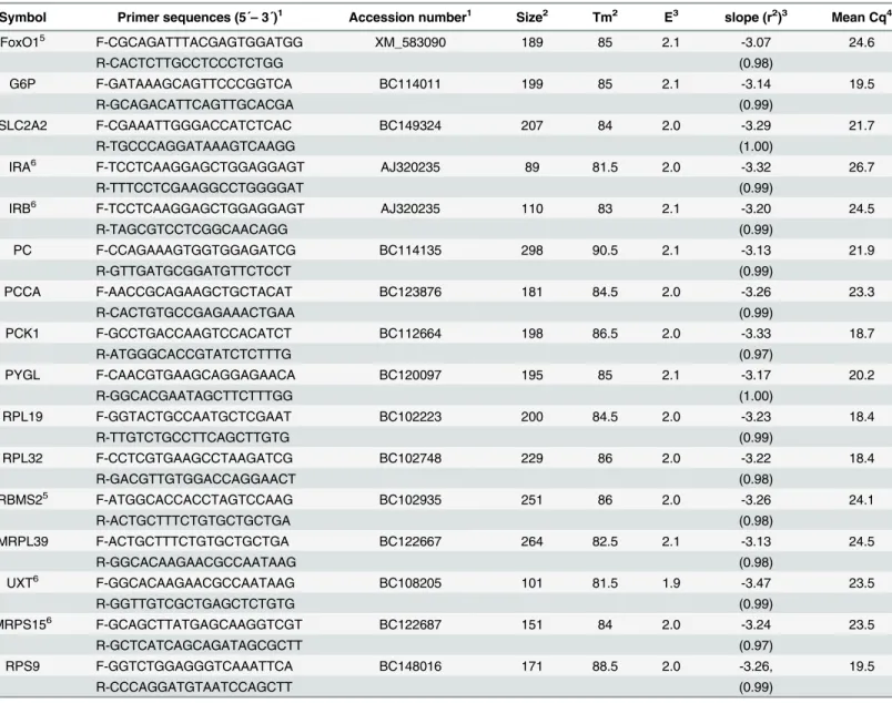
Table 1. Characteristic of primers and the real-time PCR conditions.
Symbol Primer sequences (5´–3´)1 Accession number1 Size2 Tm2 E3 slope (r2)3 Mean Cq4
FoxO15 F-CGCAGATTTACGAGTGGATGG XM_583090 189 85 2.1 -3.07 24.6
R-CACTCTTGCCTCCCTCTGG (0.98)
G6P F-GATAAAGCAGTTCCCGGTCA BC114011 199 85 2.1 -3.14 19.5
R-GCAGACATTCAGTTGCACGA (0.99)
SLC2A2 F-CGAAATTGGGACCATCTCAC BC149324 207 84 2.0 -3.29 21.7
R-TGCCCAGGATAAAGTCAAGG (1.00)
IRA6 F-TCCTCAAGGAGCTGGAGGAGT AJ320235 89 81.5 2.0 -3.32 26.7
R-TTTCCTCGAAGGCCTGGGGAT (0.99)
IRB6 F-TCCTCAAGGAGCTGGAGGAGT AJ320235 110 83 2.1 -3.20 24.5
R-TAGCGTCCTCGGCAACAGG (0.99)
PC F-CCAGAAAGTGGTGGAGATCG BC114135 298 90.5 2.1 -3.13 21.9
R-GTTGATGCGGATGTTCTCCT (0.99)
PCCA F-AACCGCAGAAGCTGCTACAT BC123876 181 84.5 2.0 -3.26 23.3
R-CACTGTGCCGAGAAACTGAA (0.99)
PCK1 F-GCCTGACCAAGTCCACATCT BC112664 198 86.5 2.0 -3.33 18.7
R-ATGGGCACCGTATCTCTTTG (0.97)
PYGL F-CAACGTGAAGCAGGAGAACA BC120097 195 85 2.1 -3.17 20.2
R-GGCACGAATAGCTTCTTTGG (1.00)
RPL19 F-GGTACTGCCAATGCTCGAAT BC102223 200 84.5 2.0 -3.23 18.4
R-TTGTCTGCCTTCAGCTTGTG (0.99)
RPL32 F-CCTCGTGAAGCCTAAGATCG BC102748 229 86 2.0 -3.22 18.4
R-GACGTTGTGGACCAGGAACT (0.98)
RBMS25 F-ATGGCACCACCTAGTCCAAG BC102935 251 86 2.0 -3.26 24.1
R-ACTGCTTTCTGTGCTGCTGA (0.98)
MRPL39 F-ACTGCTTTCTGTGCTGCTGA BC122667 264 82.5 2.1 -3.13 24.5
R-GGCACAAGAACGCCAATAAG (0.98)
UXT6 F-GGCACAAGAACGCCAATAAG BC108205 101 81.5 1.9 -3.47 23.5
R-GGTTGTCGCTGAGCTCTGTG (0.99)
MRPS156 F-GCAGCTTATGAGCAAGGTCGT BC122687 151 84 2.0 -3.24 23.5
R-GCTCATCAGCAGATAGCGCTT (0.97)
RPS9 F-GGTCTGGAGGGTCAAATTCA BC148016 171 88.5 2.0 -3.26, 19.5
R-CCCAGGATGTAATCCAGCTT (0.99)
FoxO1: Forkhead box protein O1, IRA: Insulin receptor isotype A, IRB: Insulin receptor isotype B, SLC2A2: Glucose transporter 2 (solute carrier family 2 (facilitated glucose transporter), member 2), G6P: Glucose-6-phosphatase, PCK1: Cytosolic phosphoenolpyruvate carboxykinase, PCCA: Propionyl-CoA carboxylase, PYGL: Glycogen phosphorylase, RPL19: Ribosomal protein L19: Ribosomal protein L32, RPS9: Ribosomal protein S9, UXT: Ubiquitously expressed transcript, MRPS15: Mitochondrial ribosomal protein S15, RBMS2: RNA binding motif, single strand interacting protein 2, MRPL39: Mitochondrial ribosomal protein L39
1Forward (F-) and reverse (R-) primer sequences and NIH GenBank accession number of template sequences. 2Amplicon size (base pair) and melting temperature (Tm) (°C) of PCR products
3Ampli
fication efficiency (E), slope and r2estimated from standard curves using 5 dilution series of cDNA samples corresponding to 1.25–20 ng RNA. 4Mean threshold cycle (Cq) of all the measured samples at relative
fluorescence unit of 50 and 0.1μl cDNA (5 ng RNA) input. 5Modi
fied assay conditions were used; FoxO1:125/875 nMfor F-/R- primer concentration and 10 sec at 95°C and 15 sec at 60°C for amplification step;
RBMS2: 1μMprimer concentration.
6Primer sequences were published by Neuvians et al. [27] for IRA and IRB, and by Bionaz et al. [28] for UXT and MRPS15.
approximation was used to test fixed effects. The best fit covariant structures were decided from compound symmetry, component variance, and unstructured using AIC for each vari-able. The values at the first sampling day (d-42) were integrated as covariables into the mixed model. The normality of distribution of residues was checked using the Shapiro-Wilk test
(PROC UNIVARIATE). Pearson’s correlation analysis (PROC CORR) was performed for the
correlations for tFoxO1, pFoxO1, mRNA expression of FoxO1, and for the ratio of pFoxO1/ tFoxO1 with all the other variables at each sampling day. The serum NEFA concentration, DMI, and pFoxO1/tFoxO1 were included in the correlation analysis only because the data had
already been published (NEFA and DMI) [20,21] or no significant effect of fixed factors was
found (pFoxO1/tFoxO1). Levels of significance and of relevant trend were set at P<0.05 and
0.1, respectively in the factorial analysis. In the correlation analysis, the level of significance was
set at P0.01.
Results
Serum insulin concentration
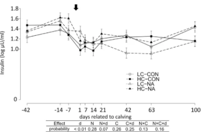
Serum insulin concentrations were significantly affected by time (Fig 1). The serum insulin
concentration increased from d-42 to d-7. At d1, the serum insulin concentration decreased and maintained the lowest levels in the whole experimental period until d14. The insulin level recovered gradually until d100 to the level at d-42.
Total protein expression (tFoxO1), extent of phosphorylation at serine
256 (pFoxO1) and mRNA expression of FoxO1
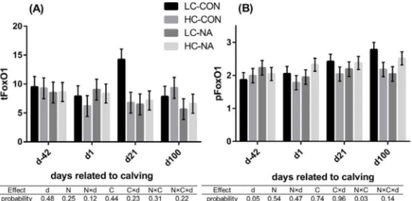
The results for the analysis of FoxO1 are shown inFig 2for tFoxO1 and pFoxO1 and in
Table 2for mRNA expression of FoxO1.The total proteinand themRNAexpression of FoxO1 were neither affected by time nor by the diets. An interaction between NA and
concen-trate levels was observed in thepFoxO1. The high-concentrate diet and NA supplementation
down-regulated the pFoxO1 when they affected separately as a single factor. However, the pFoxO1 in HC-NA was almost at the same level as that in LC-CON (LC-CON,
HC-NA>LC-NA, HC-CON).
Fig 1. Serum insulin concentration (Log10 InsulinμU/ml).Data are shown in least squares means (LSM) ±standard errors of each experimental group and sampling day. The arrow above indicate the day of calving.
The results of type 3 test for the effects of time and diets are shown in the table under the diagrams. LC-CON, HC-CON, LC-NA, HC-NA:“CON or NA”: dietary supplement of nicotinic acid (0 or 24 g/d) from d-42 to d24, “LC or HC”: 30 or 60% of concentrate proportion in the diet from d-42 to d0, increase in concentrate proportion in the diet after calving from 30 to 50% within 16 or 24 days. d: Days related to calving, N: Nicotinic acid, C: Concentrate proportion in the diet, N×d, C×d, N×C, N×C×d: interaction effect of d, N, C.
Relative quantities of mRNA of genes associated with hepatic glucose
production
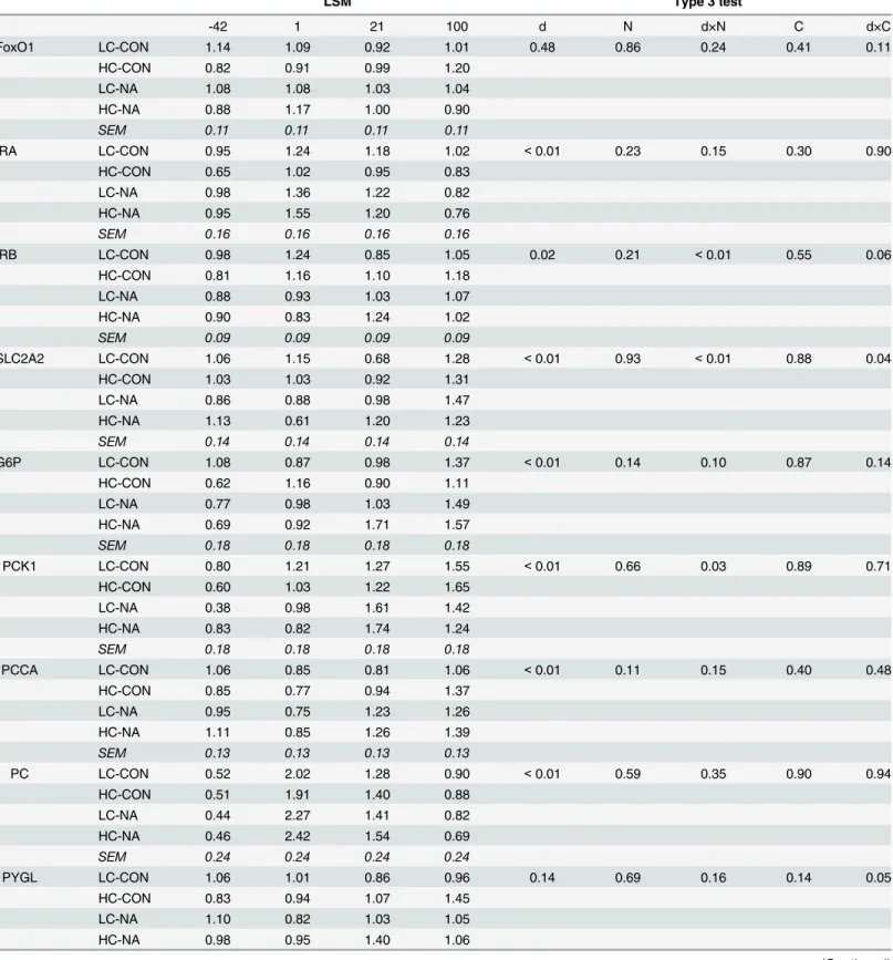
In the mRNA expression of IRB, SLC2A2, and PCK1A, an interaction of d × NA was found. For SLC2A2 and IRB, the relative quantities of mRNA at d1 was lower in NA groups compared
with the CON groups (Table 2). Moreover, for SLC2A2 and PCK1, the relative quantities of
mRNA were higher at d21 in NA groups compared with in CON groups. Similar up-regulation by NA at d21 was found for G6P, which was reflected in a trend effect of the d × NA interac-tion. Significant d × concentrate interaction effects were found in SLC2A2, IRB, and PYGL. These interaction effects were caused by the lower relative quantities of mRNA at d21 in HC groups compared with in LC groups for the affected genes SLC2A2, IRB and PLGL.
In the mRNA expression of IRA, G6P, PCCA and PC, only time had a significant effect. The relative quantities of mRNA of IRA and PC were up-regulated at d1 and down-regulated at
d21 and d100 (d-42, d100<d21<d1). A continuous up-regulation was observed in G6P
(d-42<d1<d21<d100). The relative quantity of mRNA of PCCA was down-regulated at d1
and then up-regulated at d21 and d100 (d1<d21, d-42<d100).
Correlation analysis
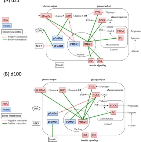
No significant correlation was found atd-42 and d1(data not shown). Atd21 the pFoxO1
(protein amount) correlated positively with DMI (0.70, P<0.001). The mRNA expression of
FoxO1correlated positively with mRNA expression of SLC2A2 (0.6, P = 0.004), IRB (0.75,
P<0.001), PC (0.59, P = 0.005). Atd100thetFoxO1(protein amount) correlated positively
with mRNA expression of PCK1 (0.64, P = 0.002),the pFoxO1(protein amount) correlated
positively with insulin (0.63, P = 0.004). The mRNA expression ofFoxO1correlated positively
with mRNA expression of SLC2A2 (0.63, P = 0.004), PCK1 (0.73, P<0.001), G6P (0.61,
P = 0.008), and PYGL (0.60, P = 0.01). Theratio of pFoxO1/tFoxO1(protein amount)
corre-lated negatively with mRNA expression of PCK1 (-0.53, P = 0.01). Graphical demonstrations
of the correlation at d21 and at d100 are presented inFig 3.
Fig 2. FoxO1 total protein expression (tFoxO1) (A) and the extent of phosphorylation of FoxO1 at serine 256 (pFoxO1) (B) in liver of cows.Data are shown in least squares means with standard errors in each group and at each time point. The results of type 3 test for the effects of time and diets are shown in the table under the diagrams. LC-CON (n = 5), HC-CON (n = 5), LC-NA (n = 5), HC-NA (n = 6):“CON or NA”: Dietary supplement of nicotinic acid (0 or 24 g/d) from d-42 to d24,“LC or HC”: 30 or 60% of concentrate proportion in the diet from d-42 to d0, increase in concentrate proportion in the diet after calving from 30 to 50% within 16 or 24 days. d: Days related to calving, N: Nicotinic acid, C: Concentrate proportion in the diet, d×N, d×C, N×C, d×N×C: Interaction effects of d, N, C, tFoxO1: Total protein expression of forkhead box protein O1, pFoxO1: Extent of phosphorylation of FoxO1 at serine 256.
Table 2. Effects of diet on the relative quantity of mRNA of investigated genes.
LSM Type 3 test
-42 1 21 100 d N d×N C d×C
FoxO1 LC-CON 1.14 1.09 0.92 1.01 0.48 0.86 0.24 0.41 0.11
HC-CON 0.82 0.91 0.99 1.20
LC-NA 1.08 1.08 1.03 1.04
HC-NA 0.88 1.17 1.00 0.90
SEM 0.11 0.11 0.11 0.11
IRA LC-CON 0.95 1.24 1.18 1.02 <0.01 0.23 0.15 0.30 0.90
HC-CON 0.65 1.02 0.95 0.83
LC-NA 0.98 1.36 1.22 0.82
HC-NA 0.95 1.55 1.20 0.76
SEM 0.16 0.16 0.16 0.16
IRB LC-CON 0.98 1.24 0.85 1.05 0.02 0.21 <0.01 0.55 0.06
HC-CON 0.81 1.16 1.10 1.18
LC-NA 0.88 0.93 1.03 1.07
HC-NA 0.90 0.83 1.24 1.02
SEM 0.09 0.09 0.09 0.09
SLC2A2 LC-CON 1.06 1.15 0.68 1.28 <0.01 0.93 <0.01 0.88 0.04
HC-CON 1.03 1.03 0.92 1.31
LC-NA 0.86 0.88 0.98 1.47
HC-NA 1.13 0.61 1.20 1.23
SEM 0.14 0.14 0.14 0.14
G6P LC-CON 1.08 0.87 0.98 1.37 <0.01 0.14 0.10 0.87 0.14
HC-CON 0.62 1.16 0.90 1.11
LC-NA 0.77 0.98 1.03 1.49
HC-NA 0.69 0.92 1.71 1.57
SEM 0.18 0.18 0.18 0.18
PCK1 LC-CON 0.80 1.21 1.27 1.55 <0.01 0.66 0.03 0.89 0.71
HC-CON 0.60 1.03 1.22 1.65
LC-NA 0.38 0.98 1.61 1.42
HC-NA 0.83 0.82 1.74 1.24
SEM 0.18 0.18 0.18 0.18
PCCA LC-CON 1.06 0.85 0.81 1.06 <0.01 0.11 0.15 0.40 0.48
HC-CON 0.85 0.77 0.94 1.37
LC-NA 0.95 0.75 1.23 1.26
HC-NA 1.11 0.85 1.26 1.39
SEM 0.13 0.13 0.13 0.13
PC LC-CON 0.52 2.02 1.28 0.90 <0.01 0.59 0.35 0.90 0.94
HC-CON 0.51 1.91 1.40 0.88
LC-NA 0.44 2.27 1.41 0.82
HC-NA 0.46 2.42 1.54 0.69
SEM 0.24 0.24 0.24 0.24
PYGL LC-CON 1.06 1.01 0.86 0.96 0.14 0.69 0.16 0.14 0.05
HC-CON 0.83 0.94 1.07 1.45
LC-NA 1.10 0.82 1.03 1.05
HC-NA 0.98 0.95 1.40 1.06
Discussion
The objective of this study was to assess the role of FoxO1 in HGP in dairy cows during the transition period. Therefore, we aimed to find indicative associations between FoxO1 expres-sion, the extent of phosphorylation of FoxO1 at serine 256, serum insulin concentration and expression of mRNA of genes associated with HGP as affected by the feeding regimen. To reveal associations between FoxO1 and metabolic status, DMI and serum NEFA concentration were also included in the correlation analysis.
We were able to show that FoxO1 mRNA and protein expression were constant during the transition period and were not affected by diet. In the extent of phosphorylation of FoxO1, an interaction of NA × concentrate was found. However, no corresponding NA × concentrate interaction effect was found in serum insulin concentration and mRNA expression of genes involved in HGP. In the correlation analysis, different correlation patterns were observed at d21 and d100.
FoxO1 expression and phosphorylation at serine 256
FoxO1 is a main mediator of effect of insulin to inhibit HGP in monogastric species [30,31].
FoxO1 has three Akt phosphorylation sites as targets of insulin signaling. Among three phos-phorylation sites, the phosphos-phorylation at serine 256 is needed for the subsequent
phosphoryla-tion of the other two sites so that it acts as a gatekeeper [32,33]. Liver-specific knockout of
FoxO1 led to more than 50% down-regulation of the transcription of G6P and PCK1 in hyper-glycemic mice. This down-regulation of G6P and PCK1 was accompanied by reducing blood
glucose level in these mice [34,35]. As to effects of diet on FoxO1 expression, Grala et al. [5]
observed ×1.2 up-regulation of FoxO1 mRNA together with up-regulations of mRNA of other gluconeogenic enzymes in the liver in dairy cows fed a restricted diet for three weeks in early lactation compared with control cows.
We observed lower insulin concentrations as well as an up-regulation of mRNA expression of several of the gluconeogenic enzymes after calving (discussed below). These results indicated an increase of HGP after calving. However, the glucose flux in the liver was not measured in this study. An up-regulation of FoxO1 and/or down-regulation of the pFoxO1 were expected. However, the amount of FoxO1 protein and mRNA, as well as that of pFoxO1 did not differ between the sampling days. This could suggest that FoxO1 expression and pFoxO1 most likely did not account for the up-regulation of gluconeogenic enzymes after calving. Similarly, Zachut
et al. [36] observed an almost equal extent of the phosphorylation of hepatic insulin receptor
Table 2. (Continued)
LSM Type 3 test
SEM 0.13 0.13 0.13 0.13
LC-CON (n = 5), HC-CON (n = 5), LC-NA (n = 5), HC-NA (n = 6):“CON or NA”: Dietary supplement of nicotinic acid (0 or 24 g/d) from d-42 to d24,“LC or HC”: 30 or 60% of concentrate proportion in the diet from d-42 to d0, increase in concentrate proportion in the diet after calving from 30 to 50% within 16 or 24 days. d: Days related to calving, N: dietary supplement of nicotinic acid, C: Concentrate proportion in the diet, d×N, d×C: Interaction effects of d, N and C. Interaction effects of N×C, d×N×C were not significant for all the variables (not shown). LSM: Least squares means, SEM: Pooled standard error of mean, FoxO1: Forkhead box protein O1, IRA: Insulin receptor isotype A, IRB: Insulin receptor isotype B, SLC2A2: Glucose transporter 2 (Solute Carrier Family 2 (Facilitated Glucose Transporter), Member 2), G6P: Glucose-6-phosphatase, PCK1: Cytosolic phosphoenolpyruvate carboxykinase, PCCA: Propionyl-CoA carboxylase, PYGL: Glycogen phosphorylase
and Akt by insulin before and after calving in high-yielding Holstein dairy cows. This confirms that phosphorylation of FoxO1 postpartum did not change in this study.
Regarding the effects of the diet, we assumed that NA promotes the HGP by up-regulating the FoxO1 as well as by inhibiting the phosphorylation of FoxO1 by insulin. Moreover, we assumed that there are interactions between concentrate intake before calving and NA supple-mentation. However, results of analysis in FoxO1 indicated that there appeared to be no direct relationship between the dietary effects found in FoxO1 analysis and those on the other vari-ables. Interestingly, in rats, a NA infusion induced a significantly lower extent of
phosphoryla-tion of insulin receptor and Akt, but not FoxO1 at serine 256 in liver [19].
Fig 3. Correlations between FoxO1-related variables and other investigated variables in the hepatic metabolic pathways related to gluconeogenesis.To demonstrate correlations graphically, the scheme was adopted to one published by Aschenbach et al. [11]. Pearson’s correlation analyses were performed using the data set from d21 (A) and d100 (B) (N = 21 dairy cows for each). Variables with positive correlations were connected by green solid lines and those with negative correlations by red dotted lines. The level of significance was at least P0.01. The metabolites connected by black arrows show the representative
metabolic pathways related to gluconeogenesis (not investigated, OAA: Oxaloacetate, PEP: Phosphoenolpyruvate, Glucose-6-P: Glucose-6-phosphate). The variables other than DMI used in the correlation analysis are shown in boxes (blue, red, white for hepatic protein expression, hepatic mRNA expression, and blood metabolites, respectively). FoxO1: Forkhead box protein O1, tFoxO1: Total protein expression of FoxO1, pFoxO1: Extent of phosphorylation of FoxO1 at serine 256, pFo/tFo: pFoxO1-to-tFoxO1 ratio, PCCA: Propionyl CoA carboxylase A, PC: Pyruvate carboxylase, PCK1: Cytosolic
phosphoenolpyruvate carboxykinase, G6P: Glucose-6-phosphatase, SLC2A2: Glucose transporter 2 (solute carrier family 2 (facilitated glucose transporter), member 2), IRA, IRB: Insulin receptor isoform A and B, PYGL: Glycogen phosphorylase, liver form, DMI: Dry matter intake, NEFA: Serum concentration of non-esterified fatty acid, Insulin: Insulin serum concentration.
Our results could indicate that HGP was not regulated by FoxO1 on the levels of mRNA and protein expression, and of the phosphorylation at serine 256 in dairy cows during the tran-sition period. This interpretation may support the fact that the HGP in dairy cows was
insensi-tive to insulin [6,11]. However, it was also possible that the other posttranslational regulation
mechanisms are of more importance in the FoxO1-mediated regulation on HGP [2]. For
exam-ple, NA supplementation might increase NAD concentration and activate a NAD-dependent deacetylase (Sirtuin-1). The activated Sirtuin-1 could promote the deacetylation of FoxO1 and
thereby increase the transcriptional activity of the FoxO1 [2]. Thus, further studies are needed
on the function of FoxO1 in the regulation of HGP in dairy cows during the transition period.
Serum insulin and hepatic mRNA expression
In the serum insulin concentration and hepatic mRNA expression of genes associated with glu-cose production, time- and diet- related changes were detected. The lower insulin levels shortly
after calving [36,37] and the up-regulation of mRNA of PC, PCK1, and G6P [8–10,36,38] were
in line with the results in other studies. These changes reflected the homeorhetic adaptation of
cows [6]. The pattern of mRNA expression of PCCA was parallel to the changes of DMI
[20,21], as also observed by Graber et al. [38]. As the PCCA is needed to catalyzes the
conver-sion of propionate to succinyl-CoA [11], these time-related changes of PCCA most likely
indi-cated the adaptation of hepatic tissues to an increased proportion of propionate as
gluconeogenic precursor at d21. The increased expression of insulin receptors during the early lactation period was reported previously on both mRNA and protein levels, and was suggested
to be an indicator for negative energy balance [36,39,40].
The mechanisms for the observed NA effects on hepatic mRNA expression are unknown.
Based on studies in monogastric species, Kang et al. [41] proposed that NA could modify the
gene expression by interacting with NA receptor, by changing the profiles of blood metabolites, and by activating FoxO1. In the current study, the NA effect found on the mRNA expression could not be completely explained by the regulation of FoxO1 or by blood metabolites. It might be possible that a part of the NA-induced modification of mRNA expression in the cur-rent study occurred through the activating NA receptors, because bovine liver expressed a
sub-stantial amount of NA receptor [17].
Correlation analysis
Correlations at each sampling day could be interpreted as coordinated changes of two variables for the short-time metabolic adaptation. The fact that the correlation pattern differed among
the sampling days was previously observed [42]. This result indicated the shift of regulation
mechanisms for HGP during the transition period.
Lack of any correlations between FoxO1 and other variables at d-42 and d1 confirmed that the FoxO1 was not involved in the regulation of HGP during this period. In spite of lack of any significant effects on rFoxO1, it correlated positively with the mRNA expressions of the other genes, which were significantly affected by time and diet. This was observed both at d21 and d100, and could reflect the multilevel regulation mechanism of transcriptional activity of FoxO1. The proportional impact of regulation of the FoxO1 activity at the level of mRNA was
probably too small to be detected by factorial analysis. The positive correlation for pFoxO1–
DMI found at d21 could indicate that the FoxO1 in inactive form decreased in cows with energy deficit. However, lack of any other correlations for tFoxO1 and pFoxO1 at d21 could indicate that FoxO1-related regulation was not dominant in the regulation of HGP at d21. In
contrast, the positive correlation for PCK1−tFoxO1 and, although the correlation coefficient
in accordance with the hypothesis. These could indicate that PCK1 was up-regulated by FoxO1 protein, and that this up-regulation was inhibited by insulin-induced phosphorylation of FoxO1.
Altogether, the results in the correlation analysis could suggest that the HGP could be regu-lated by FoxO1 and insulin in dairy cows only in the mid lactation, and not in the period of late pregnancy and early lactation.
Conclusion
The results indicated that the main regulation of HGP did not take place on the levels of mRNA and protein expression of FoxO1 or on the phosphorylation of FoxO1 at serine 256 in dairy cows from d-42 to d21. However, at d100 FoxO1 appeared to be responsible for at least a part of the regulation mechanism of HGP. The less dominance of FoxO1 as a mediator of insu-lin-effect on HGP could support the insensitivity of HGP to the suppressive effect of insulin in dairy cows. However, the possibility was not ruled out that the posttranslational modification of FoxO1 plays a more important role in the regulation of HGP. Therefore, further study is needed to clarify the roles of FoxO1 in the regulation of HGP in dairy cows during the transi-tion period.
Supporting Information
S1 Fig. Signals of total protein of FoxO1 (a), phosphorylated FoxO1 at serine 256 (b) and
negative control (c) in Western Blot analysis.Thirtyμg of reduced and denatured proteins
isolated from liver were transferred to nitrocellulose membrane. Membrane was blocked with 10% skimmed milk-TBST for 1h at room temperature and incubated with primary antibodies diluted at 1:1000 (a) or 1:200 (b) in 5% skimmed milk-TBST or in 5% skimmed milk without primary antibody (c) over night at 4°C, followed by incubation with secondary antibodies diluted at 1:20000 5% skimmed milk-TBST at room temperature for 1h. Arrows indicate the molecular weight of 70 kD which positive signals are supposed to have (a, b). The signals appearing at 100 kD in a and c were considered to be unspecific as indicated by non-primary antibody control (c).
(TIFF)
S2 Fig. Representative signals of total protein of FoxO1 and phosphorylated FoxO1 at
ser-ine 256 (70 kD) of cows from each experimental group and sampling time.Thirtyμg of
reduced and denatured proteins isolated from liver were transferred to nitrocellulose mem-brane. Membrane was blocked with 10% skimmed milk-TBST for 1h at room temperature and incubated with primary antibodies diluted at 1:1000 (tFoxO1) or 1:200 (pFoxO1) in 5% skimmed milk-TBST over night at 4°C, followed by incubation with secondary antibodies diluted at 1:20000 5% skimmed milk-TBST at room temperature for 1h. Signals of beta-actin (45 kD) are presented as internal controls. tFoxO1: total protein of FoxO1, pFoxO1:
phosphor-ylated FoxO1 at serine 256, LC-CON, HC-CON, LC-NA, HC-NA:“CON or NA”: Nicotinic
acid (0 or 24 g/d) from d-42 to d24,“LC or HC”: 30 or 60% of concentrate proportion in the
diet from d-42 to d0, increase of concentrate proportion in the diet after calving from 30 to 50% within 16 or 24 days, control: A control sample for inter membrane controls, d: Days related to calving.
(TIFF)
S3 Fig. Agarose gel electrophoresis of PCR products.About 7μl of PCR products were
RPS9, 7: RPL32, 8: RPL19, 9: Marker; 10: PC, 11: PCCA, 12: PCK1, 13: G6P, 14: SLC2A2, 15: CPT1 (172 bp, not measured in this study), 16: PYGL, 17: IRB, 18: IRA, 19: FoxO1, 20: Empty. (TIFF)
S1 Table. Results of the sequencing of PCR products and BLAST analysis1.FoxO1:
Fork-head box protein O1, IRA: Insulin receptor isotype A, IRB: Insulin receptor isotype B, SLC2A2: Glucose transporter 2 (Solute Carrier Family 2 (Facilitated Glucose Transporter), Member 2), G6P: Glucose-6-phosphatase, PCK1: Cytosolic phosphoenolpyruvate carboxykinase, PCCA: Propionyl-CoA carboxylase, PYGL: Glycogen phosphorylase, RPL19: Ribosomal protein L19: Ribosomal protein L32, RPS9: Ribosomal protein S9, UXT: Ubiquitously expressed transcript, MRPS15: Mitochondrial ribosomal protein S15, RBMS2: RNA binding motif, single strand
interacting protein 2, MRPL39: Mitochondrial ribosomal protein L39.1Analysis performed
using Basic Local Alignment Search Tool“BLASTN 2.2.31+”, an online program offered by
National Center for Biotechnology Information (Bethesda, USA) [24] and the database Bos
taurus Annotation Release 104 RNAs. (PDF)
Acknowledgments
The authors would like to acknowledge the co-workers of the Institute of Animal Nutrition of the Friedlich-Loeffler-Institute (FLI) in Braunschweig, Germany for performing the experi-ment. Moreover, we wish to thank Dr. M. Piechotta from the Clinic for Cattle, University of Veterinary Medicine Hannover for performing the insulin assay and Mrs Kathrin Hansen, Department of Physiology, University of Veterinary Medicine Hannover, for performing the Western Blot experiments.
Author Contributions
Conceived and designed the experiments: JR SD KH. Performed the experiments: AK LL RT UM SD JR KH. Analyzed the data: AK KH RT UM SD. Contributed reagents/materials/analy-sis tools: KH UM JR SD. Wrote the paper: AK KH UM SD JR.
References
1. Wang Y, Zhou Y, Graves DT. FOXO Transcription Factors: Their Clinical Significance and Regulation. BioMed Res Int. 2014; 2014: 1–13. doi:10.1155/2014/925350
2. Calnan DR, Brunet A. The FoxO code. Oncogene. 2008; 27: 2276–2288. doi:10.1038/onc.2008.21 PMID:18391970
3. Jitrapakdee S. Transcription factors and coactivators controlling nutrient and hormonal regulation of hepatic gluconeogenesis. Int J Biochem Cell Biol. 2012; 44: 33–45. doi:10.1016/j.biocel.2011.10.001 PMID:22004992
4. Oh K-J, Han H-S, Kim M-J, Koo S-H. CREB and FoxO1: two transcription factors for the regulation of hepatic gluconeogenesis. BMB Rep. 2013; 46: 567–574. doi:10.5483/BMBRep.2013.46.12.248PMID: 24238363
5. Grala TM, Kay JK, Phyn CVC, Bionaz M, Walker CG, Rius AG, et al. Reducing milking frequency during nutrient restriction has no effect on the hepatic transcriptome of lactating dairy cattle. Physiol Geno-mics. 2013; 45: 1157–1167. doi:10.1152/physiolgenomics.00134.2013PMID:24104205
6. Hayirli A. The role of exogenous insulin in the complex of hepatic lipidosis and ketosis associated with insulin resistance phenomenon in postpartum dairy cattle. Vet Res Commun. 2006; 30: 749–774. doi: 10.1007/s11259-006-3320-6PMID:17004039
8. Agca C, Greenfield RB, Hartwell JR, Donkin SS. Cloning and characterization of bovine cytosolic and mitochondrial PEPCK during transition to lactation. Physiol Genomics. 2002; 11: 53–63. doi:10.1152/ physiolgenomics.00108.2001PMID:12388798
9. Greenfield RB, Cecava MJ, Donkin SS. Changes in mRNA expression for gluconeogenic enzymes in liver of dairy cattle during the transition to lactation. J Dairy Sci. 2000; 83: 1228–1236. doi:10.3168/jds. S0022-0302(00)74989-7PMID:10877388
10. Loor JJ, Dann HM, Guretzky NAJ, Everts RE, Oliveira R, Green CA, et al. Plane of nutrition prepartum alters hepatic gene expression and function in dairy cows as assessed by longitudinal transcript and metabolic profiling. Physiol Genomics. 2006; 27: 29–41. doi:10.1152/physiolgenomics.00036.2006 PMID:16757553
11. Aschenbach JR, Kristensen NB, Donkin SS, Hammon HM, Penner GB. Gluconeogenesis in dairy cows: The secret of making sweet milk from sour dough. IUBMB Life. 2010; 62: 869–877. doi:10.1002/ iub.400PMID:21171012
12. Aschemann M, Lebzien P, Hüther L, Südekum K-H, Dänicke S. Effect of niacin supplementation on rumen fermentation characteristics and nutrient flow at the duodenum in lactating dairy cows fed a diet with a negative rumen nitrogen balance. Arch Anim Nutr. 2012; 66: 303–318. doi:10.1080/1745039X. 2012.697353PMID:22924176
13. Niehoff I-D, Hüther L, Lebzien P. Niacin for dairy cattle: a review. Br J Nutr. 2009; 101: 5–19. doi:10. 1017/S0007114508043377PMID:18702847
14. Kenéz A, Locher L, Rehage J, Dänicke S, Huber K. Agonists of the G protein-coupled receptor 109A-mediated pathway promote antilipolysis by reducing serine residue 563 phosphorylation of hormone-sensitive lipase in bovine adipose tissue explants. J Dairy Sci. 2014; 97: 3626–34. doi:10.3168/jds. 2013-7662PMID:24704242
15. Pires JAA, Pescara JB, Grummer RR. Reduction of plasma NEFA concentration by nicotinic acid enhances the response to insulin in feed-restricted Holstein cows. J Dairy Sci. 2007; 90: 4635–4642. doi:10.3168/jds.2007-0146PMID:17881684
16. Titgemeyer EC, Spivey KS, Mamedova LK, Bradford BJ. Effects of pharmacological amounts of nico-tinic acid on lipolysis and feed intake in cattle. Int J Dairy Sci. 2011; 6: 134–141. doi:10.3923/ijds.2011. 134.141
17. Titgemeyer EC, Mamedova LK, Spivey KS, Farney JK, Bradford BJ. An unusual distribution of the nia-cin receptor in cattle. J Dairy Sci. 2011; 94: 4962–4967. doi:10.3168/jds.2011-4193PMID:21943747
18. Khan M, Couturier A, Kubens JF, Most E, Mooren F-C, Krüger K, et al. Niacin supplementation induces type II to type I muscle fiber transition in skeletal muscle of sheep. Acta Vet Scand. 2013; 55: 85. doi: 10.1186/1751-0147-55-85PMID:24267720
19. Choi S, Yoon H, Oh K-S, Oh YT, Kim YI, Kang I, et al. Widespread effects of nicotinic acid on gene expression in insulin-sensitive tissues: implications for unwanted effects of nicotinic acid treatment. Metabolism. 2011; 60: 134–144. doi:10.1016/j.metabol.2010.02.013PMID:20303128
20. Kenéz Á, Tienken R, Locher L, Meyer U, Rizk A, Rehage J, et al. Changes in lipid metabolism andβ -adrenergic response of adipose tissues of periparturient dairy cows affected by an energy-dense diet and nicotinic acid supplementation. J Anim Sci. 2015; 93: 4012–4022. doi:10.2527/jas.2014-8833 PMID:26440181
21. Tienken R, Kersten S, Frahm J, Meyer U, Locher L, Rehage J, et al. Effects of an energy-dense diet and nicotinic acid supplementation on production and metabolic variables of primiparous or multiparous cows in periparturient period. Arch Anim Nutr. 2015; 69: 319–339. doi:10.1080/1745039X.2015. 1073002PMID:26305382
22. Laemmli UK. Cleavage of structural proteins during the assembly of the head of bacteriophage T4. Nature. 1970; 227: 680–685. PMID:5432063
23. Rosen S, Skaletsky HJ. Primer3 on the WWW for general users and for biologist programmers. Krawetz S, Misener S (eds) Bioinformatics Methods and Protocols: Methods in Molecular Biology. Totowa, NJ: Humana Press; 1999. pp. 365–386.
24. Zhang Z, Schwartz S, Wagner L, Miller W. A greedy algorithm for aligning DNA sequences. J Comput Biol J Comput Mol Cell Biol. 2000; 7: 203–214. doi:10.1089/10665270050081478
25. Vandesompele J, De Preter K, Pattyn F, Poppe B, Van Roy N, De Paepe A, et al. Accurate normaliza-tion of real-time quantitative RT-PCR data by geometric averaging of multiple internal control genes. Genome Biol. 2002; 3: RESEARCH0034. PMID:12184808
27. Neuvians TP, Pfaffl MW, Berisha B, Schams D. The mRNA expression of insulin receptor isoforms (IR-A and IR-B) and IGFR-2 in the bovine corpus luteum during the estrous cycle, pregnancy, and induced luteolysis. Endocrine. 2003; 22: 93–100. doi:10.1385/ENDO:22:2:93PMID:14665712
28. Bionaz M, Loor JJ. Identification of reference genes for quantitative real-time PCR in the bovine mam-mary gland during the lactation cycle. Physiol Genomics. 2007; 29: 312–319. doi:10.1152/
physiolgenomics.00223.2006PMID:17284669
29. Littell RC, Henry PR, Ammerman CB. Statistical analysis of repeated measures data using SAS proce-dures. J Anim Sci. 1998; 76: 1216–1231. PMID:9581947
30. Kousteni S. FoxO1, the transcriptional chief of staff of energy metabolism. Bone. 2012; 50: 437–443. doi:10.1016/j.bone.2011.06.034PMID:21816244
31. Guo S. Molecular basis of insulin resistance: the role of IRS and Foxo1 in the control of diabetes melli-tus and its complications. Drug Discov Today Dis Mech. 2013; 10: e27–e33. doi:10.1016/j.ddmec. 2013.06.003PMID:24015152
32. Barthel A, Schmoll D, Unterman TG. FoxO proteins in insulin action and metabolism. Trends Endocrinol Metab. 2005; 16: 183–189. doi:10.1016/j.tem.2005.03.010PMID:15860415
33. Nakae J, Oki M, Cao Y. The FoxO transcription factors and metabolic regulation. FEBS Lett. 2008; 582: 54–67. doi:10.1016/j.febslet.2007.11.025PMID:18022395
34. Matsumoto M, Pocai A, Rossetti L, DePinho RA, Accili D. Impaired regulation of hepatic glucose pro-duction in mice lacking the forkhead transcription factor Foxo1 in liver. Cell Metab. 2007; 6: 208–216. doi:10.1016/j.cmet.2007.08.006PMID:17767907
35. Samuel VT, Choi CS, Phillips TG, Romanelli AJ, Geisler JG, Bhanot S, et al. Targeting foxo1 in mice using antisense oligonucleotide improves hepatic and peripheral insulin action. Diabetes. 2006; 55: 2042–2050. doi:10.2337/db05-0705PMID:16804074
36. Zachut M, Honig H, Striem S, Zick Y, Boura-Halfon S, Moallem U. Periparturient dairy cows do not exhibit hepatic insulin resistance, yet adipose-specific insulin resistance occurs in cows prone to high weight loss. J Dairy Sci. 2013; 96: 5656–5669. doi:10.3168/jds.2012-6142PMID:23871373
37. Schulz K, Frahm J, Meyer U, Kersten S, Reiche D, Rehage J, et al. Effects of prepartal body condition score and peripartal energy supply of dairy cows on postpartal lipolysis, energy balance and ketogene-sis: an animal model to investigate subclinical ketosis. J Dairy Res. 2014; 81: 257–266. doi:10.1017/ S0022029914000107PMID:24594287
38. Graber M, Kohler S, Kaufmann T, Doherr MG, Bruckmaier RM, van Dorland HA. A field study on char-acteristics and diversity of gene expression in the liver of dairy cows during the transition period. J Dairy Sci. 2010; 93: 5200–5215. doi:10.3168/jds.2010-3265PMID:20965335
39. Mendonça LGD, Litherland NB, Lucy MC, Keisler DH, Ballou MA, Hansen LB, et al. Comparison of innate immune responses and somatotropic axis components of Holstein and Montbéliarde-sired crossbred dairy cows during the transition period. J Dairy Sci. 2013; 96: 3588–3598. doi:10.3168/jds. 2012-5804PMID:23548296
40. Gross J, van Dorland HA, Schwarz FJ, Bruckmaier RM. Endocrine changes and liver mRNA abun-dance of somatotropic axis and insulin system constituents during negative energy balance at different stages of lactation in dairy cows. J Dairy Sci. 2011; 94: 3484–3494. doi:10.3168/jds.2011-4251PMID: 21700035
41. Kang I, Kim S-W, Youn JH. Effects of nicotinic acid on gene expression: potential mechanisms and implications for wanted and unwanted effects of the lipid-lowering drug. J Clin Endocrinol Metab. 2011; 96: 3048–3055. doi:10.1210/jc.2011-1104PMID:21816780