The article was published by Academy of Chemistry of Globe Publications www.acgpubs.org/RNP © Published 3/15/2013 EISSN:1307-6167
Rec. Nat. Prod
. 7:2 (2013) 80-85
Isolation and Structure Elucidation of a New Triterpenoid from
Prunus cerasoides D. Don
Liaqat Ali
*1,2and Farzana Shaheen
11
HEJ Research Institute of Chemistry, International Center for Chemical and Biological Sciences, University of Karachi, Karachi-75270, Pakistan
2
Department of Biological Sciences and Chemistry, University of Nizwa, Birkat Al-Mauz, Nizwa-616, Oman
(Received October 28, 2012; Revised December 4, 2012; Accepted January 17, 2013)
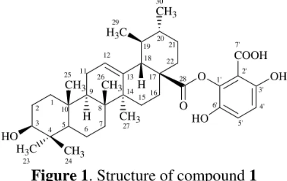
Abstract: One new metabolite, prunol (1), belonging to the pentacyclic triterpenoid skeleton was purified from the dichloromethane soluble fraction of the crude ethanolic extract of Prunus cerasoides D. Don. The structure elucidation was accomplished on the basis of one dimensional 1H and 13C NMR and two dimensional HMQC, HMBC, and COSY experiments. The molecular mass was determined by HRFAB-MS, while the major fragments were observed in the EI-MS. The comparative analysis of the NMR spectral data with the known analogues and the NOESY experiments were helpful in assigning the stereo centers in the molecule.
Keywords: Pentacyclic triterpenoids; Prunus cerasoides; ethanolic extract; NMR spectral data.
1. Introduction
Prunus is a genus of trees and shrubs, including the plums, cherries, peaches, apricots, and almonds. It is traditionally placed within the rose family, rosaceae [1]. The plant Prunus cerasoides D. Don is found in East Asia and commonly known as the Wild Himalayan Cherry. Its range extends in the Himalayas from Himachal Pradesh in India to southwest China and Burma. It grows in the forest from 1200 to 2400 meters above sea level [2–3]. The fruits and the leaves give a dark green dye. Seeds can be used in the manufacture of necklaces and rosaries. The wood is hard, strong, durable and aromatic, and branches are used as walking sticks. The branches and stems of the plant are also used for the treatment of gravel, kidney stones, asthma, thirst, leucoderma, liprosy, vomiting, and as antipyretic and refrigerant [4–5]. Because of their considerable value as both food and ornamental plants, many Prunus species have been introduced to the various parts of the World. All the members of the genus contain amygdalin and prunasin, which break down in water to form hydrocyanic acid (cyanide or prussic acid). In small amounts this exceedingly poisonous compound has been shown to stimulate respiration, and improve digestion. In excess, however, it can cause respiratory failure and even death [1]. Previously, some flavonoids and steroidal derivatives have been reported from P. cerasoides [2–3]. In the present paper, we report the isolation and structure elucidation of a new derivative of pentacyclic triterpenoids, named prunol (1).
*
2. Materials and Methods
2.1. General
Optical rotations were measured on a JASCO DIP 360 polarimeter. IR spectrum was recorded on a Bruker VECTOR 22 spectrophotometer. EI-MS and HRFAB-MS were recorded on mass spectrometers JEOL JMS HX 110. 1H and 13C NMR spectra were recorded on Bruker NMR spectrometers operating at 600 MHz (150 MHz for 13C). The chemical shift values were reported in ppm (δ) units and the coupling constants (J) were given in Hz. For TLC, pre coated aluminium sheets (silica gel 60F-254, E. Merck) were used. UV at 254 and 366 nm were used for the visualization of the TLC plates. The TLC plates were then sprayed with the cerric sulphate reagent for the visualization of the UV inactive compounds. 5-10 % MeOH:DCM was used as solvent system.
2.2. Plant Material
The plant Prunus cerasoides D. Don (Rosaceae), was collected from Swat (Pakistan) in the year 2005, and was identified by Mr. Mehboob ur Rahman, Assistnat Professor, Department of Botany, Govt Jehanzeb Post Graduate Ccollege, Saidu Sharif, Swat, K.P.K., Pakistan. A voucher specimen (CP-14) has been deposited at the herbarium of the Department.
2.3. Extraction and Isolation
The air dried and powdered aerial parts (P. cerasoides 1.6 kg) were extracted exhaustively with 80% ethanol at room temperature. The filtrate was evaporated in vacuum to yield 150 g of the residue. The residue was partitioned in different solvents on the basis of increasing polarity to get n -hexane (12 g), dichloromethane (21 g), ethyl acetate (40 g), and n-butanol (25 g) extracts.
O HO
O
COOH
OH
HO
CH3 CH3 CH3
H3C
CH3
CH3
H3C 1 2 3
12 11
10 9
8 7 6 5 4
19 18
17 16 13
15 14
20 21 22
23 24 25 26
27
28 29
30
1' 2'
3' 4' 5' 6'
7'
H
H
Figure 1. Structure of compound 1
The dichloromethane (DCM) soluble fraction was loaded on silica gel column and eluted with gradually increasing polarity solvents; sixteen sub-fractions (SF 1 ton SF 16) were obtained. Sub-fractions collected at 5-7 % MeOH/DCM (SF 11 and SF 12) were again subjected to repeated column chromatography over silica gel by using 5 % MeOH/DCM, which afforded new compound 1 (Figure 1) along with some other semi-pure fractions.
2.4. Prunol (1)
White amorphous solid: [α]D30 -56 (c 0.05, CH3OH); UV (CH3OH) λmax (log ε): 276 nm (ε =
3.44), 215 nm (ε = 2.67) nm; IR (KBr): νmax 3415, 2922, 1705, 1635, 1450, 1395 cm-1. 1H NMR (600
MHz, CD3OD): see Table 1; 13C NMR (CD3OD, 150 MHz): see Table 1; FAB-MS: m/z 609 [M + H]+;
3. Results and Discussion
3.1. Structure elucidation
Compound 1 was isolated as white amorphous powder. The UV spectrum showed the absorption maxima at 276 nm and 215 nm which indicated the presence of aromatic ring in conjugation with a carbonyl group [6]. The IR spectrum showed absorption bands for hydroxyl group (3423 cm-1), benzene ring and aromatic methine (1600 and 1458 cm-1 and 2926 cm-1), and carboxyl group (1690 and 1185 cm-1). The molecular formula of 1 was established as C37H52O7on the basis of
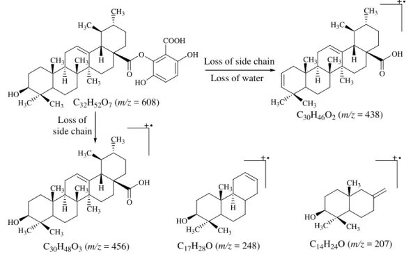
high resolution (+)-FAB-MS (m/z 609.3251 [M + H]+, calcd. 609.3247). The twelve degree of unsaturation in the molecule was attributed to benzene ring (3 + 1), C(12/13) unsaturated pentacyclic skeleton (5 + 1), and two carbonyl groups (2). The other prominent fragment ions in the EI-MS (Figure 2) at m/z 456, 438, 262, 248, 207, 203, and 133 coupled with 1H NMR spectral data (Table 1) indicated compound 1 to be a substituted phenyl ester of a pentacyclic triterpenoid [7].
O HO
O
COOH
OH
HO
CH3
CH3 CH3
H3C CH3
CH3
H3C
H
H
O OH
CH3
CH3 CH3
H3C
CH3
CH3
H3C
H
H
O OH
HO
CH3
CH3 CH3
H3C
CH3
CH3
H3C
H
H
HO
CH3
CH3
H3C
H
Loss of side chain
Loss of side chain
Loss of water
C32H52O7 (m/z = 608)
C30H46O2 (m/z = 438)
C30H48O3 (m/z = 456)
HO
CH3 CH3
H3C
C17H28O (m/z = 248) C14H24O (m/z = 207)
Figure 2. Major mass fragments in compound 1
The 1H NMR spectrum showed the presence of five tertiary methyl singlets and two secondary methyl doublets at δ 0.77, 0.85, 0.94, 0.96, 1.10, and at δ 0.87, 0.95 respectively. This indicated the ursolic acid skeleton of the molecule [8]. The proton geminal to the hydroxyl group H-C(3) was observed at δ 3.14 as a multiplet. The olefinic proton H-C(12) of the ring C appeared at δ 5.21. Two downfield doublets at δ 7.75 and 7.20 (each 1H, d, J = 8.1 Hz) indicated the presence of a substituted phenyl ring having two ortho-coupled protons. The tetra-substituted phenyl ring was further supported through 13C NMR spectrum (Table 1). The substitution pattern of the phenyl ring at C(28) was thus inferred to have two ortho-coupled protons, two OH groups and a carboxyl group. This substituted phenyl moiety with two OH groups and a carboxyl group was further confirmed through mass spectrometry by the presence of fragment ion at m/z 171 in the EI-MS (Figure 2) corresponding to the loss of benzoic acid derivative side chain. The 13C NMR spectrum (BB, and DEPT) of 1 displayed thirty seven signals, including seven methyl (C(23), C(24), C(25), C(26), C(27), C(29), and C(30)), nine methylene, nine methine and twelve quaternary carbons (Table 1). The two downfield quaternary carbons (C(28) and C(7′)) at δ 182.3 and 170.4 along with four quaternary carbons (C(1′) to C(3′) and
C(6′)) at δ 145.5, 123.4, 150.1, and 143.8, respectively, and two methine carbons (C(5′) and C(4′)) at δ
of signals at δ 125.6, and 139.2 in 13C NMR spectrum. The 1H-13C connectivities were determined through HMQC spectrum and the long-range 1H-13C HMBC correlations (Figure 3) were helpful for linking different sub-structures together for the final confirmation of structure 1. The HMBC correlations from H-C(3) to C(1), C(2), C(4), C(23), and C(24), from H-C(9) to C(5), C(8), and C(10), from H-C(12) to C(11), C(13), C(14), and C(18), from H-C(18) to C(13), C(14), C(16), and C(28), and from H-C(22) to C(28), thus indicated the relative positions of these groups in the molecule. The substitution of the phenyl ring was confirmed by the HMBC interactions from H-C(4′) to C(3′), and
C(6′); and from H-C(5′) to C(4′), and C(6′) along with the 1H-1H COSY correlations H-C(4′)↔H-C(5′)
(Figure 3).
The structure of compound
1
closely resembles to that of a reported compound,
basilol [9], with the difference in substitution pattern of the phenyl ring and pentacyclic
skeleton. The reported compound, basilol, is the 4
′-formyl substituted phenyl ester of
oleanolic acid, whereas the new compound
1
is 2-carboxy-3,6-dihydroxy substituted phenyl
ester of ursolic acid.
The relative stereochemistry of the asymmetric centers in the molecule was determined by the NOESY correlations (Figure 3) of H-C(3)↔H-C(23)↔H-C(9) and H-C(25)↔ H-C(26)↔H-C(29) and also on the basis of biogenetic considerations (Figure 4).O HO
O
COOH
OH
HO
CH3 CH3 CH3
H3C CH3
CH3
H3C
H
H
1H
1H COSY 13C HMBC 1H 1
H NOESY
1H
H
H
H H H
H
Figure 3. Important COSY, HMBC, and NOESY interactions in compound 1
Thus on the basis of the above spectral studies the structure of compound
1
was
established as 2
′
-carboxy-3
′
,6
′
-dihydroxyphenyl-3
β
-hydroxyurs-12-en-28-oate (prunol) as a
new constituent from
P. cerasoides
.
3.2 Biogenesis of compound 1
Compound 1 possesses the ursane skeleton belonging to the pentacyclic triterpene system. The pentacyclic triterpene system arises from squalene [10], which is a basic precursor for the most of steroids and terpenoids.
The dammarenyl cation is the transient product of the folding of squalene oxide on to a cyclase enzyme in a chair-chair-chair-boat conformation [11]. This cation undergoes further carbocation promoted cyclizations to form tertiary lupenyl cation with pentacyclic ring system. Ring expansion in the lupenyl cation gives rise to the oleanyl system, which on further hydride migrations and loss of proton converted into α-amyrin (Figure 4). Compound 1 possesses the identical skeleton to that of the
α-amyrin. It may have arises from esterification of ursolic acid (Figure 4), which is the oxidation product of α-amyrin at C(28) [11].
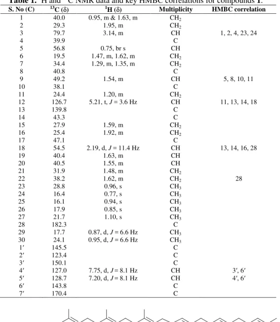
Table 1. 1H and 13C NMR data and key HMBC correlations for compounds 1.
S. No (C) 13
C (δ) 1H (δ) Multiplicity HMBC correlation 1 40.0 0.95, m & 1.63, m CH2
2 29.3 1.95, m CH2
3 79.7 3.14, m CH 1, 2, 4, 23, 24
4 39.9 C
5 56.8 0.75, br s CH
6 19.5 1.47, m, 1.62, m CH2 7 34.4 1.29, m, 1.35, m CH2
8 40.8 C
9 49.2 1.54, m CH 5, 8, 10, 11
10 38.1 C
11 24.4 1.20, m CH2
12 126.7 5.21, t, J = 3.6 Hz CH 11, 13, 14, 18
13 139.8 C
14 43.3 C
15 27.9 1.59, m CH2
16 25.4 1.92, m CH2
17 47.1 C
18 54.5 2.19, d, J = 11.4 Hz CH 13, 14, 16, 28
19 40.4 1.63, m CH
20 40.5 1.55, m CH
21 31.9 1.48, m CH2
22 38.2 1.62, m CH2 28
23 28.8 0.96, s CH3
24 16.4 0.77, s CH3
25 16.1 0.94, s CH3
26 17.9 0.85, s CH3
27 21.7 1.10, s CH3
28 182.3 C
29 17.7 0.87, d, J = 6.6 Hz CH3 30 24.1 0.95, d, J = 6.6 Hz CH3
1′ 145.5 C
2′ 123.4 C
3′ 150.1 C
4′ 127.0 7.75, d, J= 8.1 Hz CH 3′, 6′
5′ 128.7 7.20, d, J= 8.1 Hz CH 4′, 6′
6′ 143.8 C
7′ 170.4 C
O HO O COOH OH HO CH3 CH3 CH3
H3C CH3
CH3
H3C H
H
O OH
CH3 CH3 CH3
H3C CH3
CH3
H3C H
H
Esterification General cyclization scheme involving squalene epoxide
Squalene (A precursor of triterpenes and steroids)
HO
Ursolic Acid Compound 1
Oxidation
CH3
CH3 CH3 CH3
H3C CH3
CH3
H3C H
H
HO
α-amyrin A series of
Wagner Meerwein type rearrangements
CH3 CH3 CH3
CH3
H3C H
HO
H H3C
CH3
H3C
Dammarenyl cation
Acknowledgments
The authors acknowledge the efforts of Dr. Manzoor Ahmad, Assistantt Professor, University of Malakand, Swat, K.P.K., Pakistan, for the collection of the plant. One of us (L.A.) is also thankful to the Higher Education Commision (Pakistan) for providing financial support during the completion of this study.
References
[1] D. Brown (1995). Encyclopaedia of Herbs and their Uses, Dorling Kindersley, London. [2] E. Nasir and S. Ali (1973). Flora of West Pakistan, pp. 362.
[3] T. Rana, V. Chandel, V. Hallan and A. A. Zaidi (2008). Himalayan wild cherry (Prunus cerasoides D. Don): a new host of Apple chlorotic leaf spot virus, Forest Pathol.38, 73-77.
[4] R. N. Chopra, S. L. Nayar and I. C. Chopra (1956). Glossary of Indian Medicinal Plants, CSIR Publication, New Delhi, pp. 204.
[5] K. R. Kirtikar and B. D. Basu (1975). Indian Medicinal Plants vol II, M/S. Periodical Experts, New Delhi, pp. 959.
[6] G. Toker, M. Memisoglu, E. Yesilida and M. Aslan (2004). Main flavonoids of Tilia argentea DESF. Ex DC. Leaves, Turk. J. Chem.28, 745-750.
[7] J. Hussain, N. Rehman, H. Hussain, A. Al-Harrasi, L. Ali, T. S. Rizvi, M. Ahmad and Mehjabeen (2012). Analgesic, anti-inflammatory, and CNS depressant activities of new constituents of Nepeta clarkei, Fitoterapia83, 593-598.
[8] A. Numata, P. Yang, C. Takahashi, R. Fujiki, M. Nabae and E. Fujita (1989). Cytotoxic triterpenes from a Chinese medicine, goreishi, Chem. Pharm. Bull. 37, 648-651.
[9] B. S. Siddiqui, H. Aslam, S. T. Ali, S. Begum and N. Khatoon (2007). Two new triterpenoids and a steroidal glycoside from the aerial parts of Ocimum basilicum, Chem. Pharm. Bull. 55, 516-519. [10] G. D. Brown (1998). The biosynthesis of steroids and triterpenoids, Nat. Prod. Rep.15, 653-696. [11] P. M. Dewick (2001). Medicinal Natural Products; A Biosynthetic Approach, John Wiley & Sons,
England, pp. 212-236.