Original Research
Alternative Intrabladder Manometry Technique for the Indirect
Measurement of Intra-abdominal Pressure in Horses
Paulo Aléscio Canola DVM, MS
a, João Henrique Perotta DVM, MS
a,
Deborah Penteado Martins Dias DVM, PhD
a, Júlio Carlos Canola DVM, PhD
b,
Philip J. Johnson DVM, MS, MRCVS, DACVIM
c, Carlos Augusto Araújo Valadão DVM, PhD
b aVeterinary Surgery Graduate Program, São Paulo State University, School of Agrarian Sciences and Veterinary Medicine, Jaboticabal, São Paulo, Brazil bDepartment of Veterinary Clinics and Surgery, São Paulo State University, School of Agrarian Sciences and Veterinary Medicine, Jaboticabal, São Paulo, Brazil cDepartment of Veterinary Medicine and Surgery, College of Veterinary Medicine, University of Missouri, Columbia, MOa r t i c l e
i n f o
Article history:
Received 30 May 2011 Received in revised form 09 August 2011 Accepted 23 August 2011 Available online 9 February 2012
Keywords:
Equine
Intra-abdominal hypertension Abdominal Compartment syndrome Intrabladder catheter
Intravesical pressure
a b s t r a c t
Although indirect methods for estimating intra-abdominal pressure (IAP) have been extensively studied in humans, mainly for identification of IAP increments in critical care unit patients (life-threatening condition), little work has been performed in veterinary medicine, especially in horses. The standard method of acquisition of intrabladder pressure (IBP) in human subjects (fluid-filled system with no balloon distention) has been previously evaluated in horses with an unfavorable outcome. Therefore, we reex-amined the previously performed method of IBP investigation in horses by using an air-filled intrabladder manometry system in female horses. Intrabladder pressure was estimated by air-filled balloon manometry in seven healthy adult mares. The results were compared with two sets of directly acquired IAP values obtained by right paralumbar puncture using an 8-G needle in seven horses. Direct IAP values were obtained at times unrelated to IBP measurement acquisitions, and three of the seven horses used (for direct IAP measurement) were different animals from the mares used for assessment of IBP. Both sets of IAP measurements yielded subatmospheric values (IAP1¼ 10.52.2 mm Hg; IAP2¼ 11.62.0 mm Hg). Slightly subatmospheric IBP values were also recorded in some mares, but the mean (SD) IBP was 1.42.0 mm Hg. There was no correlation between IBP and IAP. These results suggest that this indirect (IBP) method for estimating IAP is not applicable for female horses.
Ó2012 Elsevier Inc. All rights reserved.
1. Introduction
A sustained elevation of intra-abdominal pressure (IAP,
>12 mm Hg) is referred to as intra-abdominal hypertension
(IAH) and is a well-recognized cause of significant morbidity and mortality in human patients [1-3]. More-over, persistently severe IAH (>20 mm Hg) results in
a rapidly fatal multiple organ dysfunction syndrome referred to as abdominal compartmental syndrome[3,4]. The importance of clinical recognition of IAH and ability to estimate IAP in at-risk human patients has been very well documented[2,5,6].
Although the importance of IAH as a potential cause of morbidity and mortality in equine patients is beginning to emerge[7,8], there have been very few published reports regarding either the clinical significance of IAH or valida-tion of clinically practical methods for the estimavalida-tion of IAP in horses [7-14]. A clinically appropriate and validated method for determination of IAP is needed for diagnosis of IAH in horses.
Poster presented at the 11th International Congress of the World Equine Veterinary Association, Guarujá, Brazil, 2009.
Corresponding author at: Paulo Aléscio Canola, DVM, MS, Jaboticabal, SP 14883-344, Brazil.
E-mail address:[email protected](P.A. Canola).
Journal of Equine Veterinary Science
j o u r n a l h o m e p a g e : w w w . j - e v s . c o m
Methods for the measurement of IAP are characterized as either direct (invasive, peritoneal cavity puncture coupled to manometry) or indirect (noninvasive, insertion of a balloon manometer into a visceral lumen). The abdomen and its constituents are considered to be noncompressive and primarilyfluidic in character, and therefore subjected to Pascal’s law[15]. As a consequence of the homogeneous transmission of pressure within the abdomen, the stomach, bladder, and rectum have all been used as sites for the indirect measurement of IAP in human patients [16]. Furthermore, indirect measurement techniques for esti-mating IAP have been extensively studied and described in the human literature, as an alternative to the more invasive, direct methods of assessment of this variable[2,5,16,17].
Intrabladder pressure (IBP) obtained by a fluid-filled intrabladder manometry system is considered to repre-sent the gold standard for indirect assessment of IAP in humans[18]. This technique consists of inserting a Foley catheter connected to afluid-filled system into the evacu-ated bladder and distending the bladder with small infusion volumes of saline[4,19-21]. Previous attempts to validate this method in horses demonstrated absence of correlation between IBP and (directly acquired) IAP[11].
Conversely, an air-filled catheter manometry system has not been investigated for purpose of IBP measurement in horses. This technique is commonly used for indirect assessment of both pleural (by esophageal pressure) and intra-abdominal (by intragastric pressure) pressures in humans [16,22-24] and in horses [9,12,13]. Ronen [25] used this method for measurement of urethral pressure in mares, with no correlation to IAP. A compliance curve developed in vitro is necessary for determination of the ideal volume for balloon manometer inflation during the in vivo application of air-filled systems[26].
To further investigate the utility of air-filled intra-bladder catheter manometry for purposes of indirectly estimating IAP in horses, we used a novel air-filled IBP-monitoring apparatus. Our system was different from the system used commonly in human clinical practice, and that had been previously evaluated in horses with an unfavor-able outcome. We hypothesized that results of our air-filled IBP-monitoring system would correlate with IAP obtained directly by paralumbar puncture.
2. Materials and Methods
2.1. In Vitro Trials
To measure IBP, a 60-cm-long catheter equipped with a 10-cm latex balloon (INAL, São Roque, Sao Paulo, Brazil) manometer at its distal extremity was constructed using polyethylene tubing (Fig. 1).
A closed system was constructed for obtaining compli-ance curves to determine the optimal saline volume with which to distend the bladder and the volume of air with which to inflate the catheter balloon manometer (so as to obtain IBP in vivo) (Fig. 2). A balloon (Del Rei Ind. de Artefatos de Látex LTDA, Ribeirão Preto, Sao Paulo, Brazil) was used to simulate the equine“bladder”in this model. Three different distending volume doses (1.0, 3.0, and 5.0 mL/kg) were evaluated[19]. The total volume for each of the tested volume doses with which the “bladder” was distended was calculated based on an assumed body weight of 400 kg (Fig. 3).
The“bladder”was subsequently distended with water using a 60-mL syringe through a three-way stopcock attached to the external access tube for inflation until the total volume calculated for each dosage (based on 400 kg of body weight) had been infused into the“bladder.”Residual air was removed from the“bladder.”The three-way stop-cock was also used to enable connection of the system to a pressure transducer (Dixtal; Manaus, Amazons, Brazil) (Fig. 4).
Following“bladder”distension (for each dosage tested), the catheter balloon manometer was sequentially dis-tended using 0.0, 0.5, 1.0, 1.5, 2.0, 3.0, 4.0, 5.0, 10.0, and 20.0 mL-aliquots of air through another three-way stop-cock, which was coupling the proximal extremity of the catheter balloon manometer to another pressure trans-ducer. Posteriorly, the pressure generated within the “bladder”by each distension volume (calculated for each dosage) was recorded after catheter balloon manometer inflation. Additionally, the“bladder”pressure registered by the catheter balloon manometer for each balloon disten-sion volume was recorded. Both pressures were recorded simultaneously. Following balloon inflation, 2 minutes transpired before data acquisition to allow for equilibration
Fig. 1.Polyethylene catheter used for intrabladder pressure measurement. Internal diameter, 0.6 cm; external diameter, 0.9 cm.
of pressures in both the “bladder” and the balloon manometer. Both pressure transducers were zeroed at the same level. For more accurate distension of the catheter balloon manometer, it was deflated following each pres-sure recording and reflated with the next volume of air to be tested, using appropriately-sized plastic syringes (Fig. 4).
2.2. Experimental Trials
2.2.1. Animals
This study was approved and supervised by the insti-tutional animal care and use committee. Seven adult mares found to be healthy and free of urinary tract disease
on physical and hematological examinations were used. The horses’ age (mean SD) and weight (mean SD) were 8.5 4.5 years and 325.0 55.0 kg, respectively. Horses were accommodated together in a paddock and fed coast-cross hay and water ad libitum, and grain (Purina; Paulínia, Sao Paulo, Brazil) was provided twice daily. Twelve hours before experimentation, each animal was weighed, accommodated separately in a stall, and maintained on the same dietary program. For purposes of restraint during experimental testing, each horse was accommodated in stocks inside an air-conditioned room (26-28C). No chemical restraint was used for assessment of IBP.
2.2.2. Instrumentation of Animals
Following placement of a tail wrap, the vulvar region was aseptically prepared for urethral catheterization. Urine was removed from the bladder by using a size 20 Levin tube (IBRAS, Campinas, Sao Paulo, Brazil). Following evacuation, the bladder was distended with 0.9% sodium chloride solution (3.0 mL/kg of body weight) through the Levin tube. This saline dosage adopted for bladder distension was selected based on results of the in vitro trials. Following distention of the bladder, the Levin tube was replaced (carefully avoiding saline leakage) with the balloon manometer catheter.
2.2.3. IBP Measurement
For IBP measurements, the same air-filled system used during in vitro trials was used as follows. The catheter balloon was inflated with 2.0 mL of air (using an identical 3.0-mL plastic syringe that had been previously used in in vitro trials) via a three-way stopcock placed at the catheter’s proximal aspect. The 2.0-mL volume used for catheter balloon inflation was also chosen based on the results of the in vitro trials. The pressure transducer sensor was zeroed at the level of the tuber ischii for each mare (Fig. 5). After a 5-minute period of adaptation, 15 noncon-secutive end-inspiratory IBP measurements were obtained for each animal[12,13].
Fig. 3.In vitro system with the“bladder”distended with 1.0 (A), 3.0 (B), and 5.0 (C) mL/kg dosages. Total distention volume for each dosage was calculated based on 400 kg of body weight.
2.2.4. IBP Versus IAP
Considering that no chemical restraint was used to diminish the stress level of the mares during IBP evalua-tion, IAP was not measured simultaneously with IBP. Not ideally, acquired IBP values were further compared with directly acquired IAP values obtained previously from seven healthy adult horses (similarly, chemical restraint was not used for IAP assessment in those horses), of which four were mares included in this study. Directly acquired IAP was measured twice (IAP1 and IAP2) in each horse. These horses were managed under identical conditions to the mares, and the same protocol for both direct IAP acquisitions was followed to minimize variability between the two IAP assessments. The mean ( SD) weights of the animals duringfirst and second IAP assessments were 271.032.0 and 298.636.7 kg, respectively.
Intra-abdominal pressures were obtained directly as follows. An 8-G needle with an air-filled extension tube attached to a pressure transducer via a three-way stopcock (closed to the atmosphere to prevent air from entering the abdomen) was aseptically inserted into the right paral-umbar fossa, 5.0 cm below the tuber coxae of each horse after appropriate injection of local anesthetic solution, taking care to avoid cecal penetration/laceration [12,13] (Fig. 6). The pressure transducer was zeroed at the level of the needle and 15 nonconsecutive end-inspiration IAP
values were recorded for each horse during both measurements (IAP1and IAP2).
2.2.5. Data Analysis
Data were analyzed using computer software (SigmaPlot for Windows 11.0; Jandel Corporation, San Rafael, CA; Microsoft Excel for Windows 2010; Microsoft Corpo-ration, Redmond, WA). The Pearson product-moment correlation coefficient was determined for purposes of comparing measured IBP values with those of IAP. StudenteNewmaneKeuls was used for determining whether there was a significant difference IBP and IAP1and between IBP and IAP2Statistical significance was set atP<.05.
3. Results
3.1. In Vitro Experiments
Using the 1.0-mL/kg distention volume, the catheter balloon manometer only registered the system pressure when inflated with 20.0 mL of air. When using the 3.0-mL/ kg distention volume, correlation between both pressures was observed only for inflating volumes between 1.0 and 5.0 mL. When using the 5.0-mL/kg distention volume, correlation was obtained only for inflating volumes between 3.0 and 20.0 mL (Table 1). Although not significant (P ¼ .121), positive correlation was observed between distention dosage and pressure generated inside the system during the in vitro trials (Pearsonr¼0.982;R2¼ 0.964) (Fig. 7). Based on these results, the 3.0-mL/kg bladder distention volume was selected for use during the experimental trials, with the catheter balloon manometer inflated with 2.0 mL of air. Sterile saline volume (mean SD) used for bladder distention on experimental trials was 976.57164.42 mL.
3.2. Experimental Trials
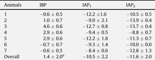
Intrabladder pressures obtained for seven mares and directly acquired IAP measurements for seven horses on two separate occasions (meanSD) are provided inTable 2. Fig. 6.Close-up of the area of placement of the 8-G needle in the rightflank (white dot), 5.0 cm below the tuber coxae (white dashed line) for direct assessment of intra-abdominal pressure (IAP).
Individual variation is evident for both IBP and IAP values. Variations in IAP (not statistically significant) are also observed between the first (IAP1) and second (IAP2) measurements for all horses. Intrabladder pressures from mares 1, 2, and 3 were compared with IAP values of three different horses (geldings) of approximately the same size and body weight. Horses 4, 5, 6, and 7 represent four mares included in both this study and the directly measured IAP group. Consequently, unlike horses 1, 2, and 3, IBP measurements for these four mares were compared with their own respective IAP values. Intrabladder pressure was significantly higher than both IAP1(P¼.001) and IAP2(P¼ .001); no statistical difference was observed between IAP1 and IAP2(Table 2). There was no correlation between IBP and IAP1(Pearsonr¼ 0.508;R2¼0.2582,P¼.244) or IAP2 (Pearsonr¼ 0.031;R2¼0.0297,P¼.947) (Fig. 8).
4. Discussion
Intrabladder pressure is regarded as the gold standard for investigating IAP in human patients[18], and afl
uid-filled intrabladder manometry system is conventionally used for this purpose[15,27]. Its utility in horses has been recently evaluated with disappointing results[7,11,28]. The adopted method was based on a system originally devel-oped for dogs[29]and later adapted for widespread use in human patients[1,30]. The original method is based on the principles of hydrostatic pressure in which IBP is trans-mitted through afluid-filled urinary catheter connected by external tubing to a pressure transducer[29,31]. Contrarily, our balloon manometry system was air-filled, similar to other studies that evaluated urinary tract pressure in horses by using much smaller balloon manometers than the one used in this study [25,32]. Air-filled balloon manometry systems have been validated for estimation of IAP via assessment of intragastric pressure (IGP) in humans [16,33-35]. However, correlation between IGP and IAP was not observed in horses[13].
The length and diameter of our manometer were based on previous publications describing measurement of esophageal pressure and IGP of horses [9,12,13,36]. Furthermore, longer balloons (such as the one used in this study) are preferred for evaluation of esophageal pressure because they provide more consistent readings [23,37]. Balloon manometers made of latex are preferable because the wall thinness is characterized by higher compliance[36].
Our balloon manometer inflation volume (2.0 mL) is also similar to the one tested by Ronen[25]. However, as described earlier, Ronen used a significantly smaller balloon than ours. As with our study, optimal inflation volumes were similarly obtained based on in vitro studies. Lower inflation volumes are preferred because excessively high volumes tend to provoke measurement errors (as a result of tissue elasticity)[25,38]. In vitro studies have concluded that for longer balloons (such as ours), volumes between 0.2 and 5.0 mL are optimal for inflation[39]. For evaluation of IBP in humans, the bladder must be distended with low volumes of saline. The saline infusion dosages tested during in vitro trials of this study were selected based on one study evaluating IBP in children [19]. According to the authors, a dosage of 1.0 mL/kg is the most efficient for measuring IBP of children, with a total infused volume of 50.0-100.0 mL. At these distention volumes, the bladder wall acts as a passive diaphragm[29,40]. However, more recent studies regarding IBP measurements in chil-dren have suggested that bladder distention volumes should not exceed 25.0 mL[4,41].
Fig. 7.Correlation between distention dosage and pressure generated inside the system during in vitro trials. (Pearsonr¼0.982;R2¼0.964,P¼.121).
Table 1
System pressures generated in vitro by three distinct distention dosages (1.0, 3.0, and 5.0 mL/kg), and respective balloon manometer pressures registered by each inflating volume of air (0.0, 0.5, 1.0, 1.5, 2.0, 3.0, 4.0, 5.0, 10.0, and 20.0 mL) for each distention dosage during in vitro trials
Volumea System Pressureb Balloon Manometer
Pressureb
Distention Dosagesc Distention Dosagesc
1.0 3.0 5.0 1.0 3.0 5.0 0.0 1.0 13.0 19.0 0.0 2.0 2.0 0.5 1.0 13.0 19.0 0.0 10.0 13.0 1.0 1.0 13.0 19.0 0.0 13.0 18.0 1.5 1.0 13.0 19.0 0.0 13.0 18.0 2.0 1.0 13.0 19.0 0.0 13.0 18.0 3.0 1.0 13.0 19.0 0.0 13.0 19.0 4.0 1.0 13.0 19.0 0.0 13.0 19.0 5.0 1.0 13.0 19.0 0.0 13.0 19.0 10.0 1.0 13.0 19.0 0.0 14.0 19.0 20.0 1.0 13.0 19.0 1.0 14.0 19.0
aVolume (mL).
bSystem pressure and balloon manometer pressure (mm Hg). cDistention dosages (mL/kg).
Table 2
Individual and overall IBP (meanSD) of seven mares compared with IAP obtained from seven horses (three geldings and four mares) in two different instances: IAP1and IAP2
Animals IBP IAP1 IAP2
1 0.60.5 12.21.6 10.50.5 2 1.00.7 9.02.1 13.90.4 3 4.60.6 12.70.8 13.70.4 4 2.90.6 9.40.5 8.80.7 5 2.90.6 12.21.8 11.30.7 6 0.70.7 9.31.4 10.00.0 7 0.60.5 8.40.6 12.81.3 Overall 1.42.0a 10.52.2 11.62.0 aDiffer from IAP
Although distention volumes ranging from 25.0 to 300.0 mL have been reported for use in adult human patients [4,18,20,21], an infusion volume of only 25.0 mL is suffi -cient, in and of itself, to increase IBP and yield misleadingly elevated values for IAP[20]. A positive correlation between bladder distention volume and IBP has also been demon-strated in horses when volumes greater than 100.0 mL were used [11]. Although not statistically significant, similarfindings were observed during our in vitro trials.
It has been reported in humans and piglets that the minimum volume for bladder infusion should be 10.0 mL [20,42]. Ideally, IBP should be measured after the bladder has been evacuated[18,20]. However, ensuring that the bladder is completely empty is not always straightforward[15].
The bladder distention volume used in this study (3.0 mL/kg) was chosen based on results of our in vitro trials. It was the only dosage at which the balloon manometer inflated with 2.0 mL was sufficiently sensitive for recording pressure in the system. Ourfinal infusion volume (976.57 164.42 mL) was considerably higher than volumes tested by other researchers[11]. Conversely, volumes up to 4 L of urine can accumulate in the adult equine bladder before the micturition reflex is triggered[43].
Based on our in vitro trials and the results of previous studies in human [4,21] and equine [11] subjects, our bladder distention volume likely overestimated IBP. However, our results were similar to those reported previously in horses when the fluid-filled manometry system was used[7,28].
Characteristics of the equine abdomen render the acquisition of consistent and repeatable IBP measurements challenging, even under controlled situations. Other factors that directly influence IBP include horse movements, breathing movements, and small variations in catheter positioning[28]. A relationship between body weight and IBP has been demonstrated in horses[11]as it is in humans [35]. Furthermore, many diverse variables influence IAP, including body position, body weight, body condition (obesity), mechanical ventilation (under general anes-thesia), intestinal motility and content, and anesthesia-induced muscle relaxation [5,10,11,15,28,35]. Considering that both sets of IAP measurements were obtained apart in all horses, we believe that uncontrolled variables such as body weight, intestinal content, and intestinal motility
surely contributed to the differences observed between both (not statistically different from one another). Discomfort associated with catheter placement and pres-ence (even after the 5-minute adaptation period), abdom-inal contractions attempting to expel the catheter (leading to increased IBP values), urination after bladder distention with saline interfering with optimal bladder distention (in which the bladder distention procedure had to be repeated), and uncertainty regarding the completeness of bladder emptying were the difficulties and limitations of the methodology encountered during this study. Stress associated with restraint, manipulation, and catheter placement could have caused elevated catecholamine levels, thus further influencing IBP measurements, because tone in the bladder wall is partially regulated by alpha- and beta-adrenergic innervation [25]. We established a 5-minute adaptation period, in the absence of sedation, following placement of the bladder catheter to reduce the influence of detrusor muscle contraction on IBP, consistent with previous studies in humans[2,15]and horses[11].
By convention, IBP is recorded at end-expiration to avoid the influence of abdominal muscle contractions on IBP recordings[2,18,44]. Our IBP recordings were obtained at end-inspiration, but the pressure equipment used in this study was not sufficiently sensitive to rapidly detect pres-sure variations during each respiratory cycle. To diminish the influence of this variable on our results, we elected to record 15 measurements for each variable studied (IBP and both sets of IAP).
Not ideally, IBP of three mares was compared with IAP of different animals. However, for the other four mares, IBP was compared with their respective IAP. We used two sets of IAP to compensate for the fact that both IBP and IAP were not obtained simultaneously. These could be considered as limitations of our study.
Considering the fact that the horses were awake during all sets of pressure acquisitions (IBP and both sets of IAP), increases in IAP or IBP were constantly observed during pressure recording as a result of increases in depth of breathing, discomfort, and frequent weight-bearing provoked by stress due to restraint and abdominal contraction during urination or attempts to expel the catheter. Variations in both IBP and IAP values associated with behavioral changes (not representative of the normal Fig. 8.Correlation between intrabladder pressure of seven mares and IAP of seven horses (three geldings and four mares) obtained at two separate times: IAP1
values at rest) were discarded, which led to the recording of nonconsecutive values for both IBP and IAP. Positioning of the pressure sensor at the level of the tuber ischii was consistent with previous descriptions[7,11].
5. Conclusion
Similar to the results of earlier studies on horses per-formed using afluid-filled system, results of this study did not demonstrate significant correlation between IBP and IAP in mares. Moreover, provocation of movements and bladder contraction by placement of the catheter resulted in difficulties obtaining meaningful results in healthy, awake mares. However, early identification of IAH would certainly be beneficial for the treatment and outcome of affected horses.
Acknowledgments
This work was funded by FAPESP (Fundação de Amparo à Pesquisa do Estado de São Paulo. Protocol 2008/00606-8).
References
[1] Malbrain ML, De Laet IE, De Waele JJ. IAH/ACS: the rationale for surveillance. World J Surg 2009;33:1110-5.
[2] Gallagher JJ. Intra-abdominal hypertension: detecting and managing a lethal complication of critical illness. AACN Adv Crit Care 2010;21:205-19.
[3] Havens JM, Soybel DI. Assessment of intra-abdominal pressure by measurement of abdominal wall tension. J Surg Res 2011;166:70-2. [4] Kimball EJ, Baraghoshi GK, Mone MC, Hansen HJ, Adams DM, Alder SC, et al. A comparison of infusion volumes in the measure-ment of intra-abdominal pressure. J Intensive Care Med 2009;24:261-8.
[5] Cheatham ML. Abdominal compartment syndrome: pathophysi-ology and definitions. Scand J Trauma Resusc Emerg Med 2009;17:10-20.
[6] Kirkpatrick AW, Keaney M, Kmet L, Ball CG, Campbell MR, Kindratsky C, et al. Intraperitoneal gas insufflation will be required for laparoscopic visualization in space: a comparison of laparoscopic techniques in weightlessness. J Am Coll Surg 2009;209:233-41. [7] Wilkins PA. Abdominal compartmental syndrome in equine
medi-cine. In: Proceedings of 23rd American College of Veterinary Internal Medicine Congress; Baltimore, MD:2005; p. 192-4. [8] Brosnahan MM, Holbrook TD, Gilliam LL, Ritchey JW, Confer AW.
Intra-abdominal hypertension in two adult horses. J Vet Emerg Crit Care 2009;19:174-80.
[9] Slocombe R, Brock K, Covelli G, Bayly WM. Effect of treadmill exer-cise on intrapleural, transdiaphragmatic and intra-abdominal pres-sures in Standardbred horses. Equine Exerc Physiol 1991;3:83-91. [10] Southwood LL, Wilkins PA. Measurement of intra-abdominal
pres-sure in normal horses. J Vet Intern Med 2005;19:488.
[11] Munsterman AS, Hanson RR. Comparison of direct and indirect methods of intra-abdominal pressure measurement in normal horses. J Vet Emerg Crit Care 2009;19:545-53.
[12] Canola PA, Perotta JH, Laskoski LM, Escobar A, Melo e Silva CA, Canola JC, et al. Effect of passive pneumoperitoneum on esophageal pressure, cardiovascular parameters and blood gas analysis in horses. Equine Vet J 2011;43:446-50.
[13] Canola PA, Perotta JH, Laskoski LM, Escobar A, Melo e Silva CA, Canola JC, et al. Use of gastric balloon manometry for estimation of intra-abdominal pressure in horses. Equine Vet J (in press). [14] Munsterman AS, Hanson RR. Evaluation of gastric pressures as an
indirect method for measurement of intraabdominal pressures in the horse. J Vet Emerg Crit Care 2011;21:29-35.
[15] Malbrain ML. Different techniques to measure intra-abdominal pressure (IAP): time for a critical re-appraisal. Intensive Care Med 2004;30:357-71.
[16] Turnbull D, Webber S, Hamnegard CH, Mills GH. Intra-abdominal pressure measurement: validation of intragastric pressure as a measure of intra-abdominal pressure. Br J Anaesth 2007;98: 628-34.
[17] Pelosi P, Quintel M, Malbrain ML. Effect of intra-abdominal pressure on respiratory mechanics. Acta Clin Belg 2007;62(Suppl 1):78-88. [18] Malbrain ML, Deeren DH. Effect of bladder volume on measured
intravesical pressure: a prospective cohort study. Crit Care 2006;10:R98.
[19] Davis PJ, Koottayi S, Taylor A, Butt WW. Comparison of indirect methods of measuring intra-abdominal pressure in children. Intensive Care Med 2005;31:471-5.
[20] De Waele JJ, Pletinckx P, Blot S, Hoste E. Saline volume in trans-vesical intra-abdominal pressure measurement: enough is enough. Intensive Care Med 2006;32:455-9.
[21] De Laet I, Hoste E, De Waele JJ. Transvesical intra-abdominal pres-sure meapres-surement using minimal instillation volumes: how low can we go? Intensive Care Med 2008;34:746-50.
[22] Benditt JO. Esophageal and gastric pressure measurements. Respir Care 2005;50:68-77.
[23] Fernandes CR. A importância da pressão pleural na avaliação da mecânica respiratória. Rev Bras Anestesiol 2006;56:287-303. [24] Behazin N, Jones SB, Cohen RI, Loring SH. Respiratory restriction and
elevated pleural and esophageal pressures in morbid obesity. J Appl Physiol 2010;108:212-8.
[25] Ronen R. Measurements of urethral pressure profile in the male horse. Equine Vet J 1994;26:55-8.
[26] Beardsmore CS, Helms P, Stocks J, Hatch DJ, Silverman M. Improved esophageal balloon technique for use in infants. J Appl Physiol 1980;49:735-42.
[27] Cheatham ML, De Waele JJ, De Laet I, De Keulenaer B, Widder S, Kirkpatrick AW, et al. The impact of body position on intra-abdominal pressure measurement: a multicenter analysis. Crit Care Med 2009;37:2187-90.
[28] Dechant JE, Nieto JE, LeJeune SS. Comparison of techniques for measurement of intra-abdominal pressure in horses. In: Proceed-ings of the International Veterinary Emergency and Critical Care Symposium; Phoenix, AZ:2008. p. 838.
[29] Kron IL, Harman PK, Nolan SP. The measurement of intra-abdominal pressure as a criterion for abdominal re-exploration. Ann Surg 1984;199:28-30.
[30] Cheatham ML, Safcsak K. Intraabdominal pressure: a revised method for measurement. J Am Coll Surg 1998;186:594-5. [31] Shuster MH, Haines T, Sekula LK, Kern J, Vazquez JA. Reliability of
intrabladder pressure measurement in intensive care. Am J Crit Care 2010;19:e29-39.
[32] Kay AD, Lavoie JP. Urethral pressure profilometry in mares. J Am Vet Med Assoc 1987;191:212-6.
[33] Engum SA, Kogon B, Jensen E, Isch J, Balanoff C, Grosfeld JL. Gastric tonometry and direct intraabdominal pressure monitoring in abdominal compartment syndrome. J Pediatr Surg 2002;37:214-8. [34] De Waele JJ, Berrevoet F, Reyntjens K, Pletinckx P, De Laet I, Hoste E.
Semicontinuous intra-abdominal pressure measurement using an intragastric compliance catheter. Intensive Care Med 2007;33:1297-300.
[35] De Keulenaer BL, De Waele JJ, Powell B, Malbrain ML. What is normal intra-abdominal pressure and how is it affected by posi-tioning, body mass and positive end-expiratory pressure? Intensive Care Med 2009;35:969-76.
[36] Hoffman AM. Clinical application of pulmonary function testing in horses. In: Lekeux P, editor. Equine respiratory diseases. Ithaca, NY: International Veterinary Information Service (www.ivis.org); 2002. B0304.0802.
[37] Milic-Emili J, Mead J, Turner JM. Topography of esophageal pressure as a function of posture in man. J Appl Physiol 1964;19:212-6. [38] Lekeux P, Hajer R, Breukink HJ. Pulmonary function in calves:
technical data. Am J Vet Res 1984;45:342-5.
[39] Zin WA, Caldeira MP, Cardoso WV, Auler JO Jr, Saldiva PH. Expiratory mechanics before and after uncomplicated heart surgery. Chest 1989;95:21-8.
[40] Chiumello D, Tallarini F, Chierichetti M, Polli F, Li Bassi G, Motta G, et al. The effect of different volumes and temperatures of saline on the bladder pressure measurement in critically ill patients. Crit Care 2007;11:R82.
[41] Ejike JC, Bahjri K, Mathur M. What is the normal intra-abdominal pressure in critically ill children and how should we measure it? Crit Care Med 2008;36:2157-62.
[42] Gudmundsson FF, Viste A, Gislason H, Svanes K. Comparison of different methods for measuring intra-abdominal pressure. Inten-sive Care Med 2002;28:509-14.
[43] Toribio RE. Essentials of equine renal and urinary tract physiology. Vet Clin North Am Equine Pract 2007;23:533-61.