Factors associated with variation in intracranial
pressure in a model of intra-abdominal hypertension
with acute lung injury
Modulação da pressão intracraniana em um modelo experimental
de hipertensão abdominal e lesão pulmonar aguda
INTRODUCTION
Increased compartment pressure is an important cause of organ dysfunction and is associated with increased mortality in severely ill patients.(1-4) Multiple
compartment syndrome, i.e., increased pressure in several compartments, causing organ or system failure, is a clinical entity that has been recently described in the medical literature.(1,5) Several critical illness conditions can be
associated with increased compartment pressures, including positive pressure ventilation, infusion of large volumes of luids and variations of vascular blood content.(1)
he efects of critical illness over on intracranial pressure (ICP),
Fernando Godinho Zampieri,1,2
Juliana Roberta Almeida,3
Guilherme Pinto de Paula Schettino,3 Marcelo Park,1,3 Fabio
Santana Machado3, Luciano Cesar
Pontes Azevedo1,3
1. Intensive Care Unit, Emergency Department, Hospital das Clínicas – Faculdade de Medicina – Universidade de São Paulo – USP – São Paulo (SP), Brazil.
2. Intensive Care Unit, Hospital Alemão Oswaldo Cruz – São Paulo (SP), Brazil. 3. Research and Teaching Institute – Hospital Sírio-Libanês – São Paulo (SP), Brazil.
ABSTRACT
Objective: To evaluate the efects of hemodynamic, respiratory and metabolic changes on intracranial pressure in a model of acute lung injury and abdominal compartment syndrome.
Methods: Eight Agroceres pigs
were submitted to ive diferent clinical scenarios after instrumentation: 1) a baseline condition with low intra-abdominal pressure and healthy lungs; 2) pneumoperitoneum with 20 mmHg intra-abdominal pressure; 3) acute lung injury induced by pulmonary lavage with surfactant deactivation; 4) pneumoperitoneum with 20 mmHg intra-abdominal pressure with lung pulmonary injury and low positive end-expiratory pressure; and 5) 27 cmH2O positive end-expiratory pressure with pneumoperitoneum and acute lung injury. Respiratory and hemodynamic variables were collected. A multivariate analysis was conducted to search for variables associated with increased
intracranial pressure in the ive scenarios.
Results: Only plateau airway
pressure showed a positive correlation with intracranial pressure in the multivariate analysis. In the models with acute lung injury, plateau airway pressure, CO2 arterial pressure, end tidal CO2 and central venous pressure were positively correlated with increased intracranial pressure.
Conclusion: In a model of multiple organ dysfunction with associated clinical conditions causing increased intra-thoracic and abdominal pressure, increased intracranial pressure triggered by elevated intra-abdominal pressure is apparently caused by worsened respiratory system compliance and a reduced brain venous drainage gradient due to increased central venous pressure.
Keywords: Respiration, artificial; Acute lung injur y; Compartment syndromes; Intracranial pressure; Diseases models, animal; Intensive care units; Swine
his study was conducted at the Anesthesiology and Intensive Care Medicine Research Laboratory – Hospital Sírio-Libanês – São Paulo (SP), Brazil.
Conlicts of interest: None.
Submitted on May 8, 2011 Accepted on June 7, 2011
Corresponding author:
Fernando Godinho Zampieri
Secretaria da Disciplina de Emergências Clínicas do
Hospital das Clínicas da Faculdade de Medicina da Universidade de São Paulo Rua Dr. Enéas de Carvalho Aguiar, 255 Zip Code: 05403-000– São Paulo (SP), Brazil.
considered in an acute brain injury context, are poorly described in the medical literature. Intra-thoracic (ITP) and intra-abdominal pressures (IAP) are known to inluence ICP.(6-8) However, the magnitude of this efect
and its clinical relevance, except in acute neurological diseases, are unknown.
he objective of this study was to evaluate the efects of hemodynamic, respiratory and metabolic changes on intracranial pressure in a validated model of acute lung injury and abdominal compartment syndrome.
METHODS
Instrumentation and stabilization period
his study was conducted in the anesthesiology and intensive care medicine research laboratory at Instituto Sírio Libanês. All experimental procedures involving animals were performed in accordance with the National Institutes of Health Guide for the Care and Use of Laboratory Animals and the Brazilian Society for Neuroscience and Behavior (SBNeC) recommendations for animal care. Eight female Agroceres pigs (weighing 38 kg ± 5 kg, range 35-42 kg) were made to fast for one night with free access to water. he pigs were premedicated with midazolam (0.3 mg/kg) and acepromazine (0.5 mg/kg). Anesthesia was induced with thionembutal (12 mg/kg), and pancuronium (0.1 mg/ kg) was used for paralysis. he animals were intubated and ventilated (Evita XL; Drager, Luebeck, Germany), maintaining the following parameters throughout the study (except when otherwise mentioned): tidal volume (TV) of 8 mL/kg; positive end-expiratory pressure
(PEEP) of 5 cmH2O; low 1 L/second; inspired oxygen
fraction (FiO2) settled to maintain between 93 and
95% peripheral saturation; and a respiratory rate (RR)
required to keep PaCO2 between 35 and 50 mmHg.
Anesthesia was maintained throughout the study with midazolam (0.3 mg/kg/hour) and fentanyl (5 mcg/kg/ hour), and muscular blockade was established with pancuronium (0.2 mg/kg/hour).
he internal jugular vein was cannulated for the introduction of a pulmonary artery catheter, and the procedure was guided by observation of the typical pressure curves. he right femoral artery was cannulated to measure systemic blood pressure and to obtain samples. he stomach was emptied using a large tube.
Intracranial pressure was measured using a monitor from Camino® ICP (Camino Laboratories, San Diego, California, USA), installed within the cerebral parenchyma.
After instrumentation, the animals were stabilized for one hour without intervention. Continued ringer lactate infusion was maintained at 4 mL/kg/hour throughout the procedure.
Systemic blood pressure, central venous pressure (CVP) and pulmonary artery pressure (PAP) were measured using quartz transducers (Edwards Critical Care, Irvine, CA) continuously shown on a multi-parameter screen (DX 2020; Dixtal, São Paulo, Brazil) that also showed heart rate (HR), peripheral saturation (SatO2) and end tidal CO2
(EtCO2). Continuous mixed venous oxygen saturation
(SvO2) was maintained with a spectrophotometer, and cardiac output (CO) was measured by automated thermo dilution (Vigilance; Edwards, Irvine, CA). At the end of each phase, blood gas was obtained, and the values were recorded.
Experiment protocol and induction of lung injury
All animals were studied in distinct conditions according to the following sequence: 1) baseline with healthy lungs (low PEEP, low IAP in normal lungs); 2) pneumoperitoneum induced with an electronic CO2 pump (Storz 26430520; Karl Storz, Tuttlingen, Germany), with a 20 mmHg IAP (low PEEP, high IAP in normal lungs); 3) acute lung injury (ALI) induced by pulmonary lavage and surfactant deactivation (low PEEP, low IAP in injured lungs); 4) pneumoperitoneum with 20 mmHg IAP with ALI (low PEEP, high IAP in injured lungs); and 5) PEEP
27 cmH2O with pneumoperitoneum and injured lungs
(high PEEP, high IAP with injured lungs). All conditions were maintained for 30 minutes.
Repeated pulmonary lavage was conducted using a Tween 20 2.5% solution (to induce surfactant deactivation) diluted in normal saline solution (3 mL/kg) until the arterial oxygen pressure PaO2/FiO2 ratio reached 150 or less. During the induction of ALI, the animals were maintained with FiO2 1.0, PEEP 5 cmH2O, TV 8 mL/kg and RR between 20 and 30 inhalations per minute and an inspiratory time of 1 second.
he cardiovascular parameters measured during each phase were as follows: mean blood pressure (MBP), CVP, PAP, HR, SvO2 and CO. he respiratory variables measured were minute volume, TV, RR, peak pressure (Ppico), plateau pressure (Pplat calculated as the mean of 5 inspiratory pauses of 2 seconds each), PEEP, EtCO2,
SatO2 and FiO2. ICP was measured throughout each
phase.
Statistical analysis
Data normality was analyzed using the Shapiro-Wilk test. Simple correlations were analyzed using Pearson’s correlation test. Independent correlations related to intracranial pressure variation were analyzed using a multilinear regression model. Multiple repeated comparisons were conducted using an ANOVA for repeated measures, with Tukey’s test for the post-hoc analysis and Mauchly’s test to warrant the sphericity. To better evaluate factors related to intracranial pressure, the groups were divided into two clinical conditions: with or without ALI (conditions 1 and 2 versus 3, 4 and 5).
he small sample size could reduce the power of the multivariate analysis of this study; therefore, data eligibility for inclusion in the multivariate analysis was deined considering 1) clinical relevance, 2) physiological plausibility, 3) low co-linearity, deined as lower than a 0.8 Pearson’s coeicient and 4) low multi-colinearity (an incremental variance factor <2.5).
he independent variables were individually and
pro-gressively entered. he inal model was obtained with the lowest possible number of independent variables. SPSS 16.0 software was used, and a p<0.05 was considered signiicant.
RESULTS
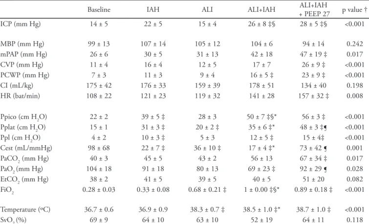
Table 1 displays the behavior of ICP during the five scenarios. Although no statistically significant difference was observed, there was a trend for higher ICP values in conditions with increased IAP. Condition 3 (ALI alone with low PEEP) did not produce increases in ICP. However, the combination of ALI and IAH (condition 4) produced a more relevant ICP increase than condition 2 (IAH alone). The use of high PEEP (condition 5) compared with low PEEP in the same condition (condition 4) showed a slight increase in ICP.
Considering the respiratory variables (Table 1), oxygenation was worst in the IAH and ALI groups, and additionally, PaCO2 was increased in the ALI with
Table 1 – Intracranial pressure, hemodynamic and respiratory data for each condition
Baseline IAH ALI ALI+IAH ALI+IAH
+ PEEP 27 p value † ICP (mm Hg) 14 ± 5 22 ± 5 15 ± 4 26 ± 8 ‡§ 28 ± 5 ‡§ <0.001
MBP (mm Hg) 99 ± 13 107 ± 14 105 ± 12 104 ± 6 94 ± 14 0.242 mPAP (mm Hg) 26 ± 6 30 ± 5 31 ± 13 42 ± 18 47 ± 19 ‡ 0.017 CVP (mm Hg) 11 ± 4 16 ± 4 12 ± 5 17 ± 7 26 ± 9 ‡ <0.001 PCWP (mm Hg) 7 ± 3 11 ± 3 9 ± 4 16 ± 5 ‡ 23 ± 9 ‡ <0.001 CI (mL/kg) 175 ± 42 176 ± 33 159 ± 39 178 ± 51 134 ± 40 0.198 HR (bat/min) 108 ± 22 121 ± 23 119 ± 32 141 ± 28 157 ± 32 ‡ 0.008
Ppico (cm H2O) 22 ± 2 39 ± 5 ‡ 28 ± 3 50 ± 7 ‡§* 56 ± 3 ‡ <0.001 Pplat (cm H2O) 15 ± 1 31 ± 3 ‡ 20 ± 2 ‡ 35 ± 6 ‡* 48 ± 3 ‡¶ <0.001 Ppl (cm H2O) 4 ± 2 10 ± 3 ‡ 5 ± 3 12 ± 5 ‡ 15 ± 4‡ <0.001 Cest (mL/mmHg) 98 ± 68 22 ± 7 ‡ 36 ± 10 ‡ 17 ± 4 ‡* 73 ± 42 ¶ 0.001 PaCO2 (mm Hg) 40 ± 3 45 ± 5 43 ± 2 56 ± 13 67 ± 34 ‡ 0.017 PaO2 (mm Hg) 104 ± 18 91 ± 18 80 ± 13 69 ± 23 ‡ 92 ± 29 ¶ 0.028 EtCO2 (mm Hg) 38 ± 2 41 ± 5 39 ± 5 40 ± 5 51 ± 20 0.082 FiO2 0.28 ± 0.03 0.33 ± 0.08 0.68 ± 0.21 ‡ 1 ± 0.00 ‡§* 0.89 ± 0.18 ‡ <0.001
Temperature (ºC) 36.7 ± 0.6 36.9 ± 0.9 38.3 ± 0.7 ‡ 38.5 ± 1.0 ‡* 38.7 ± 1.0 ‡ <0.001 SvO2 (%) 69 ± 9 64 ± 10 63 ± 10 52 ± 19 64 ± 11 0.118
hese data were reported in a previous article.(15) † ANOVA for repeated measures.‡ Tukey post hoc, p < 0.05 versus baseline.§ Tukey post hoc,
IAH group. ALI plus IAH produced an even lower
pO2/FiO2 ratio, which was only partially resolved
when PEEP was increased to 27 cmH2O.
Table 1 shows the behavior of the respiratory mechanics variables. The increased plateau pressure in all pathologic scenarios is notable and demonstrates that our ALI and IAH model effectively reproduced a common bedside clinical condition.
Hemodynamic variables are shown in table 1. Both cardiac output and mean blood pressure were constant
throughout the experiment. CVP was elevated in the studied conditions, reaching values close to 25 mmHg.
Table 2 shows an analysis of all conditions regarding variations in ICP in which Pplat remains associated with ICP, without categorizing the conditions as with or without ALI. Table 3 shows the results of the multivariate analysis. In the models without ALI, only Pplat was correlated with ICP (R = 0.625). However,
in the models with ALI, EtCO2 (R = 0.192), PaCO2
(R = 0.481), CVP (R = 0.646) and Pplat (R = 0.761) were correlated with ICP.
DISCUSSION
Critically ill patients frequently have severe systemic disease requiring support for multiple organs. he impact of a given intervention on a system may cause harm to other organs, minimizing or even neutralizing eventual beneicial efects.(1) For example, mechanical ventilation
can improve oxygenation in several clinical scenarios. However, mechanic ventilation, especially with high airway pressures, can be associated with respiratory(9,10) and
systemic complications, including severe hemodynamic disorders,(11) increased IAP(12) and increased ICP.(13)
Recently, several works have highlighted the impact of intra-abdominal pressure on critical disease pathophysiology, especially its correlation with renal disease.(14) he association between ALI and IAH is a
clinical challenge, as higher airway pressures may be required to expand the lungs because of abdominal cavity restriction.(15) Concomitantly, a higher intra-thoracic
pressure may worsen IAH, perpetuating a progressive cycle that may worsen the patient’s conditions.
Table 3 – Intracranial pressure changes in conditions either associated or not associated with acute lung injury
Conditions not associated with ALI Conditions associated with ALI
Univariate analysis Multivariate analysis& Univariate analysis Multivariate analysis*
R p value Beta standard p value R p value Beta standard p value CVP 0.479 0.060 --- --- 0.646 0.001 0.400 0.016 MBP 0.098 0.719 --- --- -0.184 0.389 --- ---CI -0.019 0.943 --- --- 0.104 0.628 --- ---Pplat 0.625 0.010 1.048 <0.001 0.761 <0.001 0.371 0.030 PEEP --- # --- # --- --- 0.432 0.035 ---
---IAP 0.613 0.012 --- --- 0.720 <0.001 --- ---PaCO2 -0.007 0.978 --- --- 0.481 0.020 0.775 0.001 EtCO2 -0.230 0.391 --- --- 0.192 0.370 0.691 0.002 Temp 0.322 0.224 --- --- -0.069 0.750 ---
---Variables eligible for multivariate analysis were sequentially entered (enter mode). CVP – central venous pressure; MBP – mean blood pressure; CI – cardiac index; Pplat – plateau pressure; PEEP – positive end-expiratory pressure; IAP – intra-abdominal pressure; EtCO2 – end tidal CO2; Temp - temperature. # A constant PEEP was maintained. & R2 = 0.339. * R2 = 0.779. No incremental variance factor was >2.5.
Table 2 – Variables associated with modulation of intracranial pressure
Univariate analysis Multivariate analysis*
R p value Beta standard p value CVP 0.638 <0.001 0.228 0.128 MBP -0.097 0.551 --- ---CI -0.004 0.980 --- ---Pplat 0.753 <0.001 0.535 0.002 PEEP 0.442 <0.001 # #
IAP 0.685 <0.001 # #
PaCO2 0.465 0.003 0.110 0.387 EtCO2 0.174 0.283 --- ---Temp 0.254 0.114 ---
---Variables eligible for multivariate analysis were sequentially entered into the model (enter mode). CVP – central venous pressure; MBP – mean blood pressure; CI – cardiac index; Pplat – plateau pressure; PEEP – positive end-expiratory pressure; IAP – intra-abdominal pressure; EtCO2 – end tidal CO2; Temp - Temperature. * R2 = 0.594.
# hese values were removed from the model due to multi-colinearity with a variance incremental factor >2.5. he decision to remove the variables was based on clinical simplicity rational and a higher determinant coeicient (R2). In this model, there was no incremental
The magnitude of pressure’s influence on thoracic and abdominal compartments with intracranial pressure in critical disease has been poorly studied. The use of PEEP in patients with increased ICP may
cause harmful ICP increases,(13) although finding
this is controversial.(16) Intra-abdominal pressure can
apparently affect intracranial pressure, likely because of increased venous and intra-thoracic pressures.(7,8)
Peritoneostomy has been described as a rescue therapy for refractory intracranial hypertension patients.(17,18)
This study aimed at evaluating whether intra-thoracic and abdominal pressure changes, in addition to oxygenation parameters, could cause an increase in intracranial pressure values in a clinically relevant model of multiple organ dysfunction.
Conditions associated with IAH were those that more greatly influenced ICP. The use of high PEEP (condition 5) had little effect on ICP, suggesting that PEEP alone was not a relevant determinant of the ICP increase observed in our model. Furthermore, this finding suggests that the use of a PEEP similar to the IAP does not cause a significant ICP increase; this increase could be related to improved compliance of the involved compartments.
In multivariate analysis of conditions not associated with ALI, only Pplat was related to ICP, suggesting that the increase in intra-thoracic pressure was the most important determinant of ICP in these conditions. Increased Pplat then likely reflects worsened respiratory system compliance induced by the increased abdominal pressure.
In conditions associated with ALI, the multivariate
analysis showed that Pplat, CVP, EtCO2 and PaCO2
were all associated with increased ICP. Increased CO2 pressures (reflected by PaCO2 and EtCO2) likely reflect the increased dead space fraction caused by ALI.
Plasma CO2 tension is also known to be relevant to
ICP. It could be anticipated that conditions coursing
with higher CO2 tensions (as ALI) would show an
even stronger relationship. Pplat was related with ICP. This result can be explained by the reasons previously mentioned. The correlation between CVP and ICP may suggest that increased venous pressures caused by increased thoracic and abdominal pressures may have limited the cerebral venous drainage, subsequently increasing ICP.
On the basis of these results, the first and most important conclusion that we may draw is that intracranial pressure is influenced in a critical illness model. As described in table 1, the association between
IAH and ALI produced an increase of up to 15 mmHg ICP. In humans, ICP increases of this magnitude are clinically relevant, as increased ICP is associated with worsened neurologic prognosis and even fatal neurologic events, such as brain herniation.
The main hypothesis to draw from this study is that Pplat is the mechanism by which IAH causes increases in ICP. The direct influence of IAP over the venous system also cannot be disregarded. This finding supports the current theory that body compartments are interconnected. Increased ICP during IAH is therefore a result of the transmission of abdominal pressure to the chest and then from the chest to the head. Increased venous pressure could perhaps reduce the pressure gradient for cerebral venous drainage. Our findings agree with the current
literature(6-8) and broaden the knowledge of body
compartments.
Our study has some limitations. First, this is a purely physiological study, and although it is based on a validated model of multiple organ dysfunction, its applicability in a real-life clinical setting can be questioned. Second, the small sample of animals, along with multiple clinical conditions, limits the power of our statistical analysis. However, this study aimed to produce hypotheses for future studies rather than a definitive hypothesis for such a complex condition.
CONCLUSION
In a model of multiple organ dysfunction with clinical conditions associated with increased thoracic and abdominal pressure, the increase in intracranial pressure is apparently caused by worsened respiratory system compliance. The increased central venous pressure in ALI conditions may also correlate with increased ICP, likely reducing the pressure gradient for cerebral venous drainage.
RESUMO
Objetivo: Avaliar o efeito de alterações hemodinâmicas, respiratórias e metabólicas sobre a pressão intracraniana em um modelo de lesão pulmonar aguda e síndrome comparti-mental abdominal.
Métodos: Oito porcos Agroceres foram submetidos,
pulmo-nar e desativação de surfactante; 4) pneumoperitôneo com pressão intra-abdominal de 20 mm Hg na vigência de lesão pulmonar aguda e com PEEP baixo; e 5) PEEP ajustado a 27 cm H2O na vigência de pneumoperitôneo e lesão pul-monar aguda. Variáveis respiratórias e hemodinâmicas fo-ram coletadas. Análise multivariada foi realizada buscando as variáveis associadas com elevação da pressão intracrania-na nos cinco cenários estudados.
Resultados: Após a análise multivariada, nas situações não associadas com lesão pulmonar aguda apenas a pressão de platô das vias aéreas se correlacionou positivamente com a pressão intracraniana. Nos modelos associados com lesão pulmonar aguda, a pressão de platô de vias aéreas, a pressão arterial de CO2, o CO2 no final da expiração e a pressão
venosa central se correlacionaram positivamente com incre-mentos da pressão intracraniana.
Conclusão: Em um modelo de disfunção orgânica
múltipla com situações clínicas associadas com aumento da pressão torácica e abdominal, o incremento da pressão intra-craniana desencadeado pela elevação da pressão abdominal parece ser decorrente da piora da complacência do sistema respiratório e da redução do gradiente para drenagem venosa cerebral ocasionado pela elevação da pressão venosa central.
Descritores: Lesão pulmonar aguda; Respiração artiicial; Sindromes de compartimento; Pressão intracraniana; Modelos animais de doenças; Unidades de terapia intensiva; Suínos
REFERENCES
1. Balogh ZJ, Butcher NE. Compartment syndromes from head to toe. Crit Care Med. 2010;38(9 Suppl):S445-51. 2. Ball CG, Kirkpatrick AW. Intra-abdominal hypertension
and the abdominal compartment syndrome. Scand J Surg. 2007;96(3):197-204. Review.
3. Kirkpatrick AW, Ball CG, Nickerson D, D’Amours SK. Intraabdominal hypertension and the abdominal compartment syndrome in burn patients. World J Surg. 2009;33(6):1142-9.
4. Kirkpatrick AW, De Waele JJ, Ball CG, Ranson K, Widder S, Laupland KB. he secondary and recurrent abdominal compartment syndrome. Acta Clin Belg Suppl. 2007;(1):60-5.
5. Scalea TM, Bochicchio GV, Habashi N, McCunn M, Shih D, McQuillan K, Aarabi B. Increased intra-abdominal, intrathoracic, and intracranial pressure after severe brain injury: multiple compartment syndrome. J Trauma. 2007;62(3):647-56; discussion 656.
6. Citerio G, Vascotto E, Villa F, Celotti S, Pesenti A. Induced abdominal compartment syndrome increases intracranial pressure in neurotrauma patients: a prospective study. Crit Care Med. 2001;29(7):1466-71.
7. Bloomield GL, Ridings PC, Blocher CR, Marmarou A, Sugerman HJ. A proposed relationship between increased intra-abdominal, intrathoracic, and intracranial pressure. Crit Care Med. 1997;25(3):496-503.
8. Bloomield GL, Ridings PC, Blocher CR, Marmarou A, Sugerman HJ. Efects of increased intra-abdominal pressure upon intracranial and cerebral perfusion pressure before and after volume expansion. J Trauma. 1996;40(6):936-41; discussion 941-3.
9. Bouferrache K, Vieillard-Baron A. Acute respiratory distress syndrome, mechanical ventilation, and right ventricular function. Curr Opin Crit Care. 2011;17(1):30-5.
10. Kolobow T, Moretti MP, Fumagalli R, Mascheroni D,
Prato P, Chen V, Joris M. Severe impairment in lung function induced by high peak airway pressure during mechanical ventilation. An experimental study. Am Rev Respir Dis. 1987;135(2):312-5.
11. Connery LE, Deignan MJ, Gujer MW, Richardson MG. Cardiovascular collapse associated with extreme iatrogenic PEEPi in patients with obstructive airways disease. Br J Anaesth. 1999;83(3):493-5.
12. Burchard KW, Ciombor DM, McLeod MK, Slothman GJ, Gann DS. Positive end expiratory pressure with increased intra-abdominal pressure. Surg Gynecol Obstet. 1985;161(4):313-8.
13. Caricato A, Conti G, Della Corte F, Mancino A, Santilli F, Sandroni C, et al. Efects of PEEP on the intracranial system of patients with head injury and subarachnoid hemorrhage: the role of respiratory system compliance. J Trauma. 2005;58(3):571-6.
14. Mufarrej FA, Abell LM, Chawla LS. Understanding Intra-Abdominal Hypertension: From the Bench to the Bedside. J Intensive Care Med. 2011 Apr 27. [Epub ahead of print]. 15. da Silva Almeida JR, Machado FS, Schettino GP, Park
M, Azevedo LC. Cardiopulmonary efects of matching positive end-expiratory pressure to abdominal pressure in concomitant abdominal hypertension and acute lung injury. J Trauma. 2010;69(2):375-83.
16. McGuire G, Crossley D, Richards J, Wong D. Efects of varying levels of positive end-expiratory pressure on intracranial pressure and cerebral perfusion pressure. Crit Care Med. 1997;25(6):1059-62.
17. Joseph DK, Dutton RP, Aarabi B, Scalea TM. Decompressive laparotomy to treat intractable intracranial hypertension after traumatic brain injury. J Trauma. 2004;57(4):687-93; discussion 693-5.