CARBOXYLATE SUBSTITUTION PATTERN AS STRUCTURAL
DIRECTIVE FOR THE FINAL PRODUCTS: SYNTHESIS, STRUCTURE
AND PROPERTIES OF [Fe
4Ca
2O
2(
μ
2-HCCl
2COO)
10(
μ
3-HCCl
2COO)
2(THF)
6]
Denis Prodius
a, Valeriu Mereacre
a, Maria Gdaniec
b, Sergiu Shova
c,g, Yurii A. Simonov
c, Nicolae
Stanica
d, Ion Geru
e, Andrea Caneschi
f, Constantin Turta
a*
aInstitute of Chemistry of the Academy of Sciences of Moldova, 3 Academy str., MD-2028, Chisinau, Moldova bFaculty of Chemistry, A.Mickiewicz University, Poznan, Poland
cInstitute of Applied Physics, Academy of Sciences of Moldova, Chisinau, Moldova dInstitute of Physical Chemistry “Murgulescu”, Romanian Academy, Bucharest, Romania
eThe Metrology and Analytical Methods of Research Center, Academy of Sciences of Moldova, Chisinau, Moldova f Department of Chemistry and INSTM Research Unit, University of Florence, Florence, Italy
gDepartment of Chemistry, State University of Moldova, Chisinau, Moldova * Fax: (373 22) 739954; Tel: (373 22) 739755; E-mail: [email protected]
Abstract: A novel hexanuclear iron-calcium-oxo complex has been synthesized and characterized by different physico-chemical methods and X-ray single crystal structural analysis: [Fe4Ca2O2(μ2-HCCl2COO)10(μ3-HCCl2COO)2(THF)6]. The molecular structure shows that there are two types of coordination for COO- anions: bidentate and tridentate.
The corresponding variable temperature susceptibility measurement shows that in the complex there exists an antiferromagnetic interaction (|J12| = |J34| = -71.86 cm-1). The iron(III) high spin state (5/2) is proved by Mössbauer
spectroscopy.High magnetic EPR measurements of 1 indicates the presence of S=0 ground state with low-lying S=1 excited state centred around g = 2.0054 ±0.0001.
Keywords:Iron(III) calcium(II) heterocluster; crystal structure; antiferromagnetic interaction. Introduction
Polynuclear carboxylates of 3d transition metals have been attracting renewed interest because of their intramolecular magnetic exchange interactions [1, 2] and their application as the simple models of oligonuclear active sites in metalloproteins [3-6]. The unambiguous attribution of the individual site in the trimer to one or another metal in these complexes is impeded by at least two factors: i) the close atomic numbers of the Fe and M metals and ii) the statistical distribution of the clusters in the crystal structure. The extensive investigations of other complexes with dichloroacetic acid have been reported [7-9]. However the information about {M3O} core containing bivalent metal that does not belong to 3d elements, proved by X-ray crystallography is still absent.
Here we presnt a novel hexanuclear iron-calcium-oxo complex [Fe4Ca2O2(μ2-HCCl2COO)10(μ3 -HCCl2COO)2(THF)6] (1) which is aggregated from two μ3-oxo fragments. The detailed crystal structure and magnetic property were discussed.
Results and discussion
X-ray diffraction revealed that crystal 1 at 293 K has a molecular structure consisted of neutral complexes, the charge balance of which is in agreement with formation of [Fe4Ca2O2(μ2-HCCl2COO)10(μ3-HCCl2COO)2(THF)6] species. Molecular structure of 1 at 293 K is depicted in fi gure 1. Two Ca2+ and four Fe3+ metal ions are associated into hexanuclear cluster through two μ3-oxo atoms (Ca1-O1 2.417(3), Fe1-O1 1.840(1), Fe2-O(1) 1.827(1) (Å) for 293 K; Ca1-O1 2.410(3), Fe1-O1 1.833(3), Fe2-O(1) 1.831(3) (Å), average values for A and B independent complexes at 130 K) and twelve dichloroacetate ligands. Ten carboxylates act as 1: 1:μ bidentate bridging units while two others as bridging tridentate 1: 2:μ units. The molecular structure of 1 could be described as a combination of two centrosymmetrically related trinuclear [Fe2CaO(CHCl2COO)6(THF)3] entities, which closely resemble published [Fe2CaO(CHCl2COO)6(THF)4] [10]. The metal···metal separation within the {Fe4Ca2(μ3-O)2} core exhibits the following values: Ca1···Fe1 3.635(2), Ca1···Fe2 3.704(2), Fe1···Fe2 3.204(2), Ca1···Ca1’ 3.957(2) (Å) for 293 K; Ca1···Fe1 3.632(2), Ca1···Fe2 3.676(2), Fe1···Fe2 3.203(2), Ca1···Ca1’ 3.918(2) (Å) in average for A and B independent complexes at 130 K. Each Fe3+ ions has a similar octahedral coordination completed by six oxygen atoms: four from bidentate bridging carboxylate groups in equatorial plane; two another from THF molecule and μ3-O in apical position. The coordination number of Ca2+ ion reaches the value 8 due to the coordination of the oxygen atom (Ca1-O2 2.450(3) Å for 293 K; 2.457(3)av Å for 130 K) from tridentate 1: 2:μ carboxylate unit.
phase transition in this temperature range. In the frame of the same space group (P1 ), the lowering of the temperature from 293 to 130 K leads to the doubling of the unit cell volume with two crystallographic independent molecules. The molecular structures of two complexes at 130 K are quite similar and slightly differ only by the orientation of the THF ligands coordinated to Ca atoms.
Fig. 1. Molecular structure of [Fe4Ca2(μ3-O)2 (CHCl2COO)12(THF)6] at 293 K. Chlorine atoms are omitted for clarity. The thermal ellipsoids are drawn at the 40% probability level.
The thermal decomposition of 1 in air is a multistage process. The elimination of THF molecules starts at 50oC. The
fi rst endothermic process is characterized by a maximum at ~70 oC and it ends at ~80 oC with ~6.5 mass loss, which corresponds to the elimination of two THF molecules per formula unit from of the crystal. The second endothermic process showed the maximum at 220 oC on the DTG curve. The total mass loss in these processes is ~68% that corresponds to removal of all THF molecules and partial decomposition of the dichloroacetate groups. An unexpected endothermal process has been observed in the range 410-455 oC. The peak registered was not accompanied by any mass change. Probably, it results from a phase transition of the reaction product. However, this statement requires additional confi rmation. According to DTG data, the residual sample after decomposition consists in about ~19% of the initial sample mass and most probably corresponds to CaO·Fe2O3.
The Mossbauer spectrum (MS) of 1 was measured at 295 K. The room temperature 57Fe MS of 1 shows two symmetrical central absorption lines which were assigned to one doublet with the parameters: δFe = 0.41 mm/s; ∆EQ = 0.53 mm/s. They indicate the presence of Fe(III) ions in the high spin state (S=5/2) with the rather symmetrical electron density distribution.
The magnetic properties of compounds 1 has been measured in the range of temperatures 300–2.0 K. The substance is characterized by a gradual reduction of the χ MT product with the lowering of temperature (Fig. 2) thus indicating an antiferromagnetic interaction between the iron (III) ions. Lower than ~ 30 K the plateau exists.
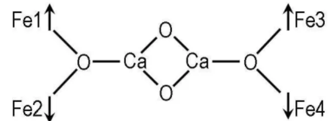
For interpretation of magnetic properties of 1 the HDVV model for Fe4 was used with the following scheme of interaction:
The original least-squares fi tting computer program FDHM [11, 12] with a Full Diagonalisation of Hamiltonian Matrix approach was employed to fi t (χT)exp.vs. T, in order to obtain the exchange couplings constants. The program uses the spin Hamiltonian operator:
H
total=
H
HDVV+
H
Z(1)
H
HDVV=
-2
J
(
S
Fe1S
Fe2+
S
Fe3S
Fe4) -2
J
´(
S
Fe1S
Fe3+
S
Fe2S
Fe4) -2
J
´´(
S
Fe1S
Fe4+
S
Fe2S
Fe3)
(2)where
H
Z=
g
B,
4
1
B
iz i
∑
=
S
(3)2 / 5
=
i
S is the spin operator of the i-th ion, Ji are the exchange parameters, B is the Bohr magneton, g is the electronic g-factor for the tetranuclear compound, B is the magnetic fi eld strength. The magnetic susceptibility data of 1 was calculated from the spin-coupled wave function by using a simplifi ed form of Van Vleck equation. The better fi tting was obtained at the following values of exchange parameters: J = J12 = J34 = - 71.86 cm-1; J’ = J
13 = J24 = + 0.18 cm -1; J” = J
14 = J23 = - 2.46 cm-1, ρ
paramagn. impur. = 2.5 % and Curie_Weiss = 2.46 K, and Fqualit._factor = 1.46·10 -5.
High magnetic EPR measurements of 1 indicates the presence of S=0 ground state with low-lying S=1 excited state centred around g = 2.0054 ±0.0001 (Fig.3). This result is in a good agreement with the above mentioned scheme of
Fig. 3. W – band powder EPR spectrum of 1 at 4 K.
exchange interactions leading to antiferromagnetic coupling between iron(III) ions in a high spin state: Fe1-Fe2 and Fe3-Fe4, respectively. As the individual iron(III) ions are in a high spin state (S=5/2) that has been shown by our investigations of the Mossbauer spectrum of 1, the existence of a ground state spin singlet for 1 is a simple consequence of D5/2⊗D5/2direct product of the rotation grouprepresentations
(D5/2⊗D5/2 = o S
s
D
)
5
0
∑
=
Experimental
Synthesis. The title compound was synthesised by the reaction of Fe(NO3)3·9H2O with 2 equivalents of Ca(CHCl2COO)2⋅6H2O in methanol, followed by evaporation of solvent at room temperature [13]. Dissolving in tetrahydrofuran (THF) the obtained solid gives a red-orange solution which crystals of [Fe4Ca2(μ3-O)2(CHCl2COO)12(THF)6] are obtained from. A brown-orange crystalline material has been separated after 2 weeks, washed with heptane and air dried. The yield is high (~ 87%). Calc. (Found) for C48H60Cl24Fe4Ca2O32: C, 25.03(24.94); H, 2.63(2.47); Fe, 9.70(9.92); Ca, 3.48(3.37)%.
The Mossbauer spectra were acquired using a constant acceleration spectrometer with symmetrical waveform. 57Co (1.0 MBq) source at room temperature has been used.
Variable temperature susceptibility studies were recorded with an Oxford Instruments Vibrating Sample Magnetometer (VSM) working between 0 and 12 T and in the 1.5–350 K temperature range.
EPR measurements.Polycrystalline powder EPR spectra were recorded at frequencies ranging from 90–270 GHz at the high-fi eld electron magnetic resonance facility at the National High Magnetic Field Laboratory in Tallahassee (USA), as described elsewhere [14, 15]. The sample’s temperature was varied from 300 to 4 K.
Crystal data for 1: (130 K) C48H60Ca2Fe4Cl24O32, Mr = 2303.32, triclinic, space group P1 , a = 13.475(3),
b = 14.678(3), c = 24.152(5) (Å), α = 84.99(3), β = 79.21(3)°, γ = 63.57(3) (°), V = 4202.0(15) Å3, Z = 2,
ρcalcd = 1.820 g/cm3, MoK
λ radiation (λ = 0.71073 Å, μ = 1.639 mm-1), R = 0.0615 (F2 > 2σ), Rw = 0.1518 (for 14773 data and 994 refi ned parameters); (293 K) C48H60Ca2Fe4Cl24O32, Mr = 2303.32, triclinic, space group P1 , a = 13.611(3), b = 14.919(3), c = 12.784(3) (Å), α = 81.28(3), β = 69.73(3)°, γ = 63.55(3) (°), V = 2180.0(8) Å3, Z = 1,
ρcalcd = 1.754 g/cm-3, MoK
λ radiation (λ = 0.71073 Å, μ = 1.580 mm-1), R = 0.0618 (F2 > 2σ), Rw = 0.1581 (for 8532 data and 466 refi ned parameters).
X-ray diffraction data were collected with a Kuma KM4CCD diffractometer using graphite-monochromated
Mo-Kα radiation. The crystal was placed 60 mm from the CCD detector. For both temperatures, more than hemisphere of reciprocal space was covered by combination of six sets of exposures; each set had a different ϕ-angle (0, 90, 180, 270, 45, 135) and each exposure of 30s covered 0.75° in ω. Coverage of the unique set is 99.2% complete up to 2Θ = 52°. The unit cell determination and data integration were carried out using the CrysAlis package of Oxford Diffraction. Intensity data were corrected for the Lorentz and polarization effects. The structure was solved by direct methods and refi ned with the SHELX program by full-matrix least-squares techniques. Non-hydrogen atoms were refi ned with anisotropic displacement parameters. H atoms were placed at calculated positions and refi ned as riding atoms in the subsequent least squares model refi nements. For better fi tting of the electron density, the positions of some of the –CHCl2 groups and one THF ligand were refi ned taking into account disordered models in the combination with the available tools (PART, DFIX and SADI) in SHELXL97 [16, 17]. The atoms belonging to the minor component of the disordered fragment were refi ned only isotropically. CCDC 692342 (130 K), CCDC 692959 (293 K) contain the supplementary crystallographic data for this structure. These data can be obtained free of charge via www.ccdc.cam.ac.uk/conts/retrieving.html (or from the Cambridge CB21EZ, UK; fax: (+44) 1223-336-033; or [email protected]
Conclusions
In conclusion the technique of synthesis of new hexanuclear bis(μ3-oxo) carboxylates containing {Fe4Ca2O2} core is developed. Mossbauer spectrum of this complex at room temperature shows one doublet characteristic for Fe(III) positions in a high spin state (S=5/2). In the range of temperature 300-20 K between iron (III) ions there is an antiferromagnetic exchange interaction in the two isosceles triangular skeletons with the following values of exchange integral (J = J12 = J34 = -71.86 cm-1). High magnetic EPR measurements of 1 indicates the presence of S=0 ground state with low-lying S=1 excited state centred around g = 2.0054 ±0.0001.
Supporting information available.
Listing crystallographic parameters in CIF format, atomic coordinates, bond distances and angles, thermal parameters and hydrogen atom positions for the title complex are available free of charge from authors.
Acknowledgements
We thank the MRDA-CRDF (BGP III-# MOP2-3061- S-03; MTFP-017/05) and CSSDT (#08.819.05.01F) for partial funding of this work, Dr. S. Nellutla for technical assistance at EPR measurements and Prof. N.S. Dalal for helpful discussion of EPR results.
References
[1] Kahn, O. Molecular Magnetism; VCH Publishers Inc.: New York, USA, 1993, p. 225;
[2] Turnbull, M. M.; Sugimoto,T.; Thompson, L. K. Molecule-based Magnetic Materials, ACS Symposium Series, USA, 1996, p. 314;
[3] Davis, C. M.; Royer, A. C.; Vincent, J. B. Inorg. Chem. 1997, 36, 5316-5320;
[6] Ruettinger, W.E.; Dismukes,G.C. Inorg. Chem. 2000, 39, 1021-1027.
[7] Jinkwon, K.; Jin Mook, L.; Youngkyu, D. Eur. J. Inorg. Chem. 2003, 14, 2563;
[8] Soler, M.; Chandra, S.K.; Davidson, E.R.; Christou, G.; Ruiz, D.; Hendrickson, D.N. Chem.Comm. 2000, 24, 2417;
[9] Soler, M.; Chandra, S.K.; Ruiz, D.; Huffman, J.C.; Hendrickson, D.N.; Christou, G. Polyhedron 2001, 20, 1279– 1283;
[10] Turta, C.; Shova, S.; Prodius, D.; Mereacre, V.; Gdaniec, M.; Simonov Yu. “Abstracts of the XXI-th International Chugaev Conference on Coordination Chemistry”, Kiev, Ukraine, 2003, p.183;
[11] Stanica, N. Ph.D. Thesis, Romanian Academy, Institute of Physical Chemistry, 1997; [12] Stanica, N.; Lepadatu, C.; Jitaru, I. Revue Roumaine de Chimie 1998, 43(5), 433-437;
[13] Prodius, D.; Turta, C.; Mereacre, V.; Shova, S.; Gdaniec, M.; Simonov Y.; Lipkowski, J.; Kuncser, V.; Filoti, G.; Caneschi, A. Polyhedron 2006, 25, 2175–2182;
[14] Cage, B.; Hassan, A. K.; Pardi, L.; Krzystek, J.; Brunel, L. C.; Dalal, N. S. J. Magn. Res. 1997, 124, 495-498; [15] Hassan, A. K.; Pardi, L.; Krzystek, J.; Sienkiewicz, A.; Goy, P.; Rohrer, M.; Brunel, L. C. J. Magn. Res. 2000,
142, 300-312;
[16] Otwinowski, Z.; Minor, W. Processing of X-ray Diffraction Data Collected in Oscillation Mode, in Methods in Enzymology, Macromolecular Crystallography, Part A, edited by C.W. Carter & R.M. Sweet, New York: Academic Press, 1997, 276, 307;
![Fig. 1. Molecular structure of [Fe 4 Ca 2 (μ 3 -O) 2 (CHCl 2 COO) 12 (THF) 6 ] at 293 K](https://thumb-eu.123doks.com/thumbv2/123dok_br/16303733.186307/2.892.236.659.183.501/fig-molecular-structure-fe-ca-chcl-coo-thf.webp)