Hepatitis C Virus Driven AXL Expression
Suppresses the Hepatic Type I Interferon
Response
Scott A. Read1, Enoch S. Tay1, Mahsa Shahidi1, Kate S. O’Connor2, David R. Booth2, Jacob George1, Mark W. Douglas1,3*
1Storr Liver Centre, Westmead Millennium Institute, University of Sydney at Westmead Hospital, Westmead, Australia,2Centre for Immunology and Allergy Research, University of Sydney at Westmead Hospital, Westmead, Australia,3Centre for Infectious Diseases and Microbiology, Marie Bashir Institute for Infectious Diseases and Biosecurity, University of Sydney at Westmead Hospital, Westmead, Australia
Abstract
Treatment of chronic hepatitis C virus (HCV) infection is evolving rapidly with the develop-ment of novel direct acting antivirals (DAAs), however viral clearance remains intimately linked to the hepatic innate immune system. Patients demonstrating a high baseline activa-tion of interferon stimulated genes (ISGs), termed interferon refractoriness, are less likely to mount a strong antiviral response and achieve viral clearance when placed on treatment. As a result, suppressor of cytokine signalling (SOCS) 3 and other regulators of the IFN response have been identified as key candidates for the IFN refractory phenotype due to their regulatory role on the IFN response. AXL is a receptor tyrosine kinase that has been identified as a key regulator of interferon (IFN) signalling in myeloid cells of the immune sys-tem, but has not been examined in the context of chronic HCV infection. Here, we show that AXL is up-regulated following HCV infection, bothin vitroandin vivoand is likely induced by
type I/III IFNs and inflammatory signalling pathways. AXL inhibited type IFNαmediated ISG expression resulting in a decrease in its antiviral efficacy against HCVin vitro. Furthermore, patients possessing the favourableIFNL3rs12979860 genotype associated with treatment response, showed lowerAXLexpression in the liver and a stronger induction ofAXLin the blood, following their first dose of IFN. Together, these data suggest that elevated AXL expression in the liver may mediate an IFN-refractory phenotype characteristic of patients possessing the unfavourable rs12979860 genotype, which is associated with lower rates of viral clearance.
Introduction
Disruption of the innate immune response has long been considered the basis for establish-ment of chronic hepatitis C virus (HCV) infection [1]. More recently,“interferon refractori-ness”has been described in the HCV infected liver, characterized by high baseline interferon
OPEN ACCESS
Citation:Read SA, Tay ES, Shahidi M, O’Connor KS, Booth DR, George J, et al. (2015) Hepatitis C Virus Driven AXL Expression Suppresses the Hepatic Type I Interferon Response. PLoS ONE 10(8): e0136227. doi:10.1371/journal.pone.0136227
Editor:Naglaa H. Shoukry, University of Montreal Hospital Research Center (CRCHUM), CANADA
Received:April 19, 2015
Accepted:July 30, 2015
Published:August 27, 2015
Copyright:© 2015 Read et al. This is an open access article distributed under the terms of the Creative Commons Attribution License, which permits unrestricted use, distribution, and reproduction in any medium, provided the original author and source are credited.
Data Availability Statement:All relevant data are within the paper and its Supporting Information files.
Funding:Work on this manuscript was in part supported by program and project grants from the National Health and Medical Research Council of Australia (358772, 1003767, 1047417 and 1053206), the Natural Sciences and Engineering Research Council of Canada PhD scholarship PGSD3-346640-2008 and by the Robert W. Storr bequest to the Sydney Medical Foundation, University of Sydney.
stimulated gene (ISG) expression, and limited response to exogenous interferon (IFN) [2,3]. Negative regulators of the IFN signalling pathway have been obvious targets in the search for a mechanism behind these observations, including the suppressors of cytokine signalling (SOCS) proteins, protein inhibitor of activated STAT (PIAS) and ubiquitin specific peptidase 18 (USP18); all of which are up-regulated by HCV [4–6].
While the exact mechanism underlying the development of interferon refractoriness remains uncertain, simultaneous genome-wide association studies (GWASs) identified a clus-ter of single nucleotide polymorphisms (SNPs) near theIFNL3gene that predict response to IFNαand RBV treatment in genotype 1 patients [7–9]. Patients possessing the “non-responder”SNP also demonstrated the IFN refractory phenotype, suggesting a causal link. While the functional relevance of theIFNL3SNPs remain uncertain, patients possessing the favourable haplotype may produce more IFNL3 [7,8], or maynotproduce a novel intrahepatic IFN termed IFNL4, which maintains low ISG expression, and facilitates viral clearance by maintaining sensitivity to IFN stimulation [10].
AXL is a member of the TAM family of receptor tyrosine kinases, and acts as a negative reg-ulator of innate immune and inflammatory signalling, primarily in myeloid cells of the immune system [11,12]. The regulatory role of AXL in epithelial tissue is less well understood, and is of particular relevance in the HCV infected liver due to its regulation of SOCS3. Follow-ing stimulation of mouse dendritic cells with IFNα, AXL was shown to highjack IFNα signal-ling by binding to the IFNαreceptor IFNAR1 to prevent its signal transduction.
Simultaneously, AXL mediated the formation of STAT1 homodimers (rather than the ISG stimulating STAT1:STAT2 heterodimers), to induce the expression of SOCS1 and SOCS3 [12].
We have previously shown that AXL is induced by HCV infectionin vitro[13] and have subsequently chosen to examine the functional relevance of AXL up-regulation by HCV. Here we confirm that AXL is up-regulated during HCV infectionin vitroandin vivoand that AXL expression in the liver is driven primarily by type I/III IFN signalling, as well as inflammatory signalling pathways. Moreover, AXL reduces activation of the innate immune response by IFNαin hepatocytes, limiting the antiviral response to HCV. Lastly, patients possessing the theIFNL3 rs12979860 responder SNP (CC) demonstrated reduced baseline AXL expression in the liver and a stronger peripheral blood mononuclear cell (PBMC) AXL up-regulation after the first injection of IFN.
Material and Methods
Patient samples
Liver biopsies were collected from untreated patients chronically infected with HBV (n = 23), patients with HCV genotype 1/3 infection and low fibrosis (n = 31/n = 24) and HCV genotype 1 infection with high fibrosis (n = 16, Metavir score 3–4). All‘low fibrosis’samples were con-firmed histologically to have Metavir fibrosis score1 and steatosis Grade1, unless other-wise stated. Peripheral blood mononuclear cell (PBMC) RNA was obtained from 15 healthy controls, as well as 18 genotype 1 HCV patients at baseline and 12 h after the first interferon injection using PAXgene blood RNA tubes (Qiagen). All genotype 1 patients were genotyped for the rs12979860 IFNL3 SNP by Taqman genotyping as described in [14]. Ethics approval and patient consent for the research use of blood and biopsies was provided for all samples.
Cell culture and virus infection
transcribed, electroporated, and baculovirus transfection of HepG2 cells was performed as in [15]. Fugene HD (Promega) was used to transfect full length HCV (JFH1 strain) and subge-nomic replicon (SGR) RNA into Huh-7 cells to examine short term (24 h) viral RNA replication.
Cytokines
IFNαwas obtained from Roche (Roferon-A), IFNβfrom Biogen Idec (Avonex), IFNγfrom Jomar Bioscience and recombinant IFNλ3 andIL6 were obtained from R&D Systems.
Chemical inhibition, gene knockdown and overexpression
Huh-7 cells were treated for 24 h with 50μM SP600125 or 25μM BAY11-7082, to inhibit JNK or NFκB signalling respectively. To inhibit STAT mediated signalling, Huh-7 cells were trans-fected with 10 nM siRNA against STAT1 (Sigma Aldrich EHU071921) or STAT3 (Sigma Aldrich EHU122051) for 24 h, with RNAiMax (Life Technologies) or AllStars scrambled siRNA (Qiagen) as a control. AXL expression was knocked down using 25μM Mission siRNA (Sigma Aldrich EHU081461) for 24 h, prior to 24 h of IFN treatment. A stable polyclonal Huh-7 cell line overexpressing AXL was established by transfecting the pCMV6-AXL ORF plasmid (Origene) with Fugene HD (Promega) according to the manufacturers protocol, and selecting with G418 (Life Technologies). The empty vector pCMV6-Entry was maintained under G418 selection as well, and was used as a control.
GAS, ISRE, AP-1 and NF-
κ
B activity reporter assays
Firefly luciferase reporter plasmids containing the gamma-activated sequence (GAS), inter-feron-stimulated response element (ISRE), activator protein 1 (AP-1) and nuclear factor kappa-light-chain-enhancer of activated B cells (NFκB) were transiently transfected into Huh-7 using Fugene HD (Promega) as previously described [13]. IFNαwas added 48 h post-electro-poration and luciferase activity was quantified using the VICTOR plate reader and normalized to total protein content.
Real-time PCR
cDNA was synthesised from 500 ng of RNA using Promega the MMLV Reverse Transcriptase Kit according to the manufacturer’s protocol. qPCR was performed on the Rotor-Gene 3000 or 6000 platform (Qiagen) using either Taqman or SYBR green protocols to amplify the HCV 5’UTR (Applied Biosystems Pa03453408), AXL (AGCGATGTGTGGTCCTTCG, TCCCTGG CGCAGATAGTCAT), SOCS3 (CACATGGCACAAGCACAAGA, CCCTCCAACACATTC CAGGT), ISG15 (CGCAGATCACCCAGAAGATC, GCCCTTGTTATTCCTCACCA), USP18 (CAGACCCTGACAATCCACCT, AGCTCATACTGCCCTCCAGA) and Viperin (CTTTTGCTGGGAAGCTCTTG, CAGCTGCTGCTTTCTCCTCT). All samples were nor-malized to 18s (Applied Biosystems 4319413E).
Western blotting
antibodies. Probed membranes were visualized using the Supersignal West Pico chemilumines-cence kit (Pierce Endogen) and exposed to X-ray film.
Identification of transcription factor binding sites
To identify transcription factor binding sites within the AXL promoter/gene, the UCSC genome browser (http://genome.ucsc.edu) was interrogated for experimentally validated chro-matin immunoprecipitation (ChIP) data [16]. Evolutionarily conserved promoter/enhancer regions within the AXL gene were identified with ECR browser (http://ecrbrowser.dcode.org/) and transcription factor binding sites identified with rVista 2.0 (http://rvista.dcode.org/) [17,18].
Data analysis
Quantitative data was expressed as mean ± standard error of the mean (SEM). Statistical analy-sis was performed using Graphpad Prism, comparing control to treated groups. Student’s t-tests were performed to compare individual treatments.
Results
AXL expression is induced by HCV infection
in vitro
and
in vivo
and is
genotype dependent
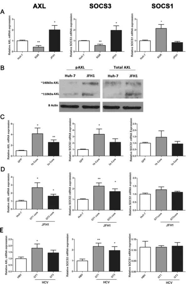
We previously demonstrated that infection of Huh-7 cells with HCV (JFH1 strain) induces the expression ofAXLas well as its downstream targetSOCS3[13]. To further confirm the mecha-nism by which AXL and SOCS3 are induced during HCV infection, we examined their expres-sion in Huh-7 cells containing a HCV subgenomic replicon (SGR), which expresses only non-structural viral proteins, and in cells containing fully replicating JFH1 virus. BothAXLand
SOCS3were up-regulated approximately 2 fold in JFH1 infected cells (minimum 2 passages, 1 week post infection, 90% infectivity), but were surprisingly down-regulated in a stable cell line of Huh-7s harbouring a genotype 2a SGR [19] (Fig 1A).SOCS1demonstrated an opposite expression pattern, being up-regulated 2 fold in cells harbouring the SGR (p<0.05). To
deter-mine whether the AXL protein is activated and thus able to induce SOCS3 in JFH1 infected Huh-7 cells, we examined AXL phosphorylation at tyrosine 779 by western blot. Interestingly, both total AXL and phosphorylated AXL were up-regulated in JFH1 infected cells (Fig 1B). Furthermore, both glycosylation states of AXL, represented by bands at approximately 110 and 140 kDa, were increased in JFH1 infected cells. These data suggest that AXL may drive SOCS3 expression in these cells, as has been shown in other models [12,20].
Because the HCV structural protein core has been shown to induce SOCS3 expressionin vitro[6], we examined AXL/SOCS3 expression in HepG2 cells over-expressing genotype 1b and 3a HCV core protein, using a baculovirus expression system. At 48 h post-electroporation, genotype 1b core significantly up-regulated bothAXLandSOCS3by 4 and 3 fold respectively, while genotype 3a core up-regulatedSOCS3alone by 2 fold (Fig 1C). Next, we examinedAXL
andSOCS3expression in Huh-7 cells harbouring chimeric HCV constructs, which express genotype 1b or 3a core in a JFH1 (genotype 2a) virus, and observed similar results. While both chimeric viruses replicated at lower levels than wild type JFH1 (S1 Fig), both induced signifi-cantAXLandSOCS3up-regulation, albeit more potently following infection with the genotype 1b chimera (Fig 1D). Neither core expression nor chimeric virus induced the expression of
HCV induced AXL expression is mediated by antiviral and inflammatory
signalling pathways
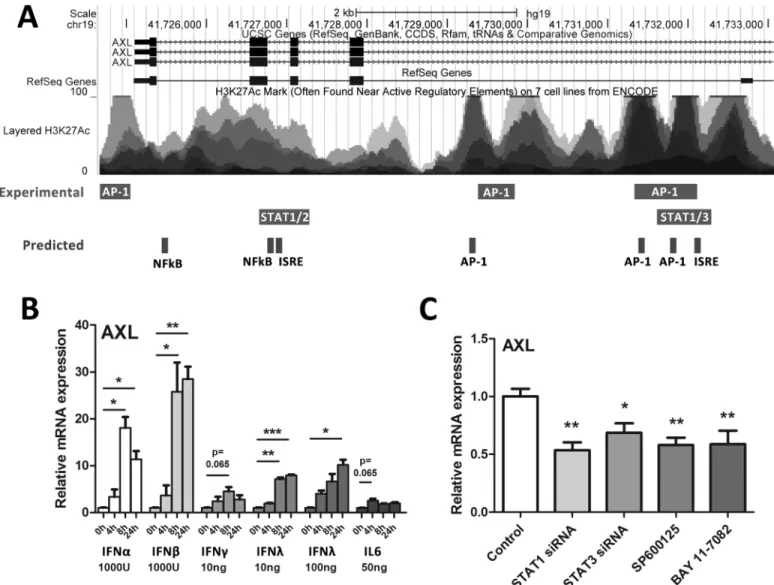
To determine cell factors that may influence AXL expression, the UCSC genome browser was used to identify experimentally validated transcription factor binding sites within theAXL pro-moter/enhancer region (S2 Fig) [16]. The ECR Browser was used to identify evolutionarily con-served regions within the promoter and introns of theAXLgene, and rVISTA was used to find conserved transcription factor binding sites, using theRattus Norvegicusgenome as a reference (S3 Fig) [17,18]. A number of transcription factors that mediate innate immune signalling, including STAT1, 2 and 3, as well as AP-1 signalling components (c-Fos, c-Jun), have been shown to bind to theAXLgene, principally at intronic sequences in the 5’region (Fig 2A). Fur-thermore, a number of evolutionarily conserved transcription factor binding sites were pre-dicted in overlapping regions, including additional NFκB binding sites.
To identify which innate immune cytokines that are typically induced by HCV can up-regu-late AXL expression, we treated Huh-7 cells with IFNsα(1000 U/ml),β(1000 U/ml),γ(10ng/ ml) andλ3 (10 and 100 ng/ml) as well as IL6 (50 ng/ml) for 24 h and measuredAXL. Interest-ingly, all cytokines tested inducedAXLexpression, albeit to differing degrees, with type I inter-feronsαandβhaving the strongest effect (Fig 2B). Potential IFNαmediated induction of other members of the TAM family of receptor tyrosine kinases (Tyro3 and Mer) was investigated, but onlyAXLwas found to be an ISG (S4 Fig).
To better understand how AXL is induced following HCV infection, we inhibited multiple innate immune and inflammatory signalling pathways in JFH1 infected Huh-7 cells, using either siRNA or chemical inhibitors. STAT1 and STAT3 siRNAs were used to inhibit IFN and IL6 induced signalling pathways; SP600125 was used to inhibit JNK and downstream AP-1 sig-nalling and BAY11-7082 was used to inhibit NFκB sigsig-nalling. The efficacy of siRNA gene knockdown and all chemical inhibitors was confirmed by STAT1/3 western blot and NFκB/ AP-1 promoter activation using luciferase reporter plasmids respectively (S5 Fig). Inhibition of STAT1 and 3, JNK or NFκB signalling each reducedAXLexpression by approximately 2 fold (Fig 2C), suggesting that HCV mediated AXL expression is complex and is likely mediated by multiple transcription factors. Expression of SOCS1 and SOCS3 was unaffected (data not shown).
AXL knockdown reduces SOCS3 expression but does not affect JFH1
replication
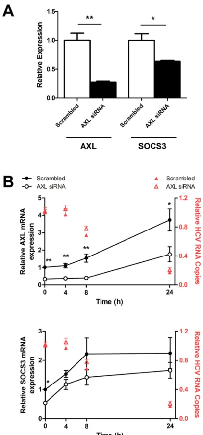
To determine the role of AXL in SOCS3 induction, as well as in the interferon response to HCV infection,AXLwas knocked down in JFH1 infected Huh-7 cells using 10nM AXL siRNA. SiRNA mediated knock down ofAXLby approximately 75% significantly reducedSOCS3
expression, by approximately 40% (Fig 3A).AXLknockdown was maintained for 24 h after 50 U/ml IFNαtreatment, whileSOCS3expression remained down-regulated, albeit modestly (Fig 3B).AXLknockdown had no effect on HCV replication, either at baseline or following IFNαtreatment (red triangles), nor did it have any effect on ISG expression (data not shown).
AXL overexpression dampens the ISG response to IFN
α
in Huh-7 cells
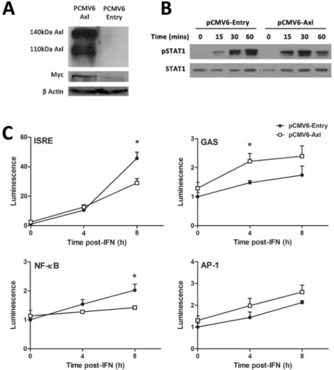
Next, AXL was over-expressed in Huh-7 cells, to determine whether it can influence STAT1 phosphorylation, promoter activation and ISG expression in response to IFNα. Strong AXL
cells transfected with baculovirus expressing HCV core protein (C), as well as in Huh-7 cells infected with core chimeric HCV viruses (D) (*p<0.05, **p<0.01). Data represents the mean and standard error of three biological replicates, each with duplicate samples.
overexpression was achieved following stable transfection with the PCMV6-AXL vector, with PCMV6-Entry as a control. This was confirmed by western blot, using antibodies against either AXL or the Myc protein tag (Fig 4A). In cells overexpressing AXL, IFNα(50 U/ml) induced a more transient activation of STAT1, characterised by stronger STAT1 phosphorylation 15 and 30 minutes post treatment, with reduced STAT1 phosphorylation at 60 minutes (Fig 4B). Fur-thermore, in AXL over-expressing Huh-7 cells, 50 U/ml IFNαinduced activation of the inter-feron stimulated response element (ISRE) and NFκB was significantly blunted 8 h post-treatment, using luciferase reporter constructs (Fig 4C). In contrast, the gamma activated sequence (GAS) activation increased almost two fold at 4 h post treatment and no change in activation of the AP-1 promoter was observed in AXL overexpressing cells. Together, these Fig 2. AXL expression is mediated by multiple innate immune signalling pathways.To identify inflammatory transcription factor binding sites within the AXL promoter/enhancer region that may mediate AXL expression, the UCSC genome browser was interrogated for experimental ChIP binding sites (A). Furthermore, the ECR browser and rVista were used to identify evolutionarily conserved regions and to predict transcription factor binding sites, respectively. Huh-7 cells were treated with a range of antiviral cytokines for 24 h then AXL expression measured by qPCR (B). IFNsα,β, andλup-regulated AXL more potently than pro-inflammatory cytokines IFNγand IL6 (average of two biological replicates in duplicate for each). (C) To inhibit potential signalling
components that induceAXL, Huh-7 cells were treated with 10nM siRNA against STAT1/STAT3 or chemical inhibitors of JNK (SP600125) or NFκB (BAY11-7082) for 24 h. Inhibition of STAT1, STAT3, JNK (SP600125) and NFκB (BAY 11–7082) all significantly reduced HCV induced AXL expression (*p<0.05,
**p<0.01,***p<0.001) (average of three biological replicates in duplicate). Data represents the mean and standard error.
Fig 3. AXL knockdown reduces SOCS3 expression but does not affect HCV replication.AXLspecific
data agree with previous reports in dendritic cells, suggesting that AXL highjacks signalling from the type I IFN receptor, thereby limiting ISRE and NFκB activation, and also produces STAT1 homodimers that bind the GAS sequence [12].
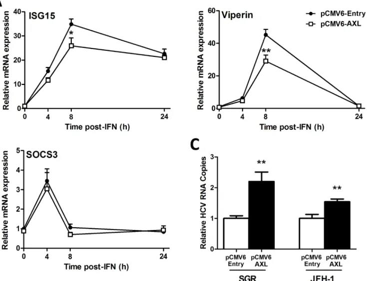
We next examined ISG expression in response to IFNαto determine whether modulation of promoter activity by AXL (Fig 4) also reduces ISG expression. Huh-7 cells over-expressing AXL were significantly less responsive to 50 U/ml IFNα, with reduced induction ofISG15and
viperin(Fig 5A). Surprisingly, AXL overexpression had no effect on expression ofSOCS1(data not shown) orSOCS3(Fig 5B), suggesting that in this model, AXL alone is likely responsible for regulation of IFNαsignalling. To determine whether AXL exerts IFN regulatory and thus
viral replication following IFNαtreatment (red triangles) (*p<0.05,**p<0.01). Data represents the mean and standard error of two biological replicates performed in duplicate.
doi:10.1371/journal.pone.0136227.g003
Fig 4. AXL overexpression dampens the response to IFNα.Stable transfection of Huh-7 cells with PCMV6-AXL was confirmed by western blot, using antibodies against AXL or the fusion Myc tag (A). Huh-7 cells overexpressing AXL induced a stronger but more transient phosphorylation of STAT1 (B), with increased phosphorylation at 15 and 30 minutes post-IFN (50 u/ml), but a strong decrease at 1 h (2 replicates). ISRE and NFκB promoter activation was decreased almost 2 fold at 8 h post-IFN treatment in cells overexpressing AXL, while GAS activation were increased 2 fold at 4 h post-IFN treatment (C). No effect on AP-1 promoter activation was observed (*p<0.05). Data represents the mean and standard error of two biological replicates performed in duplicate.
pro-viral effects, both control and AXL over-expressing Huh-7 cell lines were transfected with RNA for either full length HCV virus (JFH1 strain) or a JFH1-derived SGR, lacking structural. AXL over-expression increased viral replication over 2 fold for the SGR and 1.5 fold for JFH1 (Fig 5C), suggesting that AXL can weaken the antiviral response to HCVin vitro.
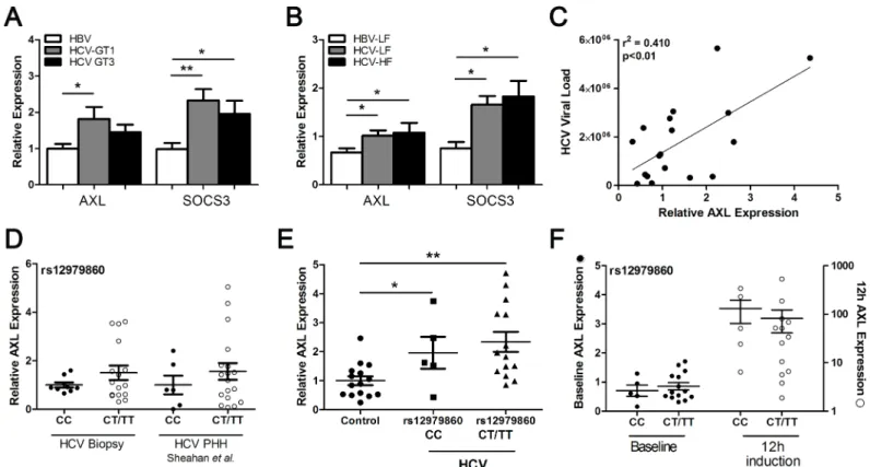
AXL and SOCS3 are up-regulated in HCV infected liver
To determine whether ourin vitroresults mimicin vivogene expression, we compared liver biopsies from patients infected with HCV genotype 1 or 3, with HBV infected livers as controls. BothAXLandSOCS3were significantly up-regulated in genotype 1 infected livers compared to HBV controls, whereasSOCS3was only significantly higher in the genotype 3 infected livers (Fig 6A). BothAXLandSOCS3were significantly increased in HCV infected livers with low (F0-2) or high (F3-4) levels of fibrosis, compared to low fibrosis HBV liver biopsies, indicating thatAXLexpression is not affected by fibrosis stage (Fig 6B). A strong correlation was also Fig 5. AXL overexpression dampens the antiviral response.Following treatment with 50 U/ml IFNα, Huh-7 cells stably expressing AXL demonstrated blunted ISG15 and viperin expression (A), but no change in SOCS3 (B), compared to control cells. Stable AXL overexpression also mediated an increase in full length JFH1 and SGR viral replication at 24 h post-transfection (C) (*p<0.05,**p<0.01). Data represents the mean and standard error of three biological replicates performed in duplicate.
observed between hepatic AXL expression and HCV viral load (Fig 6C), suggesting that increased viral replication and subsequent activation of immune signalling pathways may drive AXL expression in the liver.
We next examinedAXLexpression in liver and blood from HCV infected patients, stratified by the IFNL3 rs12979860 SNP. HepaticAXLexpression was higher in patients with the non-responder rs12979860 CT/TT genotype (Fig 6D), consistent with published data showing ele-vated hepatic ISGs in patients possessing the IFNL3 non-responder haplotype [3,14]. To exam-ine the effect of IFNL3 genotype on AXL in a PHH model, we utilized publicly available microarray data examining expression of primary human hepatocytes (PHHs) infected with HCV (Geo Dataset GSE54648). Laser capture microdissection was utilized to compare infected and infection adjacent cells, and was further stratified by IFNL3 rs12979860 genotype [21]. By analysing AXL expression in both infected and adjacent PHHs (gene expression results com-bined to simulatein vivoinfection) we found similar results asin vivoHCV infection, with ele-vatedAXLexpression in HCV infected PHH isolated from patients with the“non-responder” rs12979860 CT/TT genotype (Fig 6D).
To determine whether AXL induction is mediated directly by viral infection or by subse-quent cytokine expression, we examined AXL expression in infected, adjacent and mock infected PHHs from the same dataset. AXL expression increased in infected cells from day 1 to day 7 (S6 Fig), and was consistently higher in infected cells than either adjacent cells or mock Fig 6. HCV genotype and IFNL3 SNP modulate AXL expression.AXLandSOCS3expression were examined in liver biopsies from patients with chronic
HBV or chronic HCV infection, to look for virus specific differences in gene induction. HCV Genotype 1 infected livers demonstrated significantly higher expression of bothSOCS3andAXL(A), with no effect of liver fibrosis grade on expression of eitherSOCS3ofAXLin HCV infected livers (B).AXLexpression also demonstrated a strong correlation with viral load, supporting the direct role of HCV in AXL induction (C). The rs12979860IFNL3CC SNP correlated with
lower baseline hepaticAXLexpression in genotype 1 HCV infected livers and JFH1 infected PHHs (D). PBMC expression of AXL was up-regulated in HCV
infected individuals, and similar to hepatic expression, was lower in patients possessing the CC genotype (E). StrongerAXLup-regulation in PBMCs 12 h after the first IFN injection was also observed (F) (*p<0.05,**p<0.01).
infected controls (p<0.05 vs mock, day 3). This suggests that although cytokines may
contrib-ute to AXL expressionin vivo, AXL up-regulation occurs primarily in infected cells.
AXLexpression was also significantly elevated in peripheral blood mononuclear cells (PBMCs) from HCV infected individuals (n = 20) compared to healthy controls (n = 15) (p<0.05) (Fig 6E). Furthermore, patients possessing the responder rs12979860 CC genotype
displayed lowerAXLexpression than CT/TT carriers, in agreement with hepaticAXL expres-sion. Finally, induction ofAXL12 h after the first dose of pegylated IFN was stronger in PBMCs from patients possessing the responder CC genotype, compared with CT/TT non-responder patients (Fig 6F).
Discussion
We previously demonstrated that AXL is up-regulated following HCV infectionin vitro, and may contribute to SOCS3 expression and interferon refractoriness [13]. We now demonstrate that AXL is induced in a genotype dependent manner (GT1>3)in vitroas well asin vivo, and
is likely induced by IFNs and other inflammatory mediators. AXL knockdown showed little effect on IFN signalling, however AXL overexpression reduced Huh-7 responsiveness to IFN, as well as the antiviral response against HCV.
Our current findings build on our previous report that HCV induces AXL expressionin vitro. We have now clarified (1) that viral replication alone, in the absence of HCV structural proteins, is not sufficient to induce AXL/SOCS3 expression, and (2) that genotype 1 core pro-tein, either alone or as part of a chimeric virus, is a more potent inducer of AXL/SOCS3 than genotype 3 core (Fig 1). The similar expression pattern of AXL and SOCS3 suggests that AXL may induce SOCS3 in our model, particularly in the presence of genotype 1 core protein, and we have confirmed the association with HCV genotype in human liver tissue (Fig 6). This sug-gests that AXL may drive increased expression of SOCS3, which has been implicated in geno-type 1 mediated insulin resistance and treatment non-response [22–25].
Because type I IFN has been shown to induce AXL in myeloid cells in the blood [11,12,26], we sought to determine whether we could reproduce AXL induction in Huh-7 cells. Surpris-ingly, the inflammatory cytokines IL6 and IFNγonly modestly up-regulatedAXL, while type I/ III IFNs (β>α>λ) potently induced its expression. Knockdown of STAT1/3 or chemical inhibi-tion of JNK or NFκB signalling all reducedAXLexpression in JFH1 infected Huh-7 cells, sug-gesting that AXL induction is mediated by multiple transcription factors, similar to the IFNβ enhanceosome [27]. Because HCV induces oxidative stress, inflammation, and a strong type III immune response in the liver, it is not surprising that the activation of these transcription factors following infectionin vitroinducedAXLexpression [28–33].
Finally, we observed a trend towards lower hepaticAXLexpression and a stronger ISG response in blood PBMCs among patients with the favourable rs12979860 IFNL3 allele (CC), although this was not statistically significant, possibly due to insufficient statistical power. These data mirror previous reports, that theIFNL3non-responder haplotype is associated with higher baseline ISGs in the liver and a weaker immune response in the blood [14,34,35], but provide a novel mechanism to explain this. Because AXL expression is induced by type I and III IFNs, it is likely that the recently described IFNL4 also induces AXL. IFNL4 is only expressed in the livers of patients with the unfavourable SNP (ss469415590), so IFNL4 driven AXL could potentially reduce the antiviral response to exogenous IFN in these individuals. Nonetheless, because AXL is an ISG, it may have the ability to regulate its own expression, and possibly the expression of other negative regulators such as USP18. This adds another layer of complexity to IFN regulation, and clearly requires further study.
The consequences of HCV mediated AXL up-regulation are not limited to regulation of IFN signalling. Up-regulation of AXL is well documented in hepatocellular carcinoma (HCC), and contributes to HCC cell line proliferation, migration and invasion [36–38], through activa-tion of AKT and MAPK signalling pathways (reviewed in [39]). Moreover, AXL serves as an entry factor for a number of enveloped viruses, including the related dengue flavivirus [40–42]. If the same is true for HCV, immune-driven AXL expression in hepatocytes in the HCV infected liver could facilitate the spread of the virus into uninfected cells.
In summary, we have shown that AXL is induced by HCV infection, and is capable of regu-lating the ISG response to HCV in hepatocytes. The immunomodulatory role of AXL in the context of IFNL3/4 polymorphisms remains uncertain, but may provide a mechanism for the differences observed in hepatic and blood ISG regulation, based on the dominant IFNs in each cell type.
Supporting Information
S1 Fig. Relative HCV RNA replication in wild type JFH1 and genotype 1 and 3 core chi-mera infected Huh-7 cells.Intracellular HCV RNA content in JFH1 core chimeras was approximately 25% of wild type JFH1 virus. Reduced replication capacity is likely the result of decreased viral fitness from intergenotypic core replacement.
(TIF)
S2 Fig. Transcription factor binding to the AXL promoter and enhancer.Experimental ChIP studies have demonstrated a large degree of transcription factor binding within the 4th intron of AXL, suggesting the presence of an enhancer region.
(TIF)
S3 Fig. Conserved AXL sequence inHomo sapiensandRattus norvegicus.The 5’portion of the AXL gene is conserved in rats but not in mice as demonstrated by the ECR browser. (TIF)
S4 Fig. AXL is the only TAM receptor that is an ISG.Expression of AXL, but not TYRO3 or MER, was significantly up-regulated in Huh-7 cells 8 h after treatment with 100 U/ml IFNα, (p<0.001).
(TIF)
reporters were drastically reduced using 50μM SP600125 and 25μM BAY11-7082 respec-tively.
(TIF)
S6 Fig. AXL expression in HCV infected primary human hepatocytes.AXL expression is up-regulated following HCV infection of primary human hepatocytes, compared to adjacent uninfected cells and mock infected cells (p<0.05, infected vs mock, day 3). Data taken from
Geo Dataset GSE54648 using hepatocytes isolated by laser capture microdissection. (TIF)
Acknowledgments
We would like to thank all members of the Storr Liver Centre for their scientific insights throughout the development of this project, particularly Dr Lionel Hebbard, Dr Liang Qiao and Dr Chris Liddle.
Author Contributions
Conceived and designed the experiments: SAR EST MS KSO DRB JG MWD. Performed the experiments: SAR EST MS KSO. Analyzed the data: SAR KSO. Contributed reagents/materi-als/analysis tools: DRB JG. Wrote the paper: SAR EST MS KSO DRB JG MWD.
References
1. Horner SM, Gale M Jr (2013) Regulation of hepatic innate immunity by hepatitis C virus. Nature Medi-cine 19: 879–888. doi:10.1038/nm.3253PMID:23836238
2. Lanford RE, Guerra B, Bigger CB, Lee H, Chavez D, Brasky KM (2007) Lack of response to exogenous interferon-alpha in the liver of chimpanzees chronically infected with hepatitis C virus. Hepatology 46: 999–1008. PMID:17668868
3. Sarasin-Filipowicz M, Oakeley EJ, Duong FH, Christen V, Terracciano L, Filipowicz W, et al. (2008) Interferon signaling and treatment outcome in chronic hepatitis C. Proc Natl Acad Sci U S A 105: 7034–7039. doi:10.1073/pnas.0707882105PMID:18467494
4. Sarasin-Filipowicz M, Wang X, Yan M, Duong FH, Poli V, Hilton DJ, et al. (2009) Alpha interferon induces long-lasting refractoriness of JAK-STAT signaling in the mouse liver through induction of USP18/UBP43. Mol Cell Biol 29: 4841–4851. doi:10.1128/MCB.00224-09PMID:19564419 5. Duong FH, Filipowicz M, Tripodi M, La Monica N, Heim MH (2004) Hepatitis C virus inhibits interferon
signaling through up-regulation of protein phosphatase 2A. Gastroenterology 126: 263–277. PMID:
14699505
6. Bode JG, Ludwig S, Ehrhardt C, Albrecht U, Erhardt A, Schaper F, et al. (2003) IFN-alpha antagonistic activity of HCV core protein involves induction of suppressor of cytokine signaling-3. FASEB J 17: 488–490. PMID:12551851
7. Suppiah V, Moldovan M, Ahlenstiel G, Berg T, Weltman M, Abate ML, et al. (2009) IL28B is associated with response to chronic hepatitis C interferon-alpha and ribavirin therapy. Nat Genet 41: 1100–1104.
doi:10.1038/ng.447PMID:19749758
8. Tanaka Y, Nishida N, Sugiyama M, Kurosaki M, Matsuura K, Sakamoto N, et al. (2009) Genome-wide association of IL28B with response to pegylated interferon-alpha and ribavirin therapy for chronic hepa-titis C. Nat Genet 41: 1105–1109. doi:10.1038/ng.449PMID:19749757
9. Ge D, Fellay J, Thompson AJ, Simon JS, Shianna KV, Urban TJ, et al. (2009) Genetic variation in IL28B predicts hepatitis C treatment-induced viral clearance. Nature 461: 399–401. doi:10.1038/
nature08309PMID:19684573
10. Prokunina-Olsson L, Muchmore B, Tang W, Pfeiffer RM, Park H, Dickensheets H, et al. (2013) A variant upstream of IFNL3 (IL28B) creating a new interferon gene IFNL4 is associated with impaired clearance of hepatitis C virus. Nature Genetics 45: 164–171. doi:10.1038/ng.2521PMID:23291588
11. Sharif MN, Sosic D, Rothlin CV, Kelly E, Lemke G, Olson EN, et al. (2006) Twist mediates suppression of inflammation by type I IFNs and Axl. J Exp Med 203: 1891–1901. PMID:16831897
13. Read SA, Tay ES, Shahidi M, McLauchlan J, George J, Douglas MW (2015) The Mechanism of Inter-feron Refractoriness During Hepatitis C Virus Infection and Its Reversal with a Peroxisome Proliferator-Activated Receptor alpha Agonist. Journal of Interferon and Cytokine Research.
14. O'Connor KS, Parnell G, Patrick E, Ahlenstiel G, Suppiah V, van der Poorten D, et al. (2014) Hepatic metallothionein expression in chronic hepatitis C virus infection is IFNL3 genotype-dependent. Genes Immun 15: 88–94. doi:10.1038/gene.2013.66PMID:24335707
15. Read SA, Tay E, Shahidi M, George J, Douglas MW (2014) Hepatitis C virus infection mediates choles-teryl ester synthesis to facilitate infectious particle production. Journal of General Virology 95: 1900–
1910. doi:10.1099/vir.0.065300-0PMID:24859394
16. Kent WJ, Sugnet CW, Furey TS, Roskin KM, Pringle TH, Zahler AM, et al. (2002) The human genome browser at UCSC. Genome Res 12: 996–1006. PMID:12045153
17. Ovcharenko I, Nobrega MA, Loots GG, Stubbs L (2004) ECR Browser: a tool for visualizing and accessing data from comparisons of multiple vertebrate genomes. Nucleic Acids Res 32: W280–286.
PMID:15215395
18. Loots GG, Ovcharenko I (2004) rVISTA 2.0: evolutionary analysis of transcription factor binding sites. Nucleic Acids Res 32: W217–221. PMID:15215384
19. Kato T, Date T, Miyamoto M, Furusaka A, Tokushige K, Mizokami M, et al. (2003) Efficient replication of the genotype 2a hepatitis C virus subgenomic replicon. Gastroenterology 125: 1808–1817. PMID:
14724833
20. Zheng S, Hedl M, Abraham C (2015) TAM Receptor-Dependent Regulation of SOCS3 and MAPKs Contributes to Proinflammatory Cytokine Downregulation following Chronic NOD2 Stimulation of Human Macrophages. J Immunol 194: 1928–1937. doi:10.4049/jimmunol.1401933PMID:25567680 21. Sheahan T, Imanaka N, Marukian S, Dorner M, Liu P, Ploss A, et al. (2014) Interferon lambda alleles
predict innate antiviral immune responses and hepatitis C virus permissiveness. Cell Host Microbe 15: 190–202. doi:10.1016/j.chom.2014.01.007PMID:24528865
22. Persico M, Capasso M, Persico E, Svelto M, Russo R, Spano D, et al. (2007) Suppressor of cytokine signaling 3 (SOCS3) expression and hepatitis C virus-related chronic hepatitis: Insulin resistance and response to antiviral therapy. Hepatology 46: 1009–1015. PMID:17668875
23. Huang Y, Feld JJ, Sapp RK, Nanda S, Lin JH, Blatt LM, et al. (2007) Defective hepatic response to interferon and activation of suppressor of cytokine signaling 3 in chronic hepatitis C. Gastroenterology 132: 733–744. PMID:17258724
24. Hui JM, Sud A, Farrell GC, Bandara P, Byth K, Kench JG, et al. (2003) Insulin resistance is associated with chronic hepatitis C virus infection and fibrosis progression [corrected]. Gastroenterology 125: 1695–1704. PMID:14724822
25. Pazienza V, Clement S, Pugnale P, Conzelman S, Foti M, Mangia A, et al. (2007) The hepatitis C virus core protein of genotypes 3a and 1b downregulates insulin receptor substrate 1 through genotype-spe-cific mechanisms. Hepatology 45: 1164–1171. PMID:17465001
26. Scutera S, Fraone T, Musso T, Cappello P, Rossi S, Pierobon D, et al. (2009) Survival and migration of human dendritic cells are regulated by an IFN-alpha-inducible Axl/Gas6 pathway. J Immunol 183: 3004–3013. doi:10.4049/jimmunol.0804384PMID:19657094
27. Yie J, Senger K, Thanos D (1999) Mechanism by which the IFN-beta enhanceosome activates tran-scription. Proceedings of the National Academy of Sciences of the United States of America 96: 13108–13113. PMID:10557281
28. You LR, Chen CM, Lee YH (1999) Hepatitis C virus core protein enhances NF-kappaB signal pathway triggering by lymphotoxin-beta receptor ligand and tumor necrosis factor alpha. J Virol 73: 1672–1681.
PMID:9882379
29. Lin W, Tsai WL, Shao RX, Wu G, Peng LF, Barlow LL, et al. (2010) Hepatitis C virus regulates trans-forming growth factor beta1 production through the generation of reactive oxygen species in a nuclear factor kappaB-dependent manner. Gastroenterology 138: 2509–2518, 2518 e2501. doi:10.1053/j.
gastro.2010.03.008PMID:20230822
30. Qadri I, Iwahashi M, Capasso JM, Hopken MW, Flores S, Schaack J, et al. (2004) Induced oxidative stress and activated expression of manganese superoxide dismutase during hepatitis C virus replica-tion: role of JNK, p38 MAPK and AP-1. Biochem J 378: 919–928. PMID:14670077
31. Soo HM, Garzino-Demo A, Hong W, Tan YH, Tan YJ, Goh PY, et al. (2002) Expression of a full-length hepatitis C virus cDNA up-regulates the expression of CC chemokines MCP-1 and RANTES. Virology 303: 253–277. PMID:12490388
33. Park H, Serti E, Eke O, Muchmore B, Prokunina-Olsson L, Capone S, et al. (2012) IL-29 is the dominant type III interferon produced by hepatocytes during acute hepatitis C virus infection. Hepatology 56: 2060–2070. doi:10.1002/hep.25897PMID:22706965
34. Urban TJ, Thompson AJ, Bradrick SS, Fellay J, Schuppan D, Cronin KD, et al. (2010) IL28B genotype is associated with differential expression of intrahepatic interferon-stimulated genes in patients with chronic hepatitis C. Hepatology 52: 1888–1896. doi:10.1002/hep.23912PMID:20931559
35. Honda M, Sakai A, Yamashita T, Nakamoto Y, Mizukoshi E, Sakai Y, et al. (2010) Hepatic ISG expres-sion is associated with genetic variation in interleukin 28B and the outcome of IFN therapy for chronic hepatitis C. Gastroenterology 139: 499–509. doi:10.1053/j.gastro.2010.04.049PMID:20434452 36. Tsou AP, Wu KM, Tsen TY, Chi CW, Chiu JH, Lui WY, et al. (1998) Parallel hybridization analysis of
multiple protein kinase genes: identification of gene expression patterns characteristic of human hepa-tocellular carcinoma. Genomics 50: 331–340. PMID:9676427
37. He L, Zhang J, Jiang L, Jin C, Zhao Y, Yang G, et al. (2010) Differential expression of Axl in hepatocel-lular carcinoma and correlation with tumor lymphatic metastasis. Mol Carcinog 49: 882–891. doi:10.
1002/mc.20664PMID:20635370
38. Lee HJ, Jeng YM, Chen YL, Chung L, Yuan RH (2014) Gas6/Axl pathway promotes tumor invasion through the transcriptional activation of Slug in hepatocellular carcinoma. Carcinogenesis 35: 769–
775. doi:10.1093/carcin/bgt372PMID:24233839
39. Linger RM, Keating AK, Earp HS, Graham DK (2008) TAM receptor tyrosine kinases: biologic functions, signaling, and potential therapeutic targeting in human cancer. Adv Cancer Res 100: 35–83. doi:10.
1016/S0065-230X(08)00002-XPMID:18620092
40. Brindley MA, Hunt CL, Kondratowicz AS, Bowman J, Sinn PL, McCray PB Jr., et al. (2011) Tyrosine kinase receptor Axl enhances entry of Zaire ebolavirus without direct interactions with the viral glyco-protein. Virology 415: 83–94. doi:10.1016/j.virol.2011.04.002PMID:21529875
41. Morizono K, Xie Y, Olafsen T, Lee B, Dasgupta A, Wu AM, et al. (2011) The soluble serum protein Gas6 bridges virion envelope phosphatidylserine to the TAM receptor tyrosine kinase Axl to mediate viral entry. Cell Host Microbe 9: 286–298. doi:10.1016/j.chom.2011.03.012PMID:21501828 42. Meertens L, Carnec X, Lecoin MP, Ramdasi R, Guivel-Benhassine F, Lew E, et al. (2012) The TIM and
TAM families of phosphatidylserine receptors mediate dengue virus entry. Cell Host Microbe 12: 544–