Herpesvirus Genome Recognition Induced
Acetylation of Nuclear IFI16 Is Essential for
Its Cytoplasmic Translocation,
Inflammasome and IFN-
β
Responses
Mairaj Ahmed Ansari, Sujoy Dutta, Mohanan Valiya Veettil, Dipanjan Dutta, Jawed Iqbal, Binod Kumar, Arunava Roy, Leela Chikoti, Vivek Vikram Singh, Bala Chandran*
H. M. Bligh Cancer Research Laboratories, Department of Microbiology and Immunology, Chicago Medical School, Rosalind Franklin University of Medicine and Science, North Chicago, Illinois, United States of America
*bala.chandran@rosalindfranklin.edu
Abstract
The IL-1βand type I interferon-β(IFN-β) molecules are important inflammatory cytokines elicited by the eukaryotic host as innate immune responses against invading pathogens and danger signals. Recently, a predominantly nuclear gamma-interferon-inducible protein 16 (IFI16) involved in transcriptional regulation has emerged as an innate DNA sensor which induced IL-1βand IFN-βproduction through inflammasome and STING activation, respectively. Herpesvirus (KSHV, EBV, and HSV-1) episomal dsDNA genome recognition by IFI16 leads to IFI16-ASC-procaspase-1 inflammasome association, cytoplasmic translo-cation and IL-1βproduction. Independent of ASC, HSV-1 genome recognition results in IFI16 interaction with STING in the cytoplasm to induce interferon-βproduction. However, the mechanisms of IFI16-inflammasome formation, cytoplasmic redistribution and STING activation are not known. Our studies here demonstrate that recognition of herpesvirus genomes in the nucleus by IFI16 leads into its interaction with histone acetyltransferase p300 and IFI16 acetylation resulting in IFI16-ASC interaction, inflammasome assembly, increased interaction with Ran-GTPase, cytoplasmic redistribution, caspase-1 activation, IL-1βproduction, and interaction with STING which results in IRF-3 phosphorylation, nuclear pIRF-3 localization and interferon-βproduction. ASC and STING knockdowns did not affect IFI16 acetylation indicating that this modification is upstream of inflammasome-assembly and STING-activation. Vaccinia virus replicating in the cytoplasm did not induce nuclear IFI16 acetylation and cytoplasmic translocation. IFI16 physically associates with KSHV and HSV-1 genomes as revealed by proximity ligation microscopy and chromatin-immunoprecipitation studies which is not hampered by the inhibition of acetylation, thus suggesting that acetylation of IFI16 is not required for its innate sensing of nuclear viral genomes. Collectively, these studies identify the increased nuclear acetylation of IFI16 as a dynamic essential post-genome recognition event in the nucleus that is common to the OPEN ACCESS
Citation:Ansari MA, Dutta S, Veettil MV, Dutta D, Iqbal J, Kumar B, et al. (2015) Herpesvirus Genome Recognition Induced Acetylation of Nuclear IFI16 Is Essential for Its Cytoplasmic Translocation, Inflammasome and IFN-βResponses. PLoS Pathog 11(7): e1005019. doi:10.1371/journal.ppat.1005019
Editor:Pinghui Feng, University of Southern California, UNITED STATES
Received:March 5, 2015
Accepted:June 9, 2015
Published:July 2, 2015
Copyright:© 2015 Ansari et al. This is an open access article distributed under the terms of the
Creative Commons Attribution License, which permits unrestricted use, distribution, and reproduction in any medium, provided the original author and source are credited.
Data Availability Statement:All relevant data are within the paper and its Supporting Information files.
Funding:BC received funding from the National Institute of Health CA 180758http://www.cancer.gov/. BC received funding from the National Institute of Health AI 113391http://www.niaid.nih.gov/Pages/ default.aspx. The funders had no role in study design, data collection and analysis, decision to publish, or preparation of the manuscript.
IFI16-mediated innate responses of inflammasome induction and IFN-βproduction during herpesvirus (KSHV, EBV, HSV-1) infections.
Author Summary
Herpesviruses establish a latent infection in the nucleus of specific cells and reactivation results in the nuclear viral dsDNA replication and infectious virus production. Host innate responses are initiated by the presence of viral genomes and their products, and nucleus associated IFI16 protein has recently emerged as an innate DNA sensor regulating inflam-matory cytokines and type I interferon (IFN) production. IFI16 recognizes the herpesvirus genomes (KSHV, EBV, and HSV-1) in the nucleus resulting in the formation of the IFI16-ASC-Caspase-1 inflammasome complex and IL-1βproduction. HSV-1 genome recogni-tion by IFI16 in the nucleus also leads to STING activarecogni-tion in the cytoplasm and IFN-β
production. However, how IFI16 initiates inflammasome assembly and activates STING in the cytoplasm after nuclear recognition of viral genome are not known. We show that herpesvirus genome recognition in the nucleus by IFI16 leads to interaction with histone acetyltransferase-p300 and IFI16 acetylation which is essential for inflammasome assem-bly in the nucleus and cytoplasmic translocation, activation of STING in the cytoplasm and IFN-βproduction. These studies provide insight into a common molecular mecha-nism for the innate inflammasome assembly and STING activation response pathways that result in IL-1βand IFN-βproduction, respectively.
Introduction
Kaposi’s sarcoma associated herpes virus (KSHV), aγ-2 herpesvirus, is etiologically associated with Kaposi’s sarcoma (KS) and primary effusion lymphoma (PEL) [1]. The hallmark of KSHV infection is the establishment of latent infection, reactivation and reinfection, and KS and PEL lesion endothelial and B cells, respectively, carry episomal KSHV latent dsDNA genome [1]. Human PEL (B) cell lines BCBL-1 and BC-3 carry>80 copies of the episomal latent KSHV genome/cell and the lytic cycle can be induced by chemicals. Purified virions from the supernatants are used forin vitroinfection of human dermal microvascular endothe-lial cells (HMVEC-d) and foreskin fibroblast cells (HFF) [2].
including transfected DNA and DNA of pox viruses replicating in the cytoplasm [4,7,8,9]. They initiate the host defenses by regulating the production of IL-1β, IL-18, IL-33 or type I interferons (IFN)α/β[7,8,9,10].
Whether innate responses recognize and respond to the presence of foreign episomal genomes of herpesviruses as well as other DNA viruses in the infected cell nuclei leading into the induction of inflammatory responses was not known initially. Our studies revealed thatin vitroKSHV infection of endothelial cells induces caspase-1 activation via the nuclear resident gamma-interferon-inducible protein-16 (IFI16) also known as interferon-inducible myeloid differentiation transcriptional activator. Colocalization of IFI16 with viral genome in the infected endothelial cell nucleus, induction of IFI16-ASC inflammasomes by UV-inactivated KSHV and the absence of induction by lentivirus vectors expressing KSHV genes demon-strated that a) KSHV genes individually do not play a role in IFI16-inflammasome activation, b) the IFI16-inflammasome is not induced against linear integrated foreign DNA, and c) episomal KSHV genome is required for IFI16-inflammasome activation [11]. When we ana-lyzed the gene expression in uninfected and infected HMVEC-d cells, a significant increase in caspase-1 gene expression from 2 to 24 h post-infection (p.i.), significant induction of the ASC gene only at 24 h p.i., a slight but not significant increase in IFI16 gene expression, and no increase in NLRP-1, NLRP3 and AIM2 genes were observed [11].
We have subsequently demonstrated that only the IFI16-inflammasome is constitutively induced in KSHV latently infected endothelial and PEL cells [12], as well as in B-lymphoma, epithelial and lymphoblastoid cells latently infected withγ-1 Epstein-Barr virus (EBV) [13]. Colocalization of IFI16 with the latent KSHV and EBV genome in the nuclei suggested that continuous sensing of latent genome results in the constitutive induction of IFI16-ASC inflam-masomes. In addition, our studies showed that IFI16 recognizes theα-herpes simplex virus type-1 (HSV-1) genome soon after its entry into the nucleus resulting in the formation of IFI16-inflammasomes [14].
The 730 aa (1–2190 bp) IFI16 protein consists of an n-terminal ASC interacting PYRIN domain (41–261 bp), 200-amino-acid HIN I (401–895 bp) and HIN II (1043–1541 bp) domains involved in the sequence independent DNA recognition, and 2 nuclear localizing signals (NLS; 296–311 and 387–407 bp) which attribute to its nuclear entry after synthesis in the cytoplasm [15]. Though IFI16 is a predominately nuclear protein, after recognizing KSHV and HSV-1 DNA duringde novoinfection, the IFI16-ASC complex initially colocalized in the infected cell nucleus and subsequently localized in the perinuclear areas [11,14]. Similarly, we observed the colocalization of IFI16 and ASC both in the nucleus and cytoplasm of cells latently infected with KSHV and EBV [12,13]. Western blot analysis ofde novoKSHV infected HMVEC-d cells showed steady levels of ASC and procaspase-1 in the nuclear frac-tions. Infected cells also showed higher levels of both ASC and procaspase-1 in the cytoplasmic fractions which demonstrated that ASC and procaspase-1 undergo subcellular redistribution upon infection. Active caspase-1 (p20) was detected in the nucleus of infected HMVEC-d cells at 2 and 8 h post-infection demonstrating that the inflammasome is activated upon sensing KSHV in the nucleus, and the majority of activated caspase-1 was subsequently detected in the cytoplasmic fractions at later times of infection probably to prevent caspase-1 mediated adverse activities in the nucleus. Detection of caspase-1 in the cytoplasm duringde novoKSHV and HSV-1 infection as well as in latently infected cells demonstrated that after recognizing viral DNA in the nucleus, the newly formed IFI16-ASC inflammasome complex is transported to the cytoplasm [11,12,13,14]. However, the mechanism behind the redistribution of this com-plex is not known.
suggested to be the factor behind the cytoplasmic STING-IRF-3 activation and IFN-β produc-tion early during infecproduc-tion [16], the mechanism of post-genome detection signaling from nucleus to cytoplasm resulting in STING activation is not known. KSHV infection induces only a moderate IFN-βresponse early duringde novoinfection which was inhibited by a variety of early lytic and latent gene products at later times of infection [17]. The role of IFI16 in
IFN-βproduction during KSHV infection is not known.
Using IFI16-EGFP constructs transfection in human osteosarcoma U2OS cells, Li et al., [15] studies showed that the two NLS motifs of IFI16 (aa 96–100 and aa 128–131) are essential for the entry of newly synthesized IFI16 in the cytoplasm to the normal cell nucleus. Using a FISH assay, they demonstrated that during HSV-1 (strain 17+) infection of U2OS cells (5 PFU/cell) containing transfected IFI16-EGFP construct, virion DNA colocalized only with full length IFI16-EGFP with intact NLS and not with mutated NLS-IFI16-EGFP that were localized in the cytoplasm. They also observed that as reported by us for KSHV [11,12], EBV [13] and HSV-1 [14], a subset of wild type IFI16 translocated to the cytoplasm. In addition, co-IP of HSV-1 DNA-protein complexes followed by qPCR with four HSV-1 primer sets (UL30, US6,RL1 and RS1) demonstrated the nuclear IFI16 interaction with viral DNA in the nucleus. Using unin-fected U2OS transunin-fected with DNA, Li et al., [15] concluded that acetylation at the NLF motifs of IFI16 results in the cytoplasmic retention of newly synthesized IFI16 by prohibiting nuclear import, and the histone acetyltransferase p300 regulated the cytoplasmic IFI16 acetylation dur-ing transfection of DNA. However, the fate of nuclear IFI16 durdur-ing HSV-1 infection, whether IFI16 undergo acetylation during HSV-1 infection, the role of p300 during viral DNA recogni-tion in the nucleus, and the mechanism behind the IFI16 redistriburecogni-tion into the cytoplasm dur-ing infection was not studied [15].
Here, we demonstrate that the presence of KSHV genome in the nucleus induces the p300 mediated acetylation of IFI16 and this modification is the driving force behind the nuclear to cytoplasmic redistribution of the IFI16-inflammasome which was facilitated by Ran-GTPase. IFI16 acetylation is required for its interaction with ASC, inflammasome assembly and func-tion. In addition, cytoplasmic redistribution of acetylated IFI16 is also essential for STIN-G-IRF-3 mediated IFN-βproduction in KSHV and HSV-1 infected cells. These studies for the first time demonstrate that IFI16 acetylation is a dynamic post-herpes viral genome recognition event required for the IFI16-mediated innate responses of inflammasome induction (KSHV, EBV and HSV-1) and IFN-βproduction (KSHV and HSV-1).
Results
IFI16 recognizes the KSHV genome in the nucleus early during
de novo
infection of HMVEC-d cells leading to its redistribution to the cytoplasm
KSHV enters HMVEC-d and HFF cells by a rapid endocytic process which is followed by the transport of genome-containing capsid to the nuclear pore vicinity, capsid disassembly and entry of the linear dsDNA into the nucleus within 15–30 min p.i., followed by the establish-ment of a latent infection [18]. Our studies have shown that IFI16 colocalized with the KSHV genome at 2 h p.i. in the nucleus of HMVEC-d cells [11]. To determine the earliest time of interaction of IFI16 with KSHV genome, HMVEC-d cells were infected with KSHV containing BrdU-labeled genome (BrdU-KSHV) and immunostained with anti-BrdU antibodies (Fig 1A;(Fig 1A, yellow arrow). These results suggested that IFI16 senses the KSHV genome soon after its entry into the nucleus duringde novoinfection with a concomitant redistribution to the cytoplasm.
To determine the kinetics of IFI16 redistribution to the cytoplasm, the cytoplasmic and nuclear fractions from uninfected cells and cells infected with KSHV for various times were analyzed by western blots (WB). Consistent with the IFA results, a very faint IFI16 band was detected at 30 min p.i. in the cytoplasm which steadily increased during the observed period of Fig 1. Colocalization of IFI16 with BrdU genome labeled KSHV in the nucleus, acetylation of IFI16 in the nucleus and cytoplasmic redistribution duringde novoKSHV infection of HMVEC-d cells. (A)HMVEC-d cells were infected for 15 and 30 min with BrdU genome labeled KSHV (30 DNA copies/ cell), processed for IFA, reacted with anti-IFI16 and anti-BrdU antibodies followed by Alexa Fluor-594/488 secondary antibodies and DAPI (blue). The boxed areas are enlarged. Red arrows: KSHV genome in the cytoplasm (green dots); yellow arrows: cytoplasmic IFI16; white arrows: colocalization of IFI16 with KSHV genome (yellow spots) in the nucleus. 60X magnification.(B)HMVEC-d cells were uninfected (UI) or infected with KSHV for different time points, nuclear and cytoplasmic fractions isolated and western blotted for IFI16. Tubulin and TBP were used as purity markers for cytoplasmic and nuclear fractions, respectively, and as loading controls.(C and D)Equal quantities of whole cell lysate (WCL) proteins from uninfected and KSHV infected (24 h) cells in NETN buffer were immunoprecipitated (IP-ed) with anti-acetylated lysine antibody and western blotted for IFI16 and tubulin (C), and cytoplasmic and nuclear proteins (D) were IP-ed with anti-acetylated lysine antibody and western blotted for IFI16, tubulin and H3. Cytoplasmic and nuclear proteins were western blotted for total H3, tubulin and TBP.(E)HMVEC-d cells were serum-starved without (UT = untreated) or with 1μM p300 inhibitor C-646 for 2 h, uninfected or infected with KSHV for 2 h, washed and incubated in complete medium in the presence or absence of inhibitor for 4 and 24 h. WCL in NETN buffer were IP-ed with anti-acetylated lysine or IFI16 antibodies and western blotted for IFI16.(F)24 h lysates from (E) were IP-ed with anti-IFI16 and western blotted for acetylated lysine.(G)HMVEC-d cells treated or untreated with C-646 for 2 h were either left uninfected (UI) or infected with KSHV for various time points, nuclear and cytoplasmic fractions isolated and western blotted for IFI16.
24 h p.i. (Fig 1B, lanes 9–12) with a corresponding decrease in the nuclear IFI16 levels (Fig 1B, lanes 4–6). TBP and tubulin proteins were used as markers of nuclear and cytoplasmic prepara-tion purity and as controls for equal loading (Fig 1B, lanes 1–12). When IFA was performed to validate the biochemical data, IFI16 was predominantly in the nucleus of uninfected cells (S1A Fig, top panel). In contrast, at 30 min p.i., few IFI16 signal spots were visible in the cytoplasm which increased steadily during the observed period of 24 h p.i. (S1A Fig, red arrows). These results demonstrated that KSHV infection induces IFI16 redistribution from the nucleus to the cytoplasm as early as 30 min p.i. with steady increase thereafter.
KSHV
de novo
infection of HMVEC-d cells induces the acetylation of
IFI16 in the nucleus of infected cells
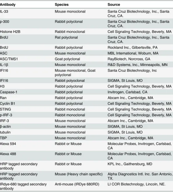
IFI16 has been shown to function as a transcriptional modulator via unknown mechanisms [19]. We theorized that acetylation of IFI16 could be one of the reasons for cytoplasmic Table 1. List of antibodies used in this study.
Antibody Species Source
IL-33 Mouse monoclonal Santa Cruz Biotechnology, Inc., Santa Cruz, CA.
p-300 Rabbit polyclonal Santa Cruz Biotechnology, Inc., Santa Cruz, CA.
Histone H2B Rabbit monoclonal Cell Signaling Technology, Beverly, MA
BrdU Rat polyclonal Santa Cruz Biotechnology, Inc., Santa
Cruz, CA.
BrdU Rabbit polyclonal Rockland Inc., Gilbertsville, PA
ASC Mouse monoclonal MBL International, Woburn, MA
ASC/TMS1 Goat polyclonal RayBiotech, Norcross, GA
IL-1β Mouse monoclonal R&D Systems, Inc., Minneapolis, MN
IFI16 Mouse monoclonal, Goat
polyclonal
Santa Cruz Biotechnology, Inc
IFI16 Rabbit polyoclonal SIGMA, St Louis, MO
H3 Rabbit polyclonal Cell Signaling Technology, Beverly, MA
Caspase-1 Rabbit polyclonal Invitrogen, Carlsbad, CA
Ran Rabbit polyclonal Abcam Inc., Cambridge, MA
Cyclin B1 Rabbit polyclonal Cell Signaling Technology, Beverly, MA STING Rabbit monoclonal Cell Signaling Technology, Beverly, MA p-IRF-3 Rabbit monoclonal Cell Signaling Technology, Beverly, MA
IRF-3 Mouse monoclonal Abcam Inc., Cambridge, MA
β-actin Mouse monoclonal SIGMA, St Louis, MO
tubulin Mouse monoclonal SIGMA, St Louis, MO
TBP Mouse monoclonal Abcam Inc., Cambridge, MA
Alexa 594 Rabbit or Mouse Molecular Probes, Invitrogen, Carlsbad, CA
Alexa 488 Rabbit or Mouse Molecular Probes, Invitrogen, Carlsbad, CA
HRP tagged secondary antibody
Rabbit or Mouse KPL Inc., Gaithersburg, MD
HRP tagged secondary antibody
Mouse (Heavy chain specific) Alpha Diagnostics Intl. Inc. San Antonio, TX.
IRdye-680 tagged secondary antibody
Anti-mouse (IRDye 680RD) LI COR Biotechnology, Lincoln, NE.
transport since acetylation of HMGB-1 (high-mobility group protein B1) protein involved in transcription/ chromatin bending has been shown to result in HMGB-1’s translocation into the cytoplasm [20]. Furthermore, IFI16 acetylation within the NLS motifs during transfection of DNA in U20S cells promoted cytoplasmic retention by blocking nuclear import of newly synthesized IFI16 [15]. However, the fate of IFI16 during nuclear DNA sensing was not studied.
To investigate the acetylation status of IFI16 during KSHV infection, uninfected and infected cell lysates were immunoprecipitated (IP-ed) with anti-acetylated lysine antibody and western blotted for IFI16. Compared to the uninfected cells, we observed a robust increase in the acetylation of IFI16 only in the infected cells (Fig 1C, lanes 1 and 2). In contrast, equal levels of acetylated tubulin were observed in both uninfected and KSHV infected cells (Fig 1C, lanes 1 and 2). The input IFI16 and loading control tubulin were of similar levels. These results sug-gested that the acetylation machinery was functional in both uninfected and infected cells and KSHV infection induced increased acetylation of IFI16.
When we next investigated the kinetics of IFI16 acetylation in the nuclear and cytoplasmic fractions by co-IP experiments, as early as 30 min p.i. an appreciable level of nuclear IFI16 acet-ylation was observed which steadily increased during the observed 24 h p.i. (Fig 1D, lanes 2–6). Correspondingly, we detected a faint band of acetylated IFI16 in the cytoplasm at 30 min p.i., with steady increase from 2 to 24 h p.i. (Fig 1D, lanes 9–12), which corroborated the results in
Fig 1B, lanes 9–12. The faint acetylated IFI16 band detected in the nucleus of uninfected cells probably represents the basal level (Fig 1D, lane 1). These detections were not due to nuclear contamination as shown by the absence of TBP and presence of tubulin in these fractions (Fig 1D). As positive control for nuclear and cytoplasmic acetylation, the proteins were IP-ed with acetylated lysine antibody and western blotted for H3 and tubulin, respectively (Fig 1D, lanes 1–12). Total H3 level was also analyzed by western blot as input control.
These results were also validated by IFA using anti-IFI16 and anti-acetylated lysine antibod-ies (S1B Fig). In the uninfected cells, IFI16 was detected in the nucleus and acetylated lysine sig-nals were observed both in the nucleus and in the cytoplasm (S1B Fig, top panel). We also observed some basal level of IFI16 and acetylated lysine colocalization in the nucleus of unin-fected cells (S1B Fig, UI, red arrow). In contrast, KSHV infection significantly increased the colocalization of acetylated lysine and IFI16 in the nucleus as well as in the cytoplasm in a time dependent manner (S1B Fig). Taken together, these results demonstrated that duringde novo
KSHV infection, IFI16 recognizes the viral genome with a concomitant increase in its acetyla-tion in the nucleus and redistribuacetyla-tion of acetylated IFI16 to the cytoplasm of the infected cells.
Acetylation inhibitor impedes the redistribution of IFI16 from the infected
cell nucleus to the cytoplasm
acetylated lysine antibody and WB for H2B showed six fold reduction in H2B acetylation by C-646 compared to the untreated KSHV (24 h) infected cells (S2G Fig, lanes 1 and 2). These results demonstrated thatde novoKSHV infection induced acetylation, which is in part due to p300, can be inhibited by C-646.
To determine the effect of C-646 on IFI16 acetylation, HMVEC-d cells were either unin-fected or inunin-fected with KSHV in the presence or absence of C-646, whole cell lysates IP-ed with anti-acetylated lysine antibody and western blotted for IFI16. Compared to untreated infected cells, C-646 treatment completely abolished the infection induced IFI16 acetylation (Fig 1E, lanes 1–6). Immunoprecipitation of IFI16 followed by WB for IFI16 demonstrated equal pull down; in addition,β-actin levels did not change due to treatment and showed equal loading (Fig 1E, lanes 1–6). IP of IFI16 and WB with anti-acetylation antibody also validated these results which showed decreased levels of acetylated IFI16 by C-646 treatment in infected cells (Fig 1F, lanes 1–3). To investigate the effect of C-646 on KSHV infection induced acetylation mediated cytoplasmic redistribution of IFI16, HMVEC-d cells were infected in the absence or presence of C-646, cytoplasmic and nuclear fractions isolated and western blotted for total IFI16. KSHV infection induced redistribution of IFI16 into the cytoplasm was abolished in C-646 treated cells (Fig 1G, lanes 7–12). Interestingly, we also observed that the nuclear IFI16 lev-els decreased at later time points by C-646 (Fig 1G, lanes 4–6) which suggested that acetylation may have a role in the stabilization of IFI16.
These results demonstrated that IFI16 acetylation during KSHV infection is dependent on p300 and acetylation is required for the redistribution of IFI16 from the nucleus to the cyto-plasm after recognition of the KSHV genome in the nucleus.
Proximity ligation assay (PLA) confirms that nuclear acetylation is
required for redistribution of IFI16 to the cytoplasm
To validate these results, we performedin situ-PLA which detects endogenous levels of pro-teins and gives the spatial distribution and localization of a single or multiple propro-teins (Fig 2A). PLA uses oligonucleotide-linked secondary antibodies and a fluorescence-based assay to detect closely associated proteins. If epitopes of a single protein or two protein epitopes are within 40 nm proximity, the antibody-linked oligonucleotides will ligate with adaptor oligonu-cleotides to form complete circles that are amplified via DNA replication and detected with fluorescent sequence-specific probes which will appear as distinct dots visible under fluorescent microscopy.
HMVEC-d cells were uninfected or infected in the presence or absence of C-646 and sub-jected to PLA using rabbit and mouse anti-IFI16 antibodies detecting different epitopes, and the detected red dots depict IFI16 (Fig 2A). IFI16 was predominantly nuclear in both untreated and C-646 treated uninfected cells (Fig 2A, top 2 panels, yellow arrows). In the absence of C-646, we observed abundant cytoplasmic IFI16 localization in KSHV infected cells at 24 h p.i. (Fig 2A, lower panels, white arrows), and an uninfected cell in the same field showed predomi-nantly nuclear IFI16 (Fig 2A, blue arrow). In contrast, while IFI16 was detected in the nucleus of C-646 treated infected cells, we did not observe IFI16 redistribution in the cytoplasm (Fig 2A, lower panels). These results demonstrated that inhibition of acetylation compromised the cytoplasmic redistribution of IFI16.
Fig 2. Proximity Ligation Assay (PLA) of IFI16’s acetylation and cytoplasmic redistribution duringde novoKSHV infection of HMVEC-d cells. (A and B)HMVEC-d cells were preincubated with C-646 for 2 h, washed, infected with KSHV for 2 h, washed and incubated in complete medium with or without C-646.(A)Cells were reacted with anti-IFI16 mouse and rabbit antibodies, washed, and subjected to PLA by incubating with species specific PLA oligo probe tagged secondary antibodies, washed, incubated with ligation mixture containing ligase, amplification solution with polymerase, and fluorescently labeled oligonucleotides. The signal, detected as a fluorescent dot at 594 nm wave length, was visualized by fluorescence microscopy. Nuclei were stained with DAPI, boxed areas are enlarged and the red dots represent IFI16. The yellow and white arrows indicate the nuclear and cytoplasmic IFI16, respectively. (B)PLA with anti-IFI16 and anti-acetylated lysine antibodies. White arrows and yellow arrows indicate cytoplasmic and nuclear acetylated IFI16,
respectively.(C)HMVEC-d cells uninfected or infected with live or UV-KSHV (30 DNA copies/cell) for 30 min or 2 h. The 2 h infected cells were further incubated for 4 or 24 h, and WCL proteins in NETN buffer were IP-ed with anti-acetylated lysine antibody and western blotted for IFI16. The bands were scanned and quantitated using FluorChemFC2 software and an AlphaImager system. Total IFI16 and tubulin were immunoblotted as input and loading controls, respectively.
cytoplasm, which increased to numerous acetylated IFI16 spots in a time dependent manner (Fig 2B, left panels, white arrows). In contrast, with C-646 treatment the acetylated IFI16 dots did not increase either in the cytoplasm or in the nucleus of infected cells (Fig 2B, lower three right panels). These studies demonstrating the reduction in cytoplasmic IFI16 redistribution by C-646 treatment validated our findings, and confirmed that IFI16 acetylation in the nucleus during KSHV infection is required for its redistribution to the cytoplasm.
Replication incompetent KSHV
de novo
infection induces IFI16
acetylation
We have previously shown that replication incompetent UV treated KSHV (UV-KSHV) enters the cells, delivers the viral DNA into the nucleus and induces the IFI16-inflammasome [11], which demonstrated that the presence of KSHV genome is the requirement for IFI16 recogni-tion and further consequences. When lysates from HMVEC-d cells infected with KSHV or UV-KSHV for 24 h were IP-ed with anti-acetylated lysine antibody and western blotted for IFI16, similar to live-KSHV infected cells, acetylation of IFI16 increased in a time dependent manner by infection with UV-KSHV (Fig 2C, lanes 1–7). These results suggested that the pres-ence of viral genome is enough to induce the IFI16 acetylation process and viral gene expres-sion is not required.
KSHV
de novo
infection of HFF cells also induces nuclear IFI16
acetylation and redistribution in the cytoplasm
We next determined whether acetylation of IFI16 and its cytoplasmic redistribution also occur in other cell types. Compared to uninfected cells, as in HMVEC-d cells, KSHV infected HFF cells (24 h p.i.) showed increased acetylation of IFI16 which was significantly inhibited by C-646 (S3A Fig, lanes 1–4), and WB for total IFI16 showed a slight reduction in C-646 treated cells (S3A Fig, lanes 1–4). In PLA analysis, infected HFF cells in the absence of the inhibitor showed robust acetylation of IFI16 and its redistribution to the cytoplasm, which was signifi-cantly abrogated by C-646 (S3B Fig). Uninfected cells showed only a basal level of acetylated IFI16 in the nucleus (S3B Fig). Evaluation of the total IFI16 levels by PLA using mouse and rab-bit anti-IFI16 antibodies revealed that IFI16 was solely nuclear in the uninfected cells (S3C Fig), while the KSHV infected cells showed IFI16 both in the nucleus and in the cytoplasm (S3C Fig). However, when the cells were treated with C-646, IFI16 was only detected in the nucleus (S3C Fig). These results demonstrated that acetylation of IFI16 is essential for its redis-tribution to the cytoplasm of KSHV infected HFF cells.
IFI16 acetylation and redistribution to the cytoplasm also occur in cells
latently infected with KSHV
IFI16 in the latently infected cells. As inde novoinfected cells, nuclear IFI16 protein levels also decreased in the presence of C-646 indicating that IFI16 stability in KSHV infected cells may be dependent upon its acetylation.
To validate these results, PLA was performed in BJAB and BCBL-1 cells using anti-IFI16 and anti-acetylated lysine antibodies (S4C Fig). Compared to the few nuclear acetylated IFI16 PLA dots in the BJAB cells (S4C Fig, upper left panel), we observed a significant increase in the acetylated IFI16 in the nucleus as well as in the cytoplasm of KSHV+ BCBL-1 cells (S4C Fig, lower left panel, yellow and white arrows, respectively). C-646 treatment resulted in a drastic reduction in acetylated IFI16 (S4C Fig, right panels). When PLA was done to examine total IFI16 and its redistribution in the absence or presence of C-646, we did not observe any cyto-plasmic IFI16 in the BJAB cells (S4D Fig, upper panels). Corroborating the biochemical data in
S4B Fig, increased nuclear and cytoplasmic IFI16 were observed in untreated BCBL-1 cells whereas IFI16 was mostly nuclear in the C-646 treated cells (S4D Fig, lower panels, yellow arrows). The KSHV latently infected endothelial (TIVE-LTC) and B (BJAB-KSHV) cells were also analyzed for IFI16 acetylation. IP of the whole cell lysates from control endothelial TIVE and BJAB, KSHV (+) TIVE-LTC and BJAB-KSHV cells with anti-acetylated antibody followed by IFI16 WB revealed significantly higher levels of acetylated IFI16 in both TIVE-LTC and BJAB-KSHV cells than in the KSHV negative control cells (S4E Fig, lanes 1–4). Equal amounts of IFI16 were detected in IP and in WB reactions (S4E Fig, lanes 1–4).
By PLA for IFI16 acetylation in the presence or absence of C-646, TIVE cells showed a mini-mal amount of acetylated IFI16 in both treated and untreated cells (S4F Fig, upper panels). In contrast, the TIVE-LTC cells showed increased levels of acetylated IFI16 both in the nucleus and in the cytoplasm (S4F Fig, lower left panel). This cytoplasmic redistribution of acetylated IFI16 was abolished by C-646 (S4F Fig, lower right panel). Total IFI16 levels in C-646 treated or untreated TIVE and TIVE-LTC cells were also analyzed by PLA using mouse and rabbit anti-IFI16 antibodies. In untreated and C-646 treated TIVE cells, IFI16 was solely nuclear (S4G Fig, upper panels). In contrast, TIVE-LTC cells showed robust IFI16 cytoplasmic redistri-bution (S4G Fig, lower left panel) which was significantly reduced by C-646 (S4G Fig, lower right panel).
Taken together, these results demonstrated that similar tode novoinfected HMVEC-d cells, p300 mediated acetylation plays an important role in the cytoplasmic redistribution of IFI16 in cells latently infected with KSHV.
IFI16 acetylation occurs during KSHV and EBV infection of primary B
cells and in B cells latently infected with EBV
As an IFI16-ASC inflammasome is formed during EBV infection of B cells and in latently infected cells, we performed PLA for IFI16 and acetylated lysine in primary human B cells infected with KSHV or EBV as well as in cells latently infected with EBV (S5 Fig). Compared to uninfected cells, both KSHV and EBV infected primary B cells showed acetylation as well as cytoplasmic redistribution of acetylated IFI16 (S5A Fig). Compared to EBV negative Ramos cells, EBV latently infected Raji (latency I) and LCL (latency III) cells showed both nuclear and cytoplasmic acetylated IFI16 (S5B Fig). These results demonstrated that acetylation of IFI16 and its cytoplasmic redistribution also occur in EBV infected cells.
Vaccinia virus infection does not induce nuclear IFI16 acetylation and
cytoplasmic translocation
in the cytoplasm. The acetylation of IFI16 was not induced by vaccinia virus infection of HMVEC-d cells (S6A Fig). Only similar levels of a few dots representing basal level of acetyla-tion were detected in the nucleus of both uninfected and vaccinia virus infected cells (S6A Fig). When mouse and rabbit antibodies were used to perform the PLA, IFI16 was predominantly detected in the nucleus of both uninfected as well as vaccinia infected HMVEC-d cells (S6B Fig). These results demonstrated that vaccinia viral DNA in the cytoplasm was not recognized by nuclear IFI16, and hence acetylation of the nuclear IFI16 and cytoplasmic translocation were not observed. These findings clearly supported our observations that the presence of nuclear KSHV, EBV and HSV-1 genomes induced the acetylation of IFI16 in the nucleus which then relocated into the cytoplasm of infected cells.
Ran-GTPase is involved in IFI16 transport from the nucleus to the
cytoplasm of KSHV infected cells
The dynamic process of exporting molecules of>50-kDa from the nucleus is initiated by exportins binding to cargo and Ran-GTP protein. The guanine-nucleotide exchange factor (GEF) of Ran that converts Ran-GDP to GTP form is in the nucleus and GTPase-activating proteins (GAPs) for Ran-GTPase are present in the cytoplasm as well as on the cytoplasmic face of the nuclear pore.
To determine whether Ran is responsible for IFI16 transport from the nucleus to the cyto-plasm, the lysates from uninfected or KSHV infected HMVEC-d cells (4 h p.i.) in the presence or absence of C-646 were IP-ed with anti-Ran-GTPase antibodies and WB for IFI16. Com-pared to the uninfected cells that showed a basal level of IFI16-RAN association (Fig 3A, lanes 1 and 2), KSHV infected cells showed robust association of IFI16 with Ran-GTPase which was inhibited by C-646 (Fig 3A, lanes 3 and 4). Comparable levels of IFI16 and Ran proteins were pulled down with their corresponding antibodies (Fig 3A, lanes 3 and 4). Higher IFI16-R-anGTP association in untreated KSHV infected cells corroborated the higher cytoplasmic redistribution of IFI16 shown in the earlier figures. When PLA was performed using anti-Ran and IFI16 antibodies, consistent with the IP results, the association between these two mole-cules increased during KSHV infection, which was abolished by C-646 (Fig 3B). These results demonstrated that acetylation enhances the association of IFI16 with Ran-GTP during infec-tion facilitating its transport to the cytoplasm and this associainfec-tion is dependent upon acetylation.
Blocking nuclear export by Leptomycin B hampers the cytoplasmic
translocation of IFI16
Blocking nuclear export by Leptomycin B did not affect nuclear IFI16
acetylation, IFI16-inflammasome formation and function
Since IFI16-ASC-procaspase-1 assembly was initiated in the nucleus, we next examined the effect of LPT on the transport of the other components of IFI16-inflammasomes. Procaspase-1 was detected in the nucleus of untreated uninfected and infected cells (Fig 3C, third panel, lanes 1 and 3). The increased cytoplasmic procaspase-1 in untreated infected cells was signifi-cantly decreased by LPT with a corresponding increase in the nucleus (Fig 3C, third panel, lanes 7 and 8, and 3 and 4). We have previously observed the presence of cleaved caspase-1 in the nucleus of infected HMVEC-d cells at 2 h and 8 h p.i. and only in the cytoplasm at 24 h p.i. [11]. Similarly, cleaved caspase-1 was detected in the infected cell cytoplasm at 24 h p.i. which was abolished by LPT treatment with a concomitant increase in the nucleus (Fig 3C, lanes 3, 4, 7 and 8).
When cell lysates of KSHV infected HMVEC-d cells in the presence or absence of LPT were analyzed by IP with anti-acetylated antibody and IFI16 WB, IFI16 was acetylated minimally in uninfected cells and to the same extent in untreated and LPT treated infected cells; however, tubulin was acetylated in both uninfected and infected samples (Fig 3D, top 2 panel, lanes 1–4). Similarly, the IFI16 and ASC association was equal in untreated and LPT treated infected cells (Fig 3D, third panel). Equal amounts of ASC and IFI16 were pulled down with their corre-sponding antibodies and their total protein levels demonstrated that these proteins were avail-able in sufficient and equal amounts in each of the experimental groups (Fig 3D, lower panels lanes 1–4).
To rule out the possibility that the detection of acetylated IFI16 is not due to the accumula-tion of newly synthesized IFI16 in the cytoplasm of KSHV infected cells, we blocked protein synthesis by using cycloheximide (CHX) at 200μg/ml which was neither toxic nor affected the KSHV infection of HMVEC-d cells (S7B, S7D, and S7F Fig). Cytoplasmic and nuclear proteins from CHX treated or untreated cells left uninfected or infected with KSHV for 4 h were isolated and subjected to western blot analysis. In the presence or absence of cycloheximide, we did not detect cytoplasmic IFI16 in the uninfected cells. In contrast, we observed similar levels of cyto-plasmic IFI16 in the infected cells in both the presence and absence of cyloheximide (Fig 3E, lanes 1 to 8). These results coupled with the LPT results suggested that the increased level of IFI16 in the cytoplasm of infected cells during KSHV infection is due to the translocation of acetylated IFI16 from the nucleus into the cytoplasm.
In PLA, nuclear IFI16 was detected in the untreated and treated uninfected cells (Fig 3F, left panels, yellow arrows). In contrast, in agreement with the biochemical findings, increased cyto-plasmic redistribution of IFI16 in KSHV infected HMVEC-d cells was detected (Fig 3F, top right panel, white arrows) which was abrogated in LPT treated cells (Fig 3F, lower right panel). In addition, the IFI16-ASC complex was observed in both the cytoplasm and nucleus of infected cells which was constrained to the nucleus of LPT treated cells (Fig 3G, right most panels). This redistribution of IFI16-ASC complex PLA spots corroborated with earlier IFA presence of 1μM C-646 for 2 h, and subjected to PLA with anti-Ran-GTPase and IFI16 antibodies. White and yellow arrows denote the cytoplasmic and nuclear association of Ran and IFI16, respectively.(C and D)HMVEC-d cells preincubated in the presence or absence of 50 nM Leptomycin B (LPT) for 2 h were uninfected (UI) or infected with KSHV for 2 h, washed, incubated for 24 h with or without LPT. (C) Nuclear or cytoplasmic fraction proteins were western blotted for IFI16, cyclin B1, caspase-1, TBP and tubulin. (D) WCL proteins were IP-ed with anti-acetylated lysine or anti-ASC antibodies and western blotted for IFI16 and tubulin. Proteins were also IP-ed for ASC or IFI16 and WB with ASC or IFI16 antibodies, respectively. Levels of total IFI16, ASC and tubulin were detected with their respective antibodies.(E)HMVEC-d cells were starved for 2 h with 200μg/ml cycloheximide (CHX), washed, infected with KSHV for 2 h, washed, incubated for 4 h with or without CHX. Cytoplasmic and nuclear fractions were subjected to western blot for IFI16, TBP and tubulin.(F and G) HMVEC-d cells in (C) were subjected to PLA. (F) Anti-IFI16 mouse and rabbit antibodies. Red dots: IFI16; yellow arrows: nuclear IFI16; white arrows: cytoplasmic IFI16. (G) Anti-IFI16 and anti-ASC antibodies. Red dots: IFI16-ASC association. Yellow arrows: nuclear IFI16-ASC interaction. White arrow: cytoplasmic IFI16-ASC interaction. Magnification: 60X.
and WB findings which demonstrated that IFI16-ASC inflammasome activation leads to redis-tribution of IFI16-ASC to the cytoplasm [11].
Taken together, these results demonstrated that a) blocking nuclear export by LPT did not interfere in the acetylation of IFI16, formation of IFI16-ASC complex or activation of caspase-1, b) blocking protein synthesis by CHX did not affect the cytoplasmic distribution of IFI16 from the nucleus, and c) the increased level of IFI16 in the cytoplasm in the infected cells was due to its redistribution from the nucleus and not due to newly translated cytoplasmic IFI16.
Inhibition of IFI16 acetylation impedes formation of the
IFI16-inflammasome
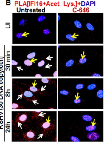
Since the redistribution of acetylated IFI16 and inflammasome activation showed a similar pat-tern in the infected cells, we sought to determine whether the acetylation of IFI16 and IFI16-in-flammasome activation are linked or independent of each other. As the association of IFI16 with the adaptor ASC is the first step in inflammasome activation, we examined these interac-tions by PLA. As shown inFig 4A and 4B, in the untreated and uninfected HMVEC-d cells, few IFI16-ASC interacting PLA dots were visible in the nucleus representing the basal level of association which was reduced by C-646 treatment (Fig 4A, top panels). In contrast, in untreated KSHV infected HMVEC-d cells, we observed a robust interaction between IFI16 and ASC both in the nucleus and in the cytoplasm (Fig 4A, yellow and white arrows, respectively, lower left panel). When the C-646 treated HMVEC-d cells were infected with KSHV, the PLA dots representing IFI16-ASC interactions in the nucleus were greatly reduced with little redis-tribution to the cytoplasm (Fig 4A, lower right panels).
Examination of IFI16 and ASC by IFA (S8A Fig) also revealed that IFI16 was predominantly in the nucleus of the uninfected cells, whilede novoKSHV infected HMVEC-d cells showed strong IFI16-ASC colocalization in the nucleus and redistribution to the cytoplasm (S8A Fig). When the cells were treated with C-646, only minimal IFI16-ASC interaction and cytoplasmic redistribution was detected (S8A Fig, third panel). Similarly, when BCBL-1 cells were examined by PLA and IFA, strong interactions between IFI16 and ASC were detected both in the nucleus and cytoplasm which were compromised by C-646 (Fig 4BandS8B Fig). Control BJAB cells did not show considerable IFI16 and ASC interaction in either untreated or C-646 treated cells (Fig 4B).
To confirm the IFI16-ASC interactions detected by PLA, cell lysates from uninfected and 4 and 24 hde novoKSHV infected HMVEC-d cells in the presence or absence of C-646 were IP-ed with ASC and western blottIP-ed for IFI16. ASC was associatIP-ed with IFI16 at 4 and 24 h p.i. but no such strong association was seen in the uninfected cells, (Fig 4C, lanes 1–3). In contrast, C-646 treatment disrupted the association between IFI16 and ASC (Fig 4C, lanes 4–6). A simi-lar amount of IFI16 was pulled down in each group either treated or not treated with C-646 (Fig 4C, lanes 1–6). A similar to primary infection, we observed increased interaction of IFI16 with ASC in the latently infected BCBL-1 cells which was greatly reduced in C-646 treated cells (Fig 4D, lanes 3 and 4). The inputs of IFI16 and ASC were similar in all groups. These results demonstrated that the presence of KSHV genome in the nucleus induced the IFI16-ASC inter-action and inflammasome formation, which are dependent upon the acetylation of IFI16 in bothde novoand latent KSHV infected cells.
de novoKSHV infected HMVEC-d cells (24 h) and in BCBL-1 cells (Figs4E, lane 2, and 6F, lane 3) and these were severely compromised by C-646 treatment (Figs4E, lane 3, and 6F, lane 4). No such aggregation was detected in the uninfected cells (Fig 4E, lane 1, and 4F, lanes 1 and 2). These results further confirmed that acetylation of IFI16 is critical for IFI16-inflammasome formation.
Inhibition of IFI16 acetylation impedes the function of the
IFI16-inflammasome
Formation of the IFI16-ASC-procaspase-1 inflammasome leads to the generation of functional caspase-1 via auto-cleavage which results in the cleavage of the pro-forms of IL-1β, IL-18 and IL-33 cytokines. Hence, we investigated the effect of C-646 on activation of caspase-1 and its downstream cytokines production. In untreated KSHV infected HMVEC-d cells, caspase-1 activation was detected at 4 and 24 h p.i., whereas, the C-646 treated counterparts did not show considerable cleavage of caspase-1 (Fig 4G, top panel, lanes 1–6). Activation of IL-1βand IL-33 was also inhibited by C-646 treatment (Fig 4G, panels 2, 3 and 4, lanes 1–6). We also observed the inhibition of procaspase-1 and pro-IL-1βcleavages by C-646 treatment in BCBL-1 cells (Fig 4H, lanes 3 and 4), and cleavage of procaspase-1 and pro-IL-1βwas not detected in BJAB cells (Fig 4H, lanes 1 and 2). The C-646 treatment did not significantly affect the viability of BJAB and BCBL-1 cells (S8C Fig). Compared to uninfected cells, increased secretion of IL-1β
was observed in KSHV infected HMVEC-d culture supernatants (18.5 pg/ml) which was sig-nificantly reduced (>5-fold) by C-646 treatment (3.8 pg/ml) (Fig 4I).
We next determined the levels of active caspase-1 in BCBL-1 cells with or without C-646 by FACS using fluorescent caspase-1 detection 660-YVAD-FMK probe (S8D and S8E Fig), and the percent active caspase-1 cell populations are shown inS8F Fig. Control BJAB cells unstained or stained with FLICA-660 did not show significant caspase-1 active cells (S8D and S8F Fig). In contrast, nearly 50% of the untreated BCBL-1 cells contained active caspase-1 which was reduced to ~18–19% in C-646 treated cells (S8E and S8F Fig).
These results confirmed that acetylation of IFI16 promotes formation of functional IFI16-ASC-procaspase-1 inflammasomes leading into active caspase-1 generation and downstream cytokine production in KSHV infected cells.
ASC is dispensable for nuclear IFI16 acetylation during
de novo
KSHV
infection of HMVEC-d cells
The recognition of viral genome by IFI16 leads into its increased interaction with ASC and inflammasome formation (Fig 4A–4D). Since reduction in IFI16 acetylation hampered IFI16-ASC association (Fig 4A–4D), we determined whether ASC plays roles in the acetylation of IFI16 and whether ASC associates with IFI16 after acetylation of IFI16. We knocked down the HMVEC-d cell ASC by Si-RNA electroporation and infected with KSHV. Knockdown effi-ciency confirmation by WB showed ~90–95% ASC reduction with no effect on IFI16 protein respectively.(C)HMVEC-d cells preincubated with or without 1μM C-646 for 2 h were washed, uninfected or infected with KSHV for 2 h, washed and incubated in complete medium with or without C-646 for 4 and 24 h. WCL proteins were IP-ed with anti-ASC antibodies and western blotted for IFI16 and ASC. Total IFI16 and ASC were used as input controls and tubulin was used as loading control.(D)WCL proteins from BJAB and BCBL-1 cells, untreated or treated with 1μM C-646 for 24 h, were IP-ed with anti-ASC antibodies and western blotted for IFI16.(E and F)HMVEC-d cells starved with or without 1μM C-646 for 2 h, washed, infected with KSHV for 2 h, washed, incubated with complete medium for 24 h in the presence or absence of 1μM C-646 (E). BJAB or BCBL-1 cells were untreated or treated with 1μM C-646 for 24 h (F). Whole cell lysates were prepared in HEPES-lysis buffer, equal amount of proteins were cross-linked using glutaraldehyde and immunoblotted for IFI16. Tubulin was used as loading control.(G)The protein samples from panel C experiments were western blotted for caspase-1, IL-1βand IL-33.(H)The protein samples from panel D experiments were western blotted for caspase-1 and IL-1β.(I) Culture supernatants of HMVEC-d cells infected with KSHV in the presence or absence of C-646 were evaluated for IL-1βby ELISA.
Fig 5. Effect of ASC knockdown on IFI16 acetylation and translocation. (A)HMVEC-d cells electroporated with Si-ASC and Si-control RNAs were uninfected or infected with KSHV for 30 min or 2 h, washed, the 2 h infected cells incubated for 4 and 24 h, and WCL prepared in NETN buffer. The knockdown efficiency was checked by immunoblotting for total ASC. WCL proteins were IP-ed with anti-acetylated lysine antibodies and western blotted for IFI16 or, IP-ed with anti-caspase-1 antibodies and WB for IFI16 and casapse-1. Total IFI16, caspase-1 and tubulin were tested as input and loading controls. (B)HMVEC-d cells uninfected or infected with KSHV for 2 h, washed, incubated with complete medium for 24 h, nuclear and cytoplasmic proteins western blotted for ASC and IFI16 or IP-ed with anti-acetylated lysine antibodies and western blotted for IFI16. TBP and tubulin were used as loading control and purity markers.
(Fig 5A, top two panels, lanes 1–8). The lysates from control and ASC knocked down cells were IP-ed with anti-acetylated lysine antibody and WB for IFI16. We observed the acetylation of IFI16 in both control and ASC knocked down cells (Fig 5A, third panel, lanes 1–8). As expected, in the absence of ASC formation of the IFI16-inflammasome complex was abrogated as shown by the absence of IFI16 in IP-reactions with anti-caspase-1 antibody and by the absence of caspase-1 activation in comparison to the Si-control KSHV infected cells. (Fig 5A, fourth, sixth and seventh panels, lanes 1–8). Caspase-1 was pulled down in all groups including ASC knocked down cells (Fig 5A, fifth panel, lanes 1–8). These results suggested that IFI16 acetylation occur independent of ASC.
We next determined whether IFI16 relocates to the cytoplasm in the absence of IFI16-ASC inflammasome formation. Western blot analysis of the cytoplasmic and nuclear fractions from Si-ASC uninfected and KSHV infected HMVEC-d cells showed efficient knockdown of ASC (Fig 5B, top panel, lanes 1–8). At 24 h p.i. in the Si-control cells, we observed the presence of IFI16 in the cytoplasm which was reduced by>2-fold in the ASC knockdown cells (Fig5B, sec-ond panel, lanes 7 and 8). No IFI16 was detected in the uninfected cell cytoplasm (Fig 5B, sec-ond panel, lanes 5 and 6). Interestingly, the nuclear IFI16 level was higher in KSHV infected ASC knockdown cells compared to the uninfected cells (Fig 5B, second panel, lanes 1–4). Since KSHV infection does not increase the IFI16 mRNA and protein levels [11], this moderate increase may be due to reduced, or lack of cytoplasmic redistribution of IFI16.
When the cytoplasmic and nuclear fractions were IP-ed with anti-acetylated lysine antibody and western blotted for IFI16, there was no change in the nuclear acetylated IFI16 levels in con-trol and ASC knockdown cells (Fig 5B, third panel, lanes 3 and 4). However, similar to the total IFI16 redistribution,>3-fold reduction in the acetylated IFI16 level was observed in the cyto-plasm of ASC knockdown cells (Fig 5B, third panel, lanes 7 and 8).
These results clearly demonstrated that in the absence of ASC, acetylation of IFI16 still takes place which is prior to inflammasome formation. The cytoplasmic redistribution of IFI16 in ASC knockdown cells must be inflammasome independent which might be attributed to cyto-plasmic export of acetylated IFI16 either alone or in complex with other proteins. However, the reduced amount of IFI16 in the cytoplasm in comparison to Si-control suggested that the IFI16-ASC inflammasome contributes to the majority of the IFI16 detected in the cytoplasm of infected cells.
KSHV infection induces increased interaction between IFI16 and histone
acetyltransferase p300 and p300 is required for KSHV induced acetylation
of IFI16
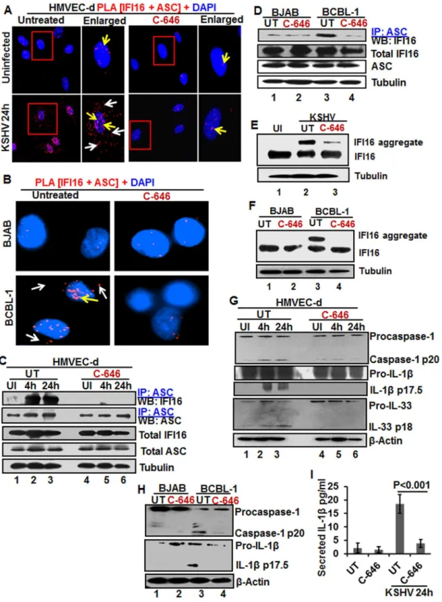
As a follow up to C-646 inhibition of KSHV induced p300 catalyzed acetylation of IFI16, we determined the interaction of p300 with IFI16. When HMVEC-d cells infected with KSHV for 24 h in the presence or absence of C-646 were IP-ed for p300 and western blotted for IFI16, we observed increased interaction of IFI16 with p300 which was reduced to basal levels with C-646 treatment (Fig 6A, lanes 1–8).
activity of p300. Increased nuclear p300 activation during infection further supports that acety-lation of IFI16 is probably mediated by increased p300 activity in the nucleus and not in the cytoplasm. Decreased activity of enzymes by the inhibitors further verified the specificities of these assays and the functionality of C-646 and TSA (Fig 6B and 6C).
Next, we knocked down p300 to validate our inhibitor studies. Efficient p300 knockdown by Si-p300 with no effect on IFI16 and ASC protein levels was observed (Fig 6D, top three pan-els, lanes 1–8). The co-IP studies of anti-acetylated lysine antibodies and IFI16 demonstrated the abrogation of IFI16 acetylation in Si-p300 KSHV infected cells while Si-control infected cells showed robust IFI16 acetylation (Fig 6D, fourth panel, lanes 1–8). Similarly, the caspase-1 and IFI16 association was detected in the control group but was abrogated in p300 knockdown infected cells, and caspase-1 was pulled down in all the tested groups (Fig 6D, fourth and fifth panels, lanes 1–8). These results further validated our findings with C-646.
In PLA studies with anti-IFI16 and anti-acetylated antibodies, very few acetylated IFI16 PLA dots were observed in the nucleus of control or p300 knockdown infected cells (Fig 6E, top panels, yellow arrows). In Si-control infected cells (24 h p.i.), a high number of acetylated IFI16 dots were visible in the nucleus and in the cytoplasm (Fig 6E, lower left panel), while only a few dots, as in uninfected cells, were detectable in the p300 knockdown KSHV infected cells (Fig 6E, lower right panel). IFI16 was solely in the nucleus of uninfected cells by total IFI16 PLA (Fig 6F, top panels, yellow arrows). Similar to the acetylated IFI16, total IFI16 was found in both the nucleus and cytoplasm of Si-control infected cells, while p300 knocked down infected cells showed only nuclear IFI16 (Fig 6F, lower panels).
When PLA was performed using anti-IFI16 and anti-ASC antibodies, the red dots repre-senting the IFI16 and ASC association were in both the nucleus and the cytoplasm of Si-control KSHV infected cells (Fig 6G, lower left panel, white and yellow arrows). In contrast, the IFI16 and ASC association was completely abrogated in p300 knockdown infected cells (Fig 6G, lower right panel).
These results further strengthened the finding that acetylation is required for the cytoplasmic redistribution of IFI16 and p300 is responsible for the acetylation of IFI16.
Acetylation of IFI16 is required for IFN-β
production in KSHV infected
cells
Besides inflammasome induction in KSHV, EBV and HSV-1 infected cells, IFI16 has also been shown to be involved in the induction of IFN-βgene through its cytoplasmic activation of the STING molecule leading into phosphorylation of the transcription factor IRF-3 which subse-quently translocates into the nucleus to stimulate the IFN-βgene promoter [21]. KSHV infec-tion induces only a moderate IFN-βresponse early duringde novoinfection and early lytic and latent gene products inhibit this response at later times of infection [17], and the role of IFI16 in IFN-βproduction during KSHV infection is not defined.
When we analyzed the role of IFI16 and its acetylation in IFN-βproduction, we detected IFN-βin the supernatants of KSHV infected HMVEC-d cells at 6 h p.i., which was significantly reduced by>4 fold by C-646 treatment (Fig 7A). A significant level of phosphorylated IRF-3 units (RFU).(D)The HMVEC-d cells electroporated with control or p300 Si-RNA were uninfected or infected with KSHV for 2 h, washed and incubated for various time points. WCL in NETN buffer were western blotted for total p300 and IFI16 or IP-ed with anti-acetylated lysine antibodies and western blotted for IFI16 or IP-ed with anti-caspase-1 antibodies and western blotted for IFI16 and caspase-1.(E, F and G)The control and p300 knockdown HMVEC-d cells were uninfected or infected with KSHV for 2 h, washed, incubated for 24 h and subjected to PLA using (E) anti-IFI16 and anti-acetylated lysine antibodies, (F) anti-IFI16 mouse and rabbit antibodies, and (G) anti-IFI16 and anti-ASC antibodies. The white and red arrows in (E) panels indicate acetylated IFI16 in the cytoplasm or nucleus, respectively. The white and red arrows in (F) panels indicate cytoplasmic and nuclear IFI16, respectively. In (G) panels, the white and red arrows indicate IFI16-ASC colocalization in the cytoplasm and nucleus, respectively.
detected in the nucleus at 6 h p.i. was reduced in C-646 treated cells (Fig 7B and 7C). Immuno-precipitation with anti-acetylated lysine antibody followed by IFI16 WB revealed the presence of acetylated IFI16 from 30 min to 24 h p.i. in KSHV infected cells, which was abolished by C-646 treatment (Fig 7D, top panel, lanes 1–8). IP reactions with anti-STING antibodies demon-strated the increased IFI16-STING interaction from 30 min to 6 h p.i. and its decrease at 24 h p.i., which was abolished by C-646 (Fig 7D, second panel, lanes 1–8). Similarly, the levels of pIRF-3 increased in untreated KSHV infected cells which were abolished in C-646 treated infected cells (Fig 7D, fourth panel, lanes 1–6).
Fig 7. Effect of IFI16 acetylation on IFN-βproduction duringde novoKSHV infection of HMVEC-d cells.HMVEC-d cells in the presence or absence of 1μM C-646 were either left uninfected or infected with KSHV (30 DNA copies/cell) for 2 h, washed and incubated with or without 1μM C-646 for 6 h.(A) Interferon-βin the culture supernatants was quantitated by ELISA.(B)Cells were examined by IFA with anti-pIRF-3 and Alexa Fluor- 488 secondary antibodies. Insets in the merged panels are enlarged. The yellow arrows indicate the pIRF-3 in the nucleus.(C)Quantitation of nuclear pIRF-3 dots.(D)WCL lysates in NETN buffer were IP-ed with anti-acetylated lysine and STING antibodies and immunoblotted for IFI16. Proteins were also immunoblotted for total IFI16, pIRF-3, total IRF-3, total STING, and tubulin was used as loading control.(E)HMVEC-d cells electroporated with control or STING Si-RNA were either left uninfected or infected with KSHV for 2 h, washed and incubated for 4 and 24 h. WCL prepared in NETN buffer were western blotted for total STING, pIRF3, total IRF3 and tubulin and IP-ed with anti-acetylated lysine antibodies and western blotted for IFI16 and tubulin or IP-ed with anti-IFI16 antibodies and western blotted for IFI16.(F)HMVEC-d cells electroporated with control or STING Si-RNA were uninfected or infected with KSHV for 2 h, washed and incubated for 6 h. Cells culture supernatants were used to measure the levels of IFN-βby ELISA.
IFI16 acetylation in
de novo
KSHV infected cells is upstream to STING
activation
Next, we knocked down STING in HMVEC-d cells to determine whether IFI16 acetylation is upstream or downstream to STING activation. Efficient knockdown was achieved by electro-poration using STING specific Si-RNA (Fig 7E, top panel). KSHV infection was not affected under these conditions as shown by the increased IFI16 acetylation which was not affected by STING knockdown (Fig 7E, second panel) which suggested that IFI16 acetylation is upstream to STING activation. Control tubulin protein was acetylated in uninfected and infected cells, and an equal amount of IFI16 was pulled down in all groups (Fig 7E, fourth panel). IRF-3 was phosphorylated post-KSHV infection which was hampered in STING knockdown cells; how-ever, total IRF-3 was detected in equal amounts in all the groups and results with tubulin showed equal loading (Fig 7E, last three panels). An increased level of IFN-βwas observed in the supernatants of HMVEC-d cells infected with KSHV which was significantly reduced by STING knockdown (Fig 7F).
These studies demonstrated that acetylation during KSHV infection induced IFI16 acetyla-tion is required for its cytoplasmic interacacetyla-tion with STING, pIRF-3 inducacetyla-tion, and IFN-β pro-duction, IFI16 acetylation is upstream to STING activation and STING does not play any role in IFI16 acetylation.
Acetylation of IFI16 is required for IFN-β
production in HSV-1 infected
cells
We and others have shown that HSV-1 infection of HFF cells also induced the IFN-βgene and secretion of IFN-βwhich was dependent upon IFI16 and IRF-3 [16,22]. We utilized C-646 to determine whether IFI16 acetylation has any role in IFN-βproduction during HSV-1 infection. C-646 did not show any cytotoxic effects on HFF cells nor did it affect the infectivity of HSV-1 (S9A and S9B Fig). At 30 min post HSV-1 infection, 20 and 16 pg/ml of IFN-βwas detected in untreated and C-646 treated supernatants, respectively (Fig 8A). At 6 h p.i., 317±16.5 pg/ml of IFN-βwas detected in untreated cells whereas significant (>67%; p<0.001) inhibition of IFN-β
production was observed in the C-646 treated cells (107±19.4 pg/ml;Fig 8A).
When we examined the phosphorylation of IRF-3 by IFA, compared to the uninfected cells, at 6 h p.i., appreciable levels of phosphorylated IRF-3 were detected in the nucleus and in the cytoplasm (Fig 8B, third panel). In contrast, C-646 treatment prior to infection reduced these levels especially in the nucleus (Fig 8B, fourth panel, and 8C).
In PLA studies, similar to KSHV infected HMVEC-d cells, we observed the cytoplasmic redistribution of acetylated IFI16 in HSV-1 infected HFF cells (6 h p.i.) which was inhibited by C-646 (Fig 8D). IFI16, which was predominantly nuclear in the uninfected HFF cells, was detected in the cytoplasm of HSV-1 infected cells which was abrogated by C-646 (Fig 8E). The observed reduction in the total as well as acetylated IFI16 levels is probably due to the degrada-tion of IFI16 by HSV-1 via its ICPO protein [14].
Fig 8. Effect of IFI16 acetylation on IFN-βproduction duringde novoHSV-1 infection in HFF cells The HFF cells uninfected or infected with HSV-1 (1 PFU/cell MOI) in the presence or absence of 1μM C-646 for 30 min or 2 h were washed and incubated with or without 1μM C-646 for 6 h. (A)IFN-β
in contrast, the IFI16 level was unchanged with C-646 which further suggested that acetylation might be facilitating the stability of IFI16.
Similar to KSHV infected cells, IFI16 acetylation was not affected by STING knockdown during HSV-1 infection (Fig 8G, lanes 1–6). Equal levels of IFI16 was pulled down in both Si-control and in STING knockdown HSV-1 infected cells, and IRF-3 was activated in Si-Si-control HSV-1 infected cells and not in STING knockdown cells (Fig 8G, lanes 1–6). HSV-1 infection induced IFN-βproduction was hampered in STING knockdown cells (Fig 8H).
Together, these results demonstrated that as in KSHV infected cells, IFI16 acetylation and its translocation to the cytoplasm in HSV-1 infected cells is also critical for its interaction with STING in the cytoplasm, subsequent STING interaction with IRF-3, phosphorylation of IRF-3, and nuclear translocation of pIRF-3 leading into IFN-βproduction.
Acetylation is not obligatory for IFI16
’
s ability to recognize nuclear viral
genomes
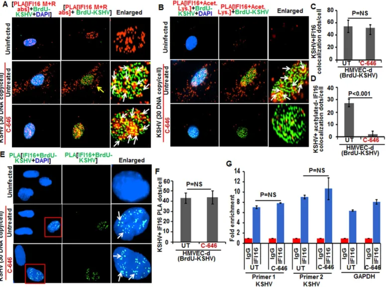
Since recognition of KSHV, HSV-1 and EBV genome by IFI16 in the nucleus of infected cells leads to inflammasome activation [11,13,14], we determined whether acetylation of IFI16 is required for its ability to sense the viral genome. Cells were infected with BrdU-KSHV for 6 h in the presence or absence of C-646, IFA was performed for BrdU followed by PLA using anti-IFI16 mouse and rabbit antibodies (Fig 9A). IFI16 was mostly nuclear in the uninfected cells. At 6 h p.i., we observed the appreciable colocalization of IFI16 with KSHV genome in the nucleus of both untreated as well as C-646 treated HMVEC-d cells (Fig 9A, enlarged panels). As before, we observed IFI16 redistribution in the cytoplasm which was absent in C-646 treated cells (Fig 9A). Increased associations of acetylated IFI16 with BrdU-KSHV were observed in untreated cells (Fig 9B, enlarged panels, and white arrows) which were completely abrogated by C-646 (Fig 9B, lower enlarged panel, and 9D). The IFI16-KSHV genome colocalization spots in untreated and C-646 treated cells were similar and the difference was not statistically significant (Fig 9C). Interestingly, the levels of acetylated IFI16 molecules associated with BrdU-KSHV were about 50% less than that of the total IFI16 associated with viral genome (Fig 9C and 9D).
To confirm the direct association of IFI16 with KSHV genome, we performed PLA and chromatin immunoprecipitation (ChIP) assays. To detect the direct binding of IFI16 with KSHV genome, we infected HMVEC-d cells with KSHV with BrdU labeled genome and per-formed the PLA using anti-BrdU and anti-IFI16 antibodies as this will give signal only when KSHV genome and IFI16 interact and are at close proximity (<40 nm). In the PLA reactions, we observed that the number of IFI16-KSHV genome colocalization spots were similar in both the untreated as well as C-646 treated KSHV infected cells (Fig 9E, white arrows, and 9F). These results further corroborated theFig 9Aresults and demonstrated that IFI16 acetylation does not play any role in viral genome recognition. Similar results were also observed in HFF cells infected with BrdU genome labeled HSV-1 (S9C and S9D Fig).
for two different locations of KSHV and with a control GAPDH primer (Fig 9G). We did not observe any significant changes in the binding of IFI16 with KSHV genome by C-646 treat-ment (Fig 9G).
These results suggested that a) IFI16 directly associates with KSHV and HSV-1 genomes, b) the acetylation of IFI16 is not required for genome recognition, c) IFI16 acetylation occurs as a dynamic post-genome recognition event, and d) post-acetylation, IFI16 probably moves away Fig 9. Effect of acetylation inhibition by C-646 on the ability of IFI16 to recognize and bind the KSHV genome. (A and B)HMVEC-d cells pre-incubated with or without 1μM C-646 for 2 h were washed, infected with BrdU genome labeled KSHV (30 DNA copies/cell) for 2 h, washed and incubated with or without 1μM C-646 for 4 h. Cells were processed, incubated with anti-BrdU antibodies, washed, and reacted with Alexa Fluor-488 (green) secondary antibodies to detect BrdU-KSHV. These slides were subjected to PLA(A)with mouse anti-IFI16 and rabbit-IFI16 antibodies or(B)with IFI16 and anti-acetylated lysine antibodies. Boxed areas are enlarged. The yellow arrows in (A) and (B) indicate the cytoplasmic IFI16 or anti-acetylated IFI16, respectively. The white arrows in (A) indicate colocalization of IFI16 PLA spots with BrdU labeled KSHV genome. The white arrows in (B) indicate colocalization of acetylated IFI16 PLA spots with BrdU labeled KSHV genome.(C and D)Bar graph of quantitation of colocalization of IFI16 or acetylated IFI16 PLA spots with BrdU labeled KSHV genome in (A) and (B) panels. At least ten fields with a minimum of 3–4 cells/field were counted.(E)The above described cells were subjected to PLA using anti-BrdU and anti-IFI16 antibodies to detect the direct association of IFI16 with BrdU-labeled KSHV genome. Boxed areas are enlarged. The PLA dots indicated by white arrows represent the association between IFI16 and BrdU-labeled KSHV genome.(F)PLA spots from 10 fields with at least 3–4 cells/field in (E) were quantitated and presented as a bar graph(G)Chromatin immunoprecipitation was performed using anti-IFI16 antibody as described in the Material and Methods. Untreated BCBL-1 cells (3 x 10⁷) or cells treated with 1μM C-646 for 24 h were cross-linked with formaldehyde and sonicated to obtain DNA fragments between 200–400 bps. q-PCR was performed with primers for two different regions on the KSHV genome and one control for GAPDH, and the results are presented as fold enrichment of immunoprecipitated DNA. The p values were calculated using Student’s T test. NS (non-significant).
from the genome for the formation of its complexes and eventually leading to its cytoplasmic translocation.
Discussion
IFI16, a member of the ALR family, has emerged as a critical sensor against both nuclear and cytoplasmic DNA with pivotal roles in inflammasome activation and IFN production [11,21]. However, how the inflammasome formed as a consequence of recognition of herpesviral genomes in the nucleus by IFI16, followed by cytoplasmic accumulation of the IFI16-ASC complex, and how HSV-1 and KSHV genome recognition in the nucleus via IFI16 lead to STING-IRF-3 activation in the cytoplasm and subsequent IFN-βproduction were not known. Our comprehensive studies for the first time demonstrate that acetylation of IFI16 after recog-nizing the viral genome occurs as a dynamic post-genome recognition event that is common to the IFI16-mediated innate responses of inflammasome induction and IFN-βproduction during herpesvirus infections.
Several molecular mechanistic steps of nuclear innate sensing by IFI16 are revealed here (Fig 10). The first step is the recognition of nuclear foreign herpes viral genomes by IFI16 which is independent of acetylation and IFI16 interaction with ASC or STING. This is followed by IFI16’s association with p300 which mediates the acetylation of IFI16. This is a key molecu-lar step common to both of the IFI16 mediated innate responses of inflammasome induction and IFN-βproduction as IFI16’s acetylation is essential for its interaction with ASC leading
into procaspase-1 interaction and activation in the nucleus, interaction with RanGTPase, cyto-plasmic translocation and IL-1βinduction during KSHV, EBV and HSV-1 infection. Cyto-plasmic translocation of acetylated IFI16 is also critical for the activation of STING resulting in the phosphorylation of IRF-3 and IFN-βproduction (Fig 10).
Crystal structures of overexpressed IFI16 proteins suggest that IFI16 binds to the sugar-phosphate backbone of dsDNA in a non-sequence specific manner with more affinity to super-helix and cruciform DNA [23,24]. Herpesviral genomes enter the nucleus as a linear, naked dsDNA with nicks and breaks and undergo rapid circularization and chromatinization [25]. Our studies demonstrating that IFI16 recognized the KSHV genome soon after its entry into the nucleus coupled with the fact that this occurs in the absence of acetylation suggests that IFI16 has evolved for rapid recognition of incoming foreign DNA (Fig 10).
Studies with overexpressed proteins suggest that DNA sensing induces filamentous clusters of IFI16 due to homotypic PYD-PYD interactions and cooperative DNA binding that might amplify signals, stabilize IFI16-dsDNA complexes and could act as danger signal [26]. Our studies show such DNA recognition by IFI16 initiates its acetylation process which is essential for the innate immune functions of both inflammasome and interferon responses executed in the cytoplasm and nucleus. Colocalization of reduced levels of acetylated IFI16 with viral genomes (Fig 9) compared to the non-acetylated IFI16 levels suggest that acetylation probably changes the affinity and structure of IFI16 resulting in a dynamic post-genome recognition event of IFI16’s disassociation from the DNA to facilitate its interaction with other proteins and transport into the cytoplasm in a continuous fashion with genomes always occupied with another IFI16 molecule as shown in the KSHV and EBV latently infected cells. This scenario is also supported by observations such as the acetylation of histone prompts its structural exten-sion and charge neutralization resulting in the weakening of DNA-histone interaction [27], and acetylation of KSHV LANA-1 resulting in its dissociation from KSHV genome [28].
and the absence of IFII6-ASC-procaspase- inflammasome formation and the translocation of acetylated IFI16 in ASC knockdown cells shown here suggested that acetylated IFI16, either alone or in combination with other yet to be identified protein(s), is also relocalized during her-pesviral infection resulting in the interaction with STING. Further studies determining whether IFI16 interacts with STING alone or in association with another protein(s) are in progress.
KSHV infection of a PMA stimulated human monocytic THP-1 cell line has been shown to result in IL-1βand IFN-βproduction by a pathway that is independent of IFI16 [29]. This dis-crepancy may be due to the fact that KSHV may be undergoing abortive infection in the PMA stimulated cells as has been shown for HSV-1 in these cells [30] and DNA released from the lysosomes is probably recognized by AIM2, c-GAS, and others to stimulate the IL-1βand Fig 10. Schematic model depicting herpesvirus infection induced IFI16 acetylation and its role in inflammasome and IFN-βproduction.Results presented here demonstrate viral DNA entry into the nucleus (1–2) leads to recognition through IFI16 (3) followed by IFI16 acetylation (4). The acetylated IFI16 forms an inflammasome complex (5) which is translocated to the cytoplasm via RanGTP. Leptomycin B treatment abrogates the acetylated IFI16 translocation (6). The inflammasome complex formation leads into the activation of caspase-1 (7) which in turn cleaves pro-IL-1βand-IL-33 (8), and IL-βis released into the culture supernatant (9). IFI16 acetylation (i) and cytoplasmic redistribution (ii) also activates STING (iii) which phosphorylates IRF-3 (iv) that moves to the nucleus leading into IFN-βgene transcription (v-vi), translation (vii) and IFN-βis secreted into the culture supernatant (viii). Use of p300 inhibitor C-646 and knockdown of p300 does not inhibit the ability of IFI16 to recognize the episomal viral DNA but blocks the acetylation of IFI16 which results in the inhibition of inflammasome formation, IFI16 translocation into the cytoplasm as well as IL-1βand IFN-βproduction.