135
Fabrication Of Gold Nanoparticles-Modified
Glassy Carbon Electrode And Its Application For
Voltammetric Detection Of Cr(III)
Santhy Wyantuti, Yeni Wahyuni Hartati, M. Lutfi Firdaus, Camellia Panatarani, Roekmiati Tjokronegoro
Abstract: A sensitive and selective differential pulse stripping voltammetric (DPSV) method is presented for the determination of trace amount Cr(III) using glassy carbon electrode (GCE) modified with gold nanoparticles (AuNPs). The method includes AuNPs fabrication and self-assembly GCE modification. This processes replaced the –H groups with –NH2 groups on the surfaces of GCE which increased the number of AuNPs attached to it. The GCE modified with AuNPS was used as electrochemical sensor of Cr(III) for the first time that gave a wide linear range from 0.5 to 75 ppb and a very low detection limit of 10 ppt (equivalent to 0.19 nM). The electrode exhibited high reproducibility in repetitive measurements with a relative standard deviation better than 2.4%. The effect of interfering ions study showed that Cu(II), Cd(II), Zn(II) Cr(VI), Ni(II) and Fe(III) ions did not have a significant effect on the measurement.
Keywords: Gold nanoparticles, glassy carbon electrode, trivalent chromium, voltammetry. ————————————————————
1
I
NTRODUCTIONChromium is a transition metal that occurs mainly in Cr(III) and Cr(VI) oxidation states in aquatic environments. Chromium compounds are naturally found in the environment due to erosion of chromium-containing rocks, precipitation, terrestrial run-off and can be introduced by volcanic eruptions. The average Cr concentration in soil is 92 ppm, in rivers 0.7 ppb and in seawater 0.2 ppb [1–4] and it can be much higher in polluted region. Increase in Cr concentration is caused by discharge of wastewater from electroplating and tanning industries, metallurgical, paint pigment and dying industries, sanitary landfill leaching and other chemical industries. Usage of Cr(III) in chemical industries is more variable than Cr(VI) [5]. Although Cr(III) is an essential nutrient, Cr(VI) is well known for its toxicity to the living things due to its higher solubility and strong oxidizing nature. Hexavalent chromium toxicity is about 1,000 times higher than Cr (III) [6]. The presence and ratio between Cr(III) and Cr(VI) depends not only on pH and oxygen concentration of the region but also on the nature and concentration of reducers, oxidation mediators and complexing agents. These co-occurrence factors are responsible for Cr(III) as the predominant species even in many oxygenated surface waters [5–7]. The dominant Cr(III) over Cr(VI) species in aquatic environments prompted us to develop a detail analytical method of Cr(III) detection in aqueous samples to improve our understanding on the biogeochemistry of Cr in environment. Various techniques for the determination of Cr(III) have been developed in recent years. The common analytical methods include separation of Cr(III) by high-performance liquid chro- matography (HPLC),
Extraction and pre-concentration step using ion exchange or chelating resin. This pretreatment often coupled with detection systems such as spectrofluorometry [8], spectrophotometry [9,10], chemiluminescene [11], atomic absorption spectrometry (AAS) [12–14], inductively coupled plasma atomic emission spectrometry (ICP-AES) [15] and inductively coupled plasma mass spectrometry (ICP-MS) [16]. However, these pretreatment and detection processes do not satisfy all requirements for routine analysis, mainly because of their complicated procedures, time consumption or the high cost of instrumentations. Electrochemical methods, in particular voltammetry, have important advantages for the determination of metals at trace concentrations such as high sensitivity and low detection limit, relative simplicity, low cost of equipment and portable option [17,18]. Analytical methods of voltammetry using film electrode for Cr(III) detection have been reported [19,20]. However, the detection limits were not low enough to analyze trace amount of Cr(III) in environmental systems. Recently, metal nanoparticle-modified electrodes have been widely used as working electrodes to improve the detection limit of chemical [21–23] and biochemical compounds [24,25] including Cr(VI) [18,26,27]. Metal nanoparticle-modified electrodes have extraordinary catalytic properties for both oxidation and reduction reactions over bulk metal electrodes. Among metal nanoparticles, gold nanoparticles (AuNPs) have unique properties such as good conductivity and better electrocatalytic ability. To the best of our knowledge, however, no report has been published on AuNPs–GCE as working electrode on voltammetric analysis of Cr(III). The purpose of this research is to fabricate AuNPs–GCE and its novel application for Cr(III) voltammetric detection.
2
E
XPERIMENTAL2.1 Reagents
Ammonium hydroxide, acetic acid, hydrochloric acid (37%), ethanol, potassium chloride, chromium trichloride, sodium citrate, sodium acetate, sodium tetrahidrobarate, hydrogen tetrachloroaurate (III) trihydrate (HAuCl4.3H2O) and aluminium
oxide were supplied by Merck. All chemicals were used without further purification. Double-distilled water was used throughout.
__________________________
Santhy Wyantuti, Yeni Wahyuni Hartati and Roekmiati Tjokronegoro are lecturers at Department of Chemistry, University of Padjadjaran, Indonesia M. Lutfi Firdaus is lecturer at Graduate School of
Science Education, University of Bengkulu, Indonesia. E-mail: [email protected]
136 2.2 Preparation of AuNPs colloids
AuNPs colloids were prepared by reduction of Au(III) using NaBH4 according to the reported procedure [28–30] with some modification. Briefly, 15 mL of HAuCl4 0.1 mM solution was mixed with double-distilled water and stirred for 5 minutes on magnetic stirrer. Then 0.5 mL sodium citrate 0.1 M was added to the mixture and stirred again for 5 minutes. Finally 290 µL NaBH4 0.1 M (pH 6.5) was added to the mixture and stirred to homogenized the mixture. Transmission electron microscopy (JEM-1400 120kV, JEOL) was used to know the size and shape of the AuNPs formed. The stability of formed AuNPs were characterized using UV-Visible spectrophotometer (Genesys 10S, Thermo Scientific).
2.3 Electrode fabrication with AuNPs modification The procedure for fabrication of GCE modified with AuNPs was adapted from previously published reports [31] with some modification. The surface of electrode was polished with 8 nm aluminum oxide powder on 1,000 mesh sandpaper. Then it was washed successively with ethanol and double-distilled water in ultrasonic bath and dried in air. Modification of electrode through self-assembly process was done by immersed the electrode into concentrated ammonium hydroxide and irradiated under UV light at wavelength 254 nm for 6 hours. The activated GCE was then dipping into AuNPs colloid for 24 hours and washed thoroughly with doubly distilled water. The surface morphology and composition of the prepared GCE modified with AuNPs was characterized using a JEOL JED-2300 Scanning Electron Microscope (SEM) with an X-ray Energy Dispersive Spectrometer (EDS).
2.4 Electrochemical measurements
The fabricated AuNPs-modified GCE was employed as the working electrode, while the counter and reference electrodes were a platinum wire and an Ag/AgCl electrode, respectively. Electrochemical measurements were carried out in a single-compartment cell using a Metrohm µAutolab type III 757 VA potentiostat at room temperature (23ºC). The detection of Cr(III) was performed in acetate buffer pH 5.0. All potentials given in this work are with respect to the Ag/AgCl saturated reference electrode (0.2 V vs. Standard hydrogen electrode or SHE).
3
R
ESULTS AND DISCUSSION3.1 Characterization of the AuNPs colloid and AuNPs-modified GCE
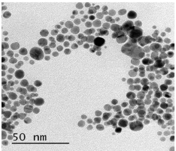
Nanoparticles of gold were synthesized at room temperature by reducing Au(III) using NaBH4. Citrate ions were used as capping agent to stabilize the formed nanoparticles. After reduction with NaBH4, the color of gold solution altered from yellow to ruby red implying that the colloidal gold nanoparticles have been formed. Further characterization by TEM confirms that the average diameter of gold nanoparticles were approximately 2-5 nm (Fig. 1) [28]. The AuNPs were also characterized by UV-Vis spectrophotometer, and then corresponding photographic images were gathered. As shown in Fig. 2, a characteristic surface plasmon resonance (SPR) band of AuNPs is observed in the spectrum at approximate of 518 nm. The resulting wavelengths characteristics and ruby red AuNPs colors were not changed even after 7 days. The prepared AuNPs were stable in the aqueous solution due to the electrostatic repulsion of the negative capping agents,
citrate ions, against the aggregation between AuNPs. Color of AuNPs colloid attributed to SPR was arising due to the collective oscillation of free conduction electrons induced by an interacting electromagnetic field.
Fig. 1. TEM image of gold nanoparticles
.
AuNPs were used further to modify the eloctorde. GCE was modified by attaching AuNPs on to the electrode surfaces. The modification process of GCE surfaces was initiated by cleaning with aluminium oxide then activating the electrode surfaces. GCE surfaces were activated in order that the attaching AuNPs distributed evenly with great amount, resulting in enhancement of GCE sensitivity to analyte. Various ways to activate GCE surface are available including by soaking GCE into concentrated ammonium hydoxide under photochemical reaction. The photochemical reaction in this study was conducted by soaking GCE into concentrated ammonium hydroxide solution then radiated with 254 nm UV for 6 hours. This processes will substitute hydrogen-group (-H) with amine-group (-NH2) at the GCE surfaces. The formation
of amine end group will enhace AuNPs attachment to the GCE surfaces. It is well known that the distributions of AuNPs on the electrode surfaces are important to perform good sensitivity of stripping voltammetry. The nature of
137 on the GCE surfaces was characterized using SEM and X-ray
EDS analysis. The shape and size distribution of AuNPs deposited at GCE surfaces were confirmed in the present study. SEM images of GCE surfaces before and after AuNPs modification and its corresponding X-ray EDS analysis show that the GCE surfaces coated with AuNPs through self-assembly process had AuNPs level of 11.5% of total glassy carbon surface (C=76,38%; O=12,07%). The percentage was higher than adsorption process (2.05%) which only soaked the electrode directly into AuNPs colloid without electrode surfaces amine-group modification. The SEM-EDS data also indicated that the AuNPs were immobilized and well-dispersed, forming a uniform monolayer on the amine-terminated activated GCE surfaces.
3.2 Application as Cr(III) sensor, effects of operational parameters
Preliminary experiments were conducted to obtain optimum condition for the application of AuNPs-GCE as Cr(III) sensor. The effect of the deposition potential on the peak current of Cr(III) stripping voltammogram was studied in the potential range from −2.0 to −1.0 V with scan rate of 0.05 V/s and amplitude modulation of 0.05 V. The highest efficiencies of 500 ppb Cr(III) accumulation in 0.2 M acetic buffer solution were obtained for a deposition potential of −1.5 V. Similar experiments were also conducted to obtain optimum deposition time and we found that deposition time of 90 seconds gives the best results. These parameters are important in stripping procedures that have a pronounced effect on both sensitivity and linear range. Therefore, the deposition potential of −1.5 V and deposition time of 90 seconds were chosen as optimum value.
3.3 Calibration curve and analytical performance Calibration curve for the determination of Cr(III) on the AuNPs-GCE was achieved by DPSV under optimum conditions. Fig. 3 shows voltammetric performance and calibration curve for different concentrations of Cr(III) at deposition potensial -1.5 V, deposition time 90 seconds, scan rate 0.05 V/s and amplitude modulation 0.05 V. The regression equation and correlation coefficient are y = 0.142 + 0.004x and r = 0.9955, respectively. The resulting calibration plots is linear over the range from 0.5 to 75 ppb. The reproducibility of the AuNPs-GCE was estimated from the response to each 1 ppb of Cr(III) at replicates measurements. This series yield a mean current response, referred to as sensitivity, of 0.14 µA ppb-1 corresponding to a relative standard deviation (RSD) of 2.4%. The detection limit is 0.01 ppb based on three times the standard deviation of the baseline, calculated according to the reference [32]. The calculated detection limit of the proposed method is better than previous reported methods including the Cr(III) determination using sophisticated ICP–MS (Table 1). Usage of AuNPs modified electrode gives at least one order of magnitude better detection limit than previous reported voltrametric methods using film electrodes. Limited publication of Cr(III) voltammetric detection compared with those of Cr(VI), in addition to its relatively poor sensitivity of non-gold electrodes lead us to infer that to obtain high quality of Cr(III) voltammetric profiles require gold or gold-modified electrodes. Previously it is known that high quality voltammetric profiles for Cr(III) oxidation were found to appear only at a gold electrode [33].
3.4 Effect of interfering ions
In order to evaluate the effect of foreign metal ions dissolved in the media, solutions containing 50 ppb of Cr(III) and each of the following metals Cd(II), Cu(II), Zn(II), Cr(VI), Ni(II) and Fe(III) all at same concentration of 50 ppb were prepared. The addition of foreign ions does not influence the current of the peak corresponding to Cr(III). The percentage variation of the
Table 1. Detection limits comparison of available Cr(III) analytical methods.
Detection Method Comment Detection
Limit (ppb) Ref.
Spectrofluorometry - 2.5 [8]
Spectrophotometry - 2 [9]
Spectrophotometry - 1.4 [10]
Chemiluminescene - 0.12 [11]
Flame AAS - 0.2 [12]
Flame AAS - 0.18 [13]
Flame AAS - 0.94 [14]
ICP-AES - 0.21 [15]
ICP-MS - 0.02 [16]
Voltammetry (Differential pulse adsorptive stripping)
Stannum film
electrode 2 [19]
Voltammetry (Squarewave adsorptive)
Bismuth film
electrode 0.1 [20]
Voltammetry (Differential pulse stripping)
AuNPs glassy carbon electrode
0.01 This
work
Fig. 3. Voltammetric performance for Cr(III) concentration variation. Inset shows corresponding callibration curve at
optimum condition.
peak current induced by the presence of interfering ions with respect to Cr(III) alone are: 1.02% for Cd, 0.88% for Cu, 0.29% for Zn, 3.70% for Cr(VI), 0.07% for Ni and 0.40% for Fe. These deviations from Cr(III) measurement at 0.9470 V and 1.360 uA remain acceptable and therefore, the accurate detection of Cr(III) is still possible under these conditions.
3.5 Analytical application
138 standard addition method. The recovery rate was 97%, which
was in good agreement with those derived from the traditional AAS method (AAnalyst 400, Perkin Elmer) after appropriate extraction. Therefore, the accuracy of the results was satisfactory, indicating that the fabricated electrochemical AuNPs-GCE sensor is practicable for the determination of Cr(III) in environmental systems.
4
C
ONCLUSIONWe have demonstrated a simple method for the fabrication of nanostructured gold modified glassy carbon electrode. After optimization of the voltammetric measurements, the prepared AuNPs-modified electrode improve the analytical performance significantly and suitable for the determination of trace amount Cr(III) with high accuracy and good reproducibility. The proposed method was applied to a real sample which gave satisfactory results.
A
CKNOWLEDGMENTThe authors gratefully acknowledge Prof. Yasuaki Einaga and Dr. Ivandini Tribidasari for valuable suggestions and discussions. This research was supported by the Ministry of Education and Culture, Indonesia through the Directorate General of Higher Education (Dirjen DIKTI).
R
EFERENCES[1] R. Rudnick, S. Gao, Composition of the continental crust, Elsevier-Pergamon, Oxford, UK, 2003.
[2] J. Gaillardet, J. Viers, B. Dupré, Trace elements in river waters, Elsevier-Pergamon, Oxford, UK, 2003.
[3] K. Bruland, Lohan MC, The oceans and marine geochemistry, Elsevier-Pergamon, Oxford, UK, 2003.
[4] Y. Sohrin, K.W. Bruland, "Global status of trace elements in the ocean", TrAC Trends Anal. Chem., vol. 30, pp. 1291–1307, 2011.
[5] R. Saha, R. Nandi, B. Saha, "Sources and toxicity of hexavalent chromium", J. Coord. Chem., vol. 64, pp. 1782–1806, 2011.
[6] D. Adriano, Trace elements in terrestrial environments: biogeochemistry, bioavailability, and risks of metals, Springer, Berlin, 2001.
[7] J. Kotaś, Z. Stasicka, "Chromium occurrence in the environment and methods of its speciation"., Environ. Pollut., vol. 107, pp. 263–83, 2000.
[8] B. Tang, T. Yue, J. Wu, Y. Dong, Y. Ding, H. Wang, "Rapid and sensitive spectrofluorimetric determination of trace amount of Cr(III) with o-vanillin-8-aminoquinoline", Talanta, vol. 64, pp. 955–60, 2004.
[9] M. Kaneko, M. Kurihara, S. Nakano, T. Kawashima, "Flow-injection determination of chromium(III) by its catalysis on the oxidative coupling of 3-methyl-2-benzothiazolinone hydrazone with N-ethyl-N-(2-hydroxy-3-sulfopropyl)-3-methoxyaniline", Anal. Chim. Acta, vol. 474, pp. 167–176, 2002.
[10]Y. Chen, I. Lee, Y. Sung, S. Wu, "Triazole functionalized gold nanoparticles for colorimetric Cr3+ sensing", Sensors Actuators B Chem. vol. 188, pp. 354–359, 2013.
[11]B. Gammelgaard, Y. Liao, O. Jøns, "Improvement on simultaneous determination of chromium species in aqueous solution by ion chromatography and chemiluminescence detection", Anal. Chim. Acta, vol. 354, pp. 107–113, 1997.
[12]R.M. Cespón-Romero, M.C. Yebra-Biurrun, M.P. Bermejo-Barrera, "Preconcentration and speciation of chromium by the determination of total chromium and chromium(III) in natural waters by flame atomic absorption spectrometry with a chelating ion-exchange flow injection system", Anal. Chim. Acta, vol. 327, pp. 37–45, 1996.
[13]K. Kiran, K.S. Kumar, B. Prasad, K. Suvardhan, R.B. Lekkala, K. Janardhanam, "Speciation determination of chromium(III) and (VI) using preconcentration cloud point extraction with flame atomic absorption spectrometry (FAAS)", J. Hazard. Mater., vol. 150, pp. 582–586, 2008.
[14]H. Bag, A.R. Turker, M. Lale, A. Tunceli, "Separation and speciation of Cr(III) and Cr(VI) with Saccharomyces cerevisiae immobilized on sepiolite and determination of both species in water by FAAS", Talanta, vol. 51, pp. 895–902, 2000.
[15]S. Hirata, Y. Umezaki, M. Ikeda, "Determination of chromium(III), titanium, vanadium, iron(III), and aluminum by inductively coupled plasma atomic emission spectrometry with an on-line preconcentrating ion-exchange column", Anal. Chem., vol. 58, pp. 2602–2606, 1986.
[16]S. Hirata, K. Honda, O. Shikino, N. Maekawa, M. Aihara, "Determination of chromium (III) and total chromium in seawater by on-line column preconcentration inductively coupled plasma mass spectrometry", Spectrochim. Acta Part B At. Spectrosc., vol. 55, pp. 1089–1099, 2000.
[17]Christopher M. A. Brett, Ana Maria Oliveira Brett, Electrochemistry: Principles, Methods, and Applications, Oxford Science Publications, UK, 1993.
[18]F.W. Campbell, R.G. Compton, "The use of nanoparticles in electroanalysis: an updated review", Anal. Bioanal. Chem., vol. 396, pp. 241–259, 2010.
[19]W.W. Zhu, N.B. Li, H.Q. Luo, "Simultaneous determination of chromium(III) and cadmium(II) by differential pulse anodic stripping voltammetry on a stannum film electrode", Talanta, vol. 72, pp. 1733– 1737, 2007.
139 Electrodes", Electroanalysis. vol. 16, pp. 1745–1754,
2004.
[21]M.N. Bui, C.A. Li, K.N. Han, X. Pham, G.H. Seong, "Simultaneous detection of ultratrace lead and copper with gold nanoparticles patterned on carbon nanotube thin film", Analyst, vol. 137, pp. 1888–1894, 2012.
[22]Y. Wei, R. Yang, X. Yu, L. Wang, J. Liu, X. Huang, "Stripping voltammetry study of ultra-trace toxic metal ions on highly selectively adsorptive porous magnesium oxide nanoflowers", Analyst, vol. 137, pp. 2183–2191, 2012.
[23]X. Dai, R.G. Compton, "Direct electrodeposition of gold nanoparticles onto indium tin oxide film coated glass: Application to the detection of arsenic(III)", Anal. Sci., vol. 22, pp. 567–570, 2006.
[24]K. Saha, S.S. Agasti, C. Kim, X. Li, V.M. Rotello, "Gold nanoparticles in chemical and biological sensing", Chem. Rev., vol. 112, pp. 2739–2779, 2012.
[25]J. Hong, W. Wang, K. Huang, W.-Y. Yang, Y.-X. Zhao, B.-L. Xiao, et al., "A highly efficient nano-cluster artificial peroxidase and its direct electrochemistry on a nano complex modified glassy carbon electrode", Anal. Sci., vol. 28, pp. 711–716, 2012.
[26]B. Liu, L. Lu, M. Wang, Y. Zi, "A study of nanostructured gold modified glassy carbon electrode for the determination of trace Cr(VI)", J. Chem. Sci., vol. 120, pp. 493–498, 2008.
[27]W. Jin, G. Wu, A. Chen, "Sensitive and selective electrochemical detection of chromium (VI) based on gold nanoparticle-decorated titania nanotube arrays", Analyst. vol. 139, pp. 235–241, 2014.
[28]G. Frens, "Controlled Nucleation for the Regulation of the Particle Size in Monodisperse Gold Suspensions", Nat. Phys. Sci., vol. 241, pp. 20–22, 1973.
[29]L. Zeng, H. Wang, X. Bo, L. Guo, "Electrochemical sensor for amino acids based on gold nanoparticles/macroporous carbon composites modified glassy carbon electrode", J. Electroanal. Chem., vol. 687, pp. 117–122, 2012.
[30]J. Fink, C.J. Kiely, D. Bethell, D.J. Schiffrin, "Self-Organization of Nanosized Gold Particles", Chem. Mater., vol. 10, pp. 922–926, 1998.
[31]R. Tian, T.N. Rao, Y. Einaga, J. Zhi, "Construction of Two-Dimensional Arrays Gold Nanoparticles Monolayer onto Boron-Doped Diamond Electrode Surfaces", Chem. Mater., vol. 18, pp. 939–945, 2006.
[32]J.N. Miller, J.C. Miller, Statistics and Chemometrics for Analytical Chemistry, Pearson/Prentice Hall, 2010.
[33]C.M. Welch, M.E. Hyde, O. Nekrassova, R.G. Compton, "The oxidation of trivalent chromium at