Structure of tick anticoagulant peptide at 1.6 Å resolution
complexed with bovine pancreatic trypsin inhibitor
ROBERT ST. CHARLES, K. PADMANABHAN,1 R.V. ARNI,2
K.P. PADMANABHAN,andA. TULINSKY
Department of Chemistry, Michigan State University, East Lansing, Michigan 48824-1322
~ReceivedOctober 1, 1999;Final RevisionNovember 8, 1999;AcceptedNovember 19, 1999!
Abstract
The structure of tick anticoagulant peptide~TAP! has been determined by X-ray crystallography at 1.6 Å resolution complexed with bovine pancreatic trypsin inhibitor~BPTI!. The TAP–BPTI crystals are tetragonal, a5b546.87,
c550.35 Å, space group P41, four complexes per unit cell. The TAP molecules are highly dipolar and form an
intermolecular helical array along the c-axis with a diameter of about 45 Å. Individual TAP units interact in a head-to-tail fashion, the positive end of one molecule associating with the distal negative end of another, and vice versa. The BPTI molecules have a uniformly distributed positively charged surface that interacts extensively through 14 hydrogen bonds and two hydrogen bonded salt bridges with the helical groove around the helical TAP chains. Comparing the structure of TAP in TAP–BPTI with TAP bound to factor Xa~Xa!suggests a massive reorganization in the N-terminal tetrapeptide and the first disulfide loop of TAP~Cys5T–Cys15T!upon binding to Xa. The Tyr1TOH atom of TAP moves 14.2 Å to
interact with Asp189 of the S1 specificity site, Arg3TCZ moves 5.0 Å with the guanidinium group forming a cation–
p-electron complex in the S4 subsite of Xa, while Lys7TNZ differs in position by 10.6 Å in TAP-BPTI and TAP-Xa,
all of which indicates a different pre-Xa–bound conformation for the N-terminal of TAP in its native state. In contrast to TAP, the BPTI structure of TAP–BPTI is practically the same as all those of previously determined structures of BPTI, only arginine and lysine side-chain conformations showing significant differences.
Keywords: bovine pancreatic trypsin inhibitor; conformational changes; factor Xa; tick anticoagulant peptide
The extrinsic and intrinsic pathways of the blood coagulation cas-cade merge at factor Xa production, the reactions thereafter ter-minating in an insoluble fibrin clot. Thus, Xa plays an important pivotal role in hemostasis. With factor Va and a phospholipid sur-face, Xa catalyzes the Ca12ion-dependent activation of
prothrom-bin to thromprothrom-bin, which is the required enzyme in the conversion of fibrinogen to fibrin monomers. Considering the strategic position of Xa relating the coagulation pathways, inhibitors of this serine protease represent potentially useful and important antithrombotic agents.
TAP is an acidic 60 residue protein produced by the soft tick
Ornithodoros moubata and is a tenacious competitive inhibitor
of Xa ~Ki 5 0.59 nM! ~Waxman et al., 1990!. Although its disulfide connectivity is like that of members of the Kunitz fam-ily of serine proteinase inhibitors ~Fig. 1!, its amino acid se-quence only resembles them remotely ~Antuch et al., 1994!. However, NMR studies have shown that TAP possesses a gen-erally similar, but distorted folding topology conserving a hexa-peptide antiparallelb-strand and about 2.5 turns ofa-helix~Antuch et al., 1994; Lim-Wilby et al., 1995!. Partial structural homology notwithstanding, the N-terminal retro-binding mechanism of TAP inhibition~van de Locht et al., 1996; Wei et al., 1998!, with the peptide chain running parallel to~Ser214–Gly218!of Xa, differs spectacularly from that of canonical Kunitz-type inhibitors like BPTI, where a substrate-like binding loop interacts in an anti-parallel b-strand way with the active site of the enzyme ~Bode & Huber, 1992!. The N-terminal retro-binding mode was first observed in hirudin-thrombin~Rydel et al., 1991!, and mimicked with synthetic inhibitors ~Tabernero et al., 1995; Mochalkin & Tulinsky, 1999!, and was also seen in the structure of the nazumamide–thrombin ~Nienaber & Amparo, 1996! and the ornithodorin–thrombin ~van de Locht et al., 1996! complexes, and most recently, in the crystal structure of Xa inhibited with
Reprint requests to: A. Tulinsky, Department of Chemistry, Michigan State University, East Lansing, Michigan 48824-1322; e-mail: tulinsky@ cem.msu.edu.
1Present address: Department of Biochemistry, Michigan State
Univer-sity, East Lansing, Michigan 48824.
2Present address: Department of Physics, IBILCE0UNESP, CP 136, Sao Jose do Rio Preto-SP, CEP 15054-000, Brazil.
Abbreviations: ADA, N-@2-acetamido#-2-iminodiacetic acid; amino acids of TAP and BPTI, distinguished by T or B subscript tagging residue num-ber; BPTI, bovine pancreatic trypsin inhibitor; MALDI, matrix-assisted laser desorption ionization mass spectroscopy; Piand Si, peptide and en-zyme site notation after Schechter and Berger~1967!; TAP, tick anticoag-ulant peptide; TAP–BPTI, TAP–BPTI complex; TAP–Xa, TAP–Xa complex; Xa, factor Xa.
TAP~Wei et al., 1998!; in the latter, TAP showed an interaction at the active site utilizing the N-terminal Tyr1T in the S1
spec-ificity pocket of Xa. Such binding renders TAP highly specific for Xa, which otherwise exhibits little or no appreciable inhibi-tory activity toward most serine proteinases including trypsin, thrombin, and other blood proteases~Waxman et al., 1990!. Other elements of the TAP structure, including some of the C-terminal
a-helix, are involved in Xa binding through a secondary binding site near the Na1 ion binding site of Xa ~Zhang & Tulinsky,
1997! in support of a two-step binding mechanism proposed on the basis of mutagenesis~Jordan et al., 1992!.
We present here a high resolution~1.6 Å!crystal structure of TAP cocrystallized with BPTI. The new model of the TAP struc-ture clarifies portions of the molecule that were poorly defined in previous work. Considerable differences in structure exist in the N-terminal 15 residues between TAP bound to Xa and TAP in the TAP–BPTI complex, indicating a different pre-Xa–bound confor-mation for the N-terminal peptide in its native state and a possible means for rationalizing the two step binding mechanism of TAP to Xa~Jordan et al., 1992; Wei et al., 1998!. Crystal lattice inter-actions are responsible for an unusual intermolecular helical pack-ing arrangement of TAP molecules along the crystallographic c-axis, with the negatively charged helical TAP groove being filled by positively charged BPTI molecules.
Results and discussion
Electron density for both TAP and BPTI was well defined in the final~2Fo2Fc!map, allowing all the residues in both molecules
to be modeled. Density was weaker for the last two of BPTI
~Gly57B–Ala58B! suggesting some flexibility at the C-terminus ~^B& ;30 Å2compared to average values of 18.5 Å2for TAP and
22.8 Å2 for BPTI!. Both termini of TAP, however, were
excep-tionally well defined. Difference~Fo2Fc!density maps were used to additionally assign 120 solvent water molecules and three clearly defined and resolved sulfate anions. The latter interact multiply and electrostatically, compensating charge through ion pairs and0or forming hydrogen bonds with nearby proton donors of arginine, lysine, or tyrosyl side chains of TAP, BPTI, and neighboring water molecules~Table 1!. The first sulfate makes an ion pair with Arg42B,
the second with Lys15B and Arg17B of symmetry related BPTI
molecules, and the last with Arg20Band Lys30T. It is noteworthy
that Lys15Bis the P1 specificity residue of BPTI when complexed
with trypsin~Bode & Huber, 1992!.
The TAP–BPTI structure
Attempts to grow crystals of TAP from solutions of the homog-enous protein proved to be unsuccessful. Because the isoelectric point of TAP is 4.65 while that of BPTI is 9.24, an attempt was made to cocrystallize an equimolar solution of the two reasoning that the basic regions of BPTI might electrostatically complement electronegative ones of TAP and thus facilitate crystallization of a complex. It was, therefore, reassuring when MALDI mass spec-trometric analysis confirmed the composition of crystals as con-taining both TAP and BPTI. Other relevant factors considered were:~1!the molecules were expected to be similar in molecular size, and~2! possibly similar in structure by virtue of their near
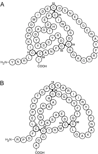
A
B
Fig. 1. Primary structures and numbering of~A!TAP and~B!BPTI.
Table 1. Hydrogen-bonded and hydrogen-bonded ion pair interactions of sulfate anions in TAP–BPTI
d ~Å!
SO4–1 O-1 N2TND2 3.01
O-1 Y49TOH 3.19
O-1 OW161 3.12
O-2 R42BN 2.79
O-2 R42BNE 3.00
O-3 OW253 2.86
O-4 R42BNH2 2.75
O-4 OW279 2.58
SO4–2 O-2 K15BNZ 2.58a
O-2 R17BNH2 3.40b
O-3 R17BNE 3.12
O-3 OW254 2.88
O-4 R17BN 2.80
O-4 OW217 2.94
SO4–3 O-1 K30TNZ 3.88b
O-2 R20BNH2 2.76
O-3 Y35BOH 2.83
O-4 R20BNH1 3.10
identical disulfide connectivity even though there is no significant level of sequence homology between the two ~Fig. 1! ~Antuch et al., 1994!. NMR structure determinations of TAP~Antuch et al., 1994; Lim-Wilby et al., 1995!and the crystal structure of TAP-Xa
~Wei et al., 1998!showed that only a two-chainb-strand and a C-terminal helix superposed on the structure of BPTI~Glu22T–
Arg27T, Cys33T–Ile38T, and Tyr52T–Cys59T, respectively, in the
present case!, although other comparably sized regions are super-posable separately.
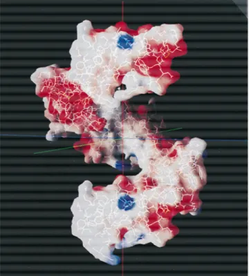
The crystal structure of TAP-BPTI consists of one TAP mol-ecule and one BPTI molmol-ecule in the asymmetric unit, with a pro-tein fraction of 60% corresponding to a tight packing arrangement consistent with the strong X-ray diffraction characteristics of the crystals ~better than 1.6 Å resolution; I0s~I! ; 4.0 at 1.6 Å, Table 2!. Based on the crystal structure, there is no single dominant surface interface between the two molecules to suggest they crys-tallized from solution as a hetero-dimeric complex. Instead, the TAP and BPTI molecules appear to have added to the growing TAP–BPTI crystal independently as monomers. The TAP mol-ecules are highly dipolar~Figs. 2, 3!, and form an intermolecular helical array along the c-axis of the crystal with a diameter of
;45 Å~Fig. 4!satisfying 41crystallographic screw axis symmetry
and making 42 contacts less than 3.6 Å between TAP molecules. They interact in a head-to-tail fashion, with the electropositive end of one molecule containing the N- and C-termini~Figs. 2, 3! as-sociating with the distal electronegative end of another, and vice versa~Fig. 4!. BPTI molecules are inclined to the helix axis and interact with the helical groove around the intermolecular TAP chains through seven molecules, making a total of 57 contacts less than 3.6 Å per TAP module, two of which are hydrogen-bonded salt bridges from different BPTI units~Arg3T, Asp3B, and Asp16T,
Arg17B!along with 14 other hydrogen bonds~Table 3!. The TAP helix is principally held together by two hydrogen-bonded salt bridges~Tyr1T, Glu22T, and its reciprocal!along with four
hydro-gen bonds~and their reciprocals!. In addition, there are 18 contacts less than 3.6 Å, and a salt bridge~Asp13T, Arg23T!between
sep-arate TAP helices, while BPTI molecules also interact with each other only sparingly~18 contacts less than 3.6 Å, including recip-rocals!four of which are between BPTI molecules intercalated in different TAP helices~Table 3!.
The structure of TAP in TAP–BPTI and TAP–Xa
The TAP structure of TAP–BPTI is only remotely similar to that of two NMR structure determinations@Protein Data Bank~PDB!code: 1TAP~Antuch et al., 1994!and 1TCP~Lim-Wilby et al., 1995!#, but resembles that of TAP in the crystal structure of the TAP–Xa complex@PDB code: KIG~Wei et al., 1998!#fairly closely. Of the sets of NMR solutions of 1TAP and 1TCP, the smallest RMS difference between optimally superposed CA positions on TAP of TAP–BPTI ~60 CA! was found to be 1.7 Å~1TAP, model 19!. Compared with the NMR structures, the TAP from the TAP–Xa crystal structure is more like that of TAP–BPTI, giving an RMS difference between superposed models of 1.4 Å~60 CA positions!, but 0.64 Å when using 50 selected CA positions. Although a crys-tallographic BPTI structure @PDB code: 4PTI ~Marquart et al., 1983!#led to the molecular replacement location of BPTI in TAP– BPTI, the NMR models failed to fix the position of TAP, whereas TAP of TAP–Xa led to a satisfactory molecular replacement solu-tion. The largest differences in structure found between TAP–BPTI and TAP–Xa are within the N-terminal segment, principally be-tween Tyr1Tand Cys15T, and in the~Asn28T–Gly31T!loop.
Sig-nificant variation within these regions is also observed between the
Table 2. Diffraction data and refinement summary statistics
Resolution~Å! 1.62
Observations~I0s~I!.1.0! 86,488
I0s~I! ~outermost range! ~1.88–1.62!Å 3.9
Independent reflections 13,056
Redundancy 6.6
Completeness~%! 93.1
Outermost range~%! 88.4
Rmerge~%! 5.4
Outermost range~%! 18.8
Protein atoms 943
Water molecules 120
Refinement range~Å! 8.0–1.62
Number reflections 12,942
Rfinal~%! 18.8
Rfree~%! 21.1
RMSDs from ideal values
Bond lengths~Å! 0.011
Bond angles~deg! 1.49
Bmain chain~Å2! 1.8
Bside chain~Å2! 3.1
NMR structures, suggesting that the segments are inherently flex-ible in TAP.
The N-terminal tripeptide of TAP binds to the active site of Xa
~Wei et al., 1998!in a retro-manner~Tabernero et al., 1995;
Nien-aber & Amparo, 1996; van de Locht et al., 1996; Mochalkin & Tulinsky, 1999!with the peptide chain running parallel to~Ser214– Gly218!of Xa, not unlike the N-terminal residues of hirudin in the hirudin–thrombin complex ~Rydel et al., 1991!. Although the
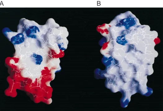
A
B
Fig. 3. Electrostatic potential surface of~A!TAP and~B!BPTI calculated using the program GRASP~Nicholls et al., 1991!. ESP. 10 kT, blue;, 28 kT, red;;0, gray.
Table 3. Intermolecular hydrogen bonds in TAP–BPTI crystals
TAP TAP
d
~Å! TAP BPTI
d
~Å! BPTI BPTI
d ~Å!
Tyr1N Glu22OE1 2.89a Arg3NH1 Asp3OD1 3.10a Leu6O Gly57O 3.01
Tyr1N Glu22OE2 3.07a Arg3NH1 Asp3OD2 2.81a Glu7OE1 Ala58OTd 3.18
Asp13OD1 Arg23NH1 3.40a,b,c Arg3NH2 Asp3OD2 2.45a Pro13O Arg17NH1 2.68b
Asp13OD1 Arg23NH2 2.94a,b Arg9NE Ala27O 3.22
Asp13OD2 Arg23NH1 3.14a,b Asp16OD1 Arg17NH1 3.59a,c
Arg27NH1 Pro40O 2.91 Asp16OD1 Arg17NH2 3.08a
Arg27NH1 His43O 2.97 Asp16OD2 Arg17NH1 2.72a
Arg27NH2 His43O 3.10 Asn18ND2 Cys14O 2.96
Gly29O Arg53NH2 2.40b Asn18ND2 Ala16O 2.89
Gly29O Arg53NH1 3.00b Tyr25OH Glu49OE1 2.56
Gly31N Glu41OE1 2.79 Asn28ND2 Asp50OD2 3.21
Gly31O Arg53NH2 3.16
Asp34OD1 Ser47OG 2.74
Asp47OD2 Asp3N 2.68
Asp47O Arg42NH1 3.16
Asp47O Arg42NH2 3.03
Tyr52OH Arg53NE 3.03
Tyr52OH Asp50OD1 2.68
Ile60O Arg39NH2 3.01
Ile60O Arg39NH1 3.03
aHydrogen-bonded salt bridge. bBetween TAP helices.
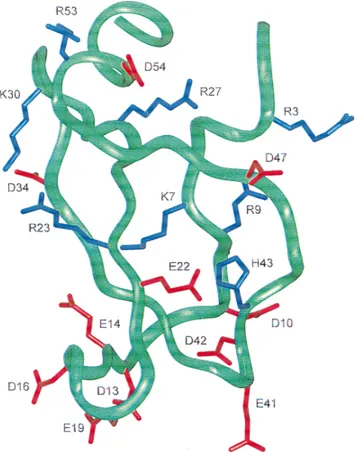
~Asn2T–Arg3T!residues of TAP–BPTI possess a nearly ideal
ex-tended backbone conformation similar to that found in TAP–Xa, the side chains of Tyr1Tand Arg3Tof the two structures are
se-verely rotated with respect to each other~Fig. 5!: Tyr1TOH swings
14.2 Å to bind with Asp189 in the S1 specificity site while the guanidino group of Arg3Tmoves to form a cation–p-electron
com-plex in the S4 subsite of Xa~Dougherty & Stauffer, 1990; Schwa-bacher et al., 1993; Lin & Johnson, 1995!. Both of these residues have been shown to be crucial for inhibitory activity. The site specific mutation of R3N leads to a 40,000-fold decrease in activ-ity, while removal of Tyr1Tproduces a 1,000-fold loss~Dunwiddie
et al., 1992!. The tyrosyl group of Tyr1T in TAP–BPTI packs
against the ~Cys5T–Cys59T! disulfide bridge with its hydroxyl
making intramolecular hydrogen bonds with Cys59TO~2.80 Å!
and the guanidinium group of Arg27TNH1~3.13 Å! ~Fig. 5!. The
latter residue is also involved in hydrogen bonds between TAP molecules within a TAP helix~Table 3!. Although Tyr1TN makes
a salt bridge with Glu22Tin the TAP helix~Table 3!, the
confor-mation of the residue appears to be the natural one of the native state by virtue of its association with~Cys5T–Cys59T!and Arg27T,
and the fact that no further movement is necessary to form the intermolecular salt bridge: Tyr1TN is already positioned optimally
for the interaction in this conformation. The conformation of the Tyr1Ttyrosyl is, moreover, similar to that of the CA–CD atoms of
Arg1B, which also interact with the~Cys5B–Cys55B!disulfide of
BPTI like the tyrosyl of TAP.
The extended ~Asn2T–Arg3T! dipeptide is part of a 310 turn
arrangement involving~Asn2T–Leu4T!, with a hydrogen bond
be-tween Asn2TO and Cys5TN. In one of the NMR ensembles~ An-Fig. 4. Electrostatic potential surface of the intermolecular TAP helix of
TAP-BPTI. Calculated using the program GRASP~Nicholls et al., 1991!. ESP.10 kT, blue;, 28 kT, red;;0, gray. Five TAP molecules of helical array shown.
tuch et al., 1994!, the N-terminal region is disordered, while the other~Lim-Wilby et al., 1995!is consistent with a 310helix similar
to that found in TAP–BPTI and TAP–Xa. A corresponding 310
structure also occurs at the N-terminus of BPTI. The N-terminal tyrosyl orientation in TAP–BPTI most likely represents a stable pre-Xa–bound conformation that changes to the more extended one when TAP interacts with the active site of Xa burying the Tyr1Tside group in the S1 specificity pocket~Wei et al., 1998!.
Crystal packing interactions could be partially responsible for the tyrosyl position in TAP–BPTI, as this region associates somewhat with a cavity of a symmetry-related TAP molecule. However, pres-ence of the 310turn in NMR solution structures, the similarity of
the conformations of the side groups of Tyr1T and Arg1B, their
intramolecular disulfide associations in both TAP and BPTI, and the hydrogen bond to Arg27Tin TAP, all rule against the possibility.
Comparing the TAP structure of TAP–BPTI with that of TAP in TAP–Xa suggests that Asn2Tmay play a more significant role in
the binding of TAP to Xa than initially thought. In addition to hydrogen bonds between Asn2T~N and OD1!and the active site of
Xa~Gln192OE1 and Gly218N, respectively! ~Wei et al., 1998!, Asn2TND2 makes another obviously important intramolecular
hy-drogen bond with Tyr49TOH ~2.86 Å!. This interaction helps to
further fix and maintain the position of the~Asn2–Leu4!310-turn.
In TAP–BPTI, the Tyr49Thydrogen bond is mediated by SO4–1 ~Table 1!, is a little longer~3.13 Å!, and not as linear. Nonetheless, its presence in TAP–BPTI stabilizes the position of the 310-turn
close to the optimal one found in the TAP–Xa bound structure: a small shift in the Asn2Tside group on binding to Xa leads to a
geometrically ideal~Asn2TND2–Tyr49TOH!hydrogen bond~
dis-tance and angle!at the expense of losing the sulfate ion interaction and slightly disrupting the better-defined 310-turn hydrogen bond
of TAP–BPTI. Although the energetic trade-off on Xa binding appears to be about equally compensated, the importance of the entropic contribution of TAP in the TAP–BPTI structure is to be noted, maintaining the proper position of the 310-turn for binding
to Xa. The key residue overseeing this role is Asn2T. A similar
stabilization of the N-terminal 310-turn in BPTI also occurs, but
through a nonpolar hydrophobic interaction between Pro2Band the ~Cys5B–Cys59B!disulfide bridge.
In addition to the changes of the N-terminal tetrapeptide, con-siderable variation in the structure of the first disulfide loop of TAP
~Cys5T–Cys15T!is also observed between TAP in TAP–BPTI and
the Xa bound structure that could be the result of a massive reor-ganization of the region accompanying binding to Xa~Fig. 5!. The largest deviations are within the Lys7Tto Asp10T segment. The
lysyl group of Lys7Tis somewhat important in the Xa complex,
binding indirectly with the cation hole~Brandstetter et al., 1996; Mochalkin & Tulinsky, 1999!of the S4 subsite~hydrogen bonded salt bridge, Lys7TNZ–Glu97OE2, 3.00 Å!, where the guanidinium
group of Arg3Talso forms a cation–p-electron complex with Tyr99,
Phe174, Trp215, and a hydrogen-bonded salt bridge with the other cation hole carboxylate oxygen of Glu97OE1~Wei et al., 1998!. The NZ atoms of Lys7Tin TAP–BPTI and TAP–Xa differ in
po-sition by 10.6 Å, while Arg3TCZ differs by 5.0 Å on binding to the
S4 site. Optimal superposition of Ty1T–Ile6T of TAP–BPTI and
TAP–Xa shows that the xvalues of CB and CG of Arg3T are
similar, while those of the remaining atoms differ. Because Arg3T
is a surface residue~Figs. 2, 3!, its terminal atoms appear to be in different positions in the two structures due to Xa binding in TAP–Xa and salt bridge formation between Arg3Tand Asp3Bin TAP-BPTI ~Table 3!, so its native conformation remains uncertain.
The differences in structure of the first disulfide loop of TAP in TAP–BPTI and TAP–Xa start at the Ile6T side group and are
followed by those of Lys7T, with the two lysyl groups projecting
in opposite directions~Fig. 5!. The Pro8Tresidue in TAP–BPTI
partially overlaps the lysyl position in the TAP–Xa complex while its~Pro8T–Trp11T! segment exhibits a type 1 b-bend, with the
proline ring and the indole of Trp11Tstacking on one another. The
position of Pro8Tis displaced by a residue in TAP–Xa, while the
side groups of Arg9Tand Asp10Tare completely different, as are
the orientations of the indole rings of Trp11T~Fig. 5!. The
differ-ences then continue on to the disulfide bridge at Cys15T. Although
some of the smaller changes in structure could be due to crystal packing interactions, in either the TAP–BPTI or TAP–Xa crystals, the main-chain differences and the large side-group re-orientations between the two must be considered conformational changes ac-companying TAP binding to Xa.
The largest differences in structure between TAP–BPTI and TAP–Xa are essentially all confined to the first 15 residues of TAP. Other lesser differences are:~1!in a small surface loop~Asn28T–
Gly31T!, due to a peptide flip between ~Asn28T–Gly29T!, ~2!
another peptide flip at Asp34T,~3!the side chain of Glu41T,~4!the
orientation of the imidazole ring of His43T, ~5! thex1angle of
Asp47T, and~6!some differences in the C-terminal helix;
differ-ences ~3! and ~4! could be due to formation of intermolecular hydrogen bonds within the TAP helix~Table 3!. The TAP molecule has a secondary binding site on Xa~Wei et al., 1998!. Of the six TAP residues that bind to the site, four display a high degree of structural fidelity compared to TAP-BPTI~including all the main-chain interactions!, whereas the side groups of Asp47T@~5!above#
and Tyr49Tare different. The Asp47Tresidue is involved in three
hydrogen bonds with BPTI in TAP–BPTI~Table 3! so that the different position of the Asp47Tside chain in TAP–Xa appears to
be due to Xa binding and the formation of a salt bridge with Arg222 and Lys224 of the secondary binding site. The latter res-idues are not only constituents of this additional Xa binding site but are also residues of the Na1ion binding site of Xa~Zhang &
Tulinsky, 1997!. The relationship between the two sites, however, could not be correlated further because the Na1 ion and water
structure of the TAP–Xa complex was not reported. Finally, the Tyr49T shift leads to a hydrogen bond with Glu146 of the Xa
secondary site, and in addition, improves the one to Asn2TND2 of
the N-terminal of TAP; the latter helps maintain the position of the
~Asn2T–Leu4T!310turn already mentioned in the TAP–Xa complex.
The general conservation of the conformation of TAP residues that interact with the secondary binding site of Xa in the two-step TAP–Xa binding mechanism~Jordan et al., 1992!suggests that the initial and rapid kinetic step in the binding of TAP to Xa most likely involves binding to the secondary site~Wei et al., 1998!. The large conformational changes in the TAP N-terminus are then prob-ably responsible for the slower, tight-binding step. Thus, the TAP– BPTI structure provides additional pertinent evidence regarding the TAP–Xa binding mechanism, clearly indicating the conforma-tional changes that occur in TAP.
The structure of BPTI
The structure of BPTI in TAP–BPTI crystals is practically the same as that of previously determined crystal structures of BPTI
~Marquart et al., 1983; Wlodawer et al., 1987a, 1987b; Lubkowski & Wlodawer, 1999!. When compared with the 4PTI coordinates
except that of the C-terminal Ala58 residue, gives an RMS dif-ference of 0.40 Å. Significantly, however, the side chains of 6 of the 10 arginine0lysine residues of BPTI in TAP–BPTI have different orientations. Three of the differences are most likely due to TAP–BPTI interactions~Arg17B, Arg42B, Arg53B; Table 3!.
Another outstanding difference is in the Glu7B side chain that
makes a hydrogen bond with the carboxylic terminal of Ala58B
of a symmetry-related BPTI molecule~Table 3!. This difference is most likely due to the presence of SO4–1 in TAP–BPTI, which
partially occludes the same region occupied by the side group of Glu7B of the BPTI structure.
Materials and methods
The TAP used for crystallization was recombinant material pro-vided by Corvas International, Inc.~San Diego, California! ~ Lim-Wilby et al., 1995!; the BPTI was purchased from Calbiochem~La Jolla, California!. Using a cocrystallization approach, crystals of a 1:1 complex of TAP–BPTI were successfully grown by the vapor diffusion hanging drop method. The protein solution was prepared by first combining equal proportions of 20 mg0mL solutions of each protein and then allowing them to stand overnight at 48C. Crystallization drops were made by mixing 2mL of the protein solution with an equal volume of well solution consisting of 0.1 M ADA buffer, pH 6.5, 1.2 M ammonium sulfate. Elongated crystals grew in about seven days that belong to the tetragonal system, space group P41or P43, with unit cell dimensions a5b546.87
and c550.35 Å. MALDI mass spectrometric analysis confirmed the composition of the crystals as containing both TAP and BPTI. Four TAP–BPTI complexes per unit cell~one per asymmetric unit!
give Vm52.06 Å30Da and a protein fraction50.60.
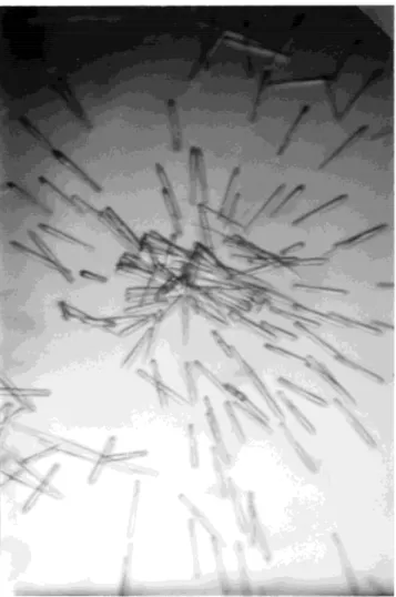
An unusual and somewhat interesting phenomenon was ob-served in growing TAP–BPTI crystals. When seeded with a BPTI single crystal, TAP–BPTI crystals grew in roughly concentric cir-cles around the seed, pointing in generally radial directions at the seed~Fig. 6!. Although only an apparent curiosity, it may be re-lated to the highly dipolar nature of TAP–BPTI crystals arising from the macroscopic dipole expected of the TAP helices in crys-tals~Fig. 4!, which originates from the electrostatic compensation of markedly dipolar TAP molecules~Figs. 2, 3!over a relatively large distance but leaves opposite electrostatic charges uncompen-sated at either end of the helix.
X-ray diffraction intensity data of a TAP–BPTI crystal were measured using a RAXIS-II image plate detector mounted on an Rigaku RU200 rotating anode X-ray generator producing CuKa
radiation with a graphite monochrometer at 50 kV0100 mA power. A TAP–BPTI crystal ~0.63 0.4 30.2 mm! sealed in a glass capillary diffracted X-rays to better than 1.6 Å resolution at room temperature@I0s~I! ;4.0 in the outermost range#. Two and a half degree oscillation frames were measured at a crystal-detector dis-tance of 60 mm and swing angle of 08, thus sweeping out diffrac-tion data to a maximum resoludiffrac-tion of 1.6 Å. A total number of 72 frames of diffraction data were recorded. The raw intensities were reduced to scaled structure amplitudes using the R-AXIS data processing software package~Higashi, 1990!, results of which are summarized in Table 2.
The crystal structure of TAP–BPTI was solved by molecular replacement using the program AMoRe~Navaza, 1994!from the CCP4 crystallographic package~Laskowski et al., 1993!. An initial self-rotation search to check for symmetry between TAP and BPTI did not produce any significant peaks in agreement with lack of
sequence and structure homology~Antuch et al., 1994!. A rotation-translation search for BPTI was then performed using the coordi-nates of a previously determined BPTI crystal structure ~PDB code: 4PTI! ~Marquart et al., 1983!. The search was carried out using the ~8.0–2.5! Å data and a 15 Å radius of integration. A translation search of the most outstanding rotation result produced a single unambiguous solution in space group P41with a
correla-tion coefficient of 0.37 and Rfactorof 51.8%. A similar solution was
found using the 8.0–3.0 Å and 8.0–2.0 Å data but was not pro-duced using the 8.0–3.5 Å resolution range. Rigid body refinement of the 8.0–2.5 Å solution resulted in a correlation of 0.44 and an
Rfactorof 50.1%.
With the BPTI molecule fixed, a rotation and translation search was carried out based on TAP models derived from two separate NMR structure determinations@PDB code: 1TAP~Antuch et al., 1994!and 1TCP~Lim-Wilby et al., 1995!#. These searches did not produce any significant solutions. Electron density maps using phases based on BPTI alone did not show continuous density of a TAP molecule and were difficult to interpret due to many breaks in the density. Although small segments of polyalanine peptides, pre-sumably corresponding to TAP structure, were introduced into cal-culations that refined to R;30% at 2.8 Å resolution, the density was still generally uninterpretable in terms of a TAP molecule.
However, using the TAP coordinates of the recently released crys-tal structure of the TAP–Xa complex ~PDB code: 1KIG! ~Wei et al., 1998!proved otherwise. A single solution in the 8.0–2.5 Å range was found using this TAP coordinate set as a search model even though it ranked 18th in the rotation solution list~correlation 0.54, Rfactor545%!. Using a modified model of TAP in which the
first four N-terminal residues~Tyr1T–Leu4T!, which bind to the
active site of Xa~Wei et al., 1998!, and the C-terminal Ile60Twere
omitted, along with 10 arginine and lysine side chains beyond the CB atom, the same rotation solution was obtained, but now ranked first in the rotation list. Concurrent rigid-body refinement of the TAP and BPTI models produced a correlation of 0.59 and an Rfactor
of 44.2%.
Positional and thermal parameter refinement of the TAP–BPTI model was conducted using the program CNS ~Brünger, 1998!. An initial simulated annealing refinement of the complex was carried out with~8.0–2.0! Å data using the slow-cool procedure in which the temperature was decreased from 2,000 to 300 K in 258 intervals. In this and all subsequent refinements, about 5% of the data set~665 reflections!was withheld for calculating the
Rfree value. The slow-cool procedure reduced the Rfactor from
46.7% ~Rfree 5 47.0%! to 32.4% ~Rfree 5 40.1%!. This was followed by 100 cycles of conjugate gradient positional refine-ment and 30 cycles of individual isotropic B refinerefine-ment conclud-ing at R530.4%~Rfree538.8%!. The corresponding~2Fo2Fc!
electron density map revealed excellent density for both mol-ecules, most of BPTI and about 304 of TAP. Manual rebuilding was required to properly fit the differently positioned first loop segment of TAP~Tyr1T–Cys15T! and a smaller loop ~Asn28T–
Gly31T! compared to the TAP–Xa structure. Extending resolu-tion to 1.6 Å, the TAP–BPTI model was improved further through several more rounds of manual adjustment and positional refine-ment~200 cycles!. During this stage, solvent molecules and three sulfate ions were fitted to the difference electron density maps. The final round of positional and individual isotropic Bfactor
re-finement in the 8.0–1.6 Å resolution range including occupan-cies of water molecules produced an R-value of 18.8% ~Rfree5
21.1%!. The Ramachandranf0c values of both proteins all fall within energetically allowed regions. The occupancies of the wa-ter molecules are all.0.62 with^B&534 Å2. The^B&value of
TAP is 18.5 Å2, whereas that of BPTI is 22.8 Å2. A summary of
refinement statistics is given in Table 2. The coordinates of TAP– BPTI have been deposited in the PDB code 1D0D.
Acknowledgments
This work has been supported by NIH Grants HL25942 and HL43229. We also thank Corvas International, Inc. for partial financial support and pro-viding samples of recombinant TAP, and Terry Brunck for numerous dis-cussions throughout the course of this work.
References
Antuch W, Guntert P, Billeter M, Hawthorne T, Grossenbacher H, Wuthrich K. 1994. NMR solution structure of the recombinant tick anticoagulant protein
~rTAP!, a factor Xa inhibitor from the tick Ornithodoros moubata. FEBS
Lett 352:251–257.
Bode W, Huber R. 1992. Natural protein proteinase inhibitors and their inter-action with proteinases. Eur J Biochem 204:433– 451.
Brandstetter H, Kuhns A, Bode W, Huber R, van der Saal W, Withersohn K, Engh RA. 1996. X-ray structure of active site-inhibited clotting factor Xa.
J Biol Chem 271:29988–29992.
Brünger AT. 1998. Crystallography and NMR system: A new software suite for macromolecular structure determination. Acta Crystallogr D54:905–921. Dougherty DA, Stauffer DA. 1990. Acetylcholine binding by a synthetic
recep-tor: Implications for biological recognition. Science 250:1558–1560. Dunwiddie CT, Neeper MP, Nutt EM, Waxman L, Smith DE, Hofmann KJ,
Lumma PK, Garsky VM, Vlasuk GP. 1992. Site-directed analysis of the functional domains in the factor Xa inhibitor tick anticoagulant peptide.
Biochemistry 31:12126–12131.
Higashi T. 1990. Auto-indexing of oscillation images. J Appl Crystallogr
23:252–257.
Jordan SP, Mao SS, Lewis SD, Shafer JA. 1992. Reaction pathways for inhi-bition of blood coagulation factor Xa by tick anticoagulant peptide.
Bio-chemistry 31:5374–5380.
Laskowski RA, MacArthur MW, Moss DS, Thorton JM. 1993. PROCHECK: A program to check the stereochemical quality of protein structures. J Appl
Crystallogr 26:283–291.
Lim-Wilby MSL, Hallenga K, de Maeyer M, Lasters I, Vlasuk GP, Brunck TK. 1995. NMR structure determination of tick anticoagulant peptide~TAP!.
Protein Sci 4:178–186.
Lin Z, Johnson ME. 1995. Proposed cation-pmediated binding by factor Xa.
FEBS Lett 370:1–5.
Lubkowski J, Wlodawer A. 1999. Decamers observed in crystals of bovine pancreatic trypsin inhibitor. Acta Crystallogr D55:335–337.
Marquart M, Walter J, Deisenhofer J, Bode W, Huber R. 1983. The geometry of the reactive site and the peptide groups in trypsin, trypsinogen and its complexes with inhibitors. Acta Crystallogr B39:480– 490.
Mochalkin I, Tulinsky A. 1999. Structures of thrombin retro-inhibited with SEL2711 and SEL2770 as they relate to factor Xa binding. Acta Crystallogr
D55:785–793.
Navaza J. 1994. AMoRe: An automated package for molecular replacement.
Acta Crystallogr A50:157–163.
Nicholls A, Sharp KA, Honig B. 1991. Protein folding and association: Insights from the interfacial and thermodynamic properties of hydrocarbons.
Pro-teins 11:281–296.
Nienaber VL, Amparo EC. 1996. A noncleavable retro-binding peptide that spans the substrate binding cleft of serine proteases. J Am Chem Soc
118:6807– 6810.
Rydel TJ, Tulinsky A, Bode W, Huber R. 1991. Refined structure of the hirudin– thrombin complex. J Mol Biol 221:583– 601.
Schechter I, Berger A. 1967. On the size of the active site in proteases. Biochem
Biophys Res Commun 27:157–162.
Schwabacher AW, Zhang S, Davy W. 1993. Directionality of the cation-peffect: A charge-mediated size selectivity in binding. J Am Chem Soc 115:6995– 6996.
Tabernero L, Chang CY, Ohringer SL, Lau WF, Iwanowicz EJ, Han WC, Wang TC, Seiler SM, Roberts DGM, Sack JS. 1995. Structure of a retro-binding peptide inhibitor complexed with humana-thrombin. J Mol Biol 246:14–20. van de Locht A, Stubbs MT, Bode W, Friedrich T, Bollschweiler C, Hoffken W, Huber R. 1996. The ornithodorin–thrombin crystal structure, a key to the TAP enigma? EMBO J 15:6011– 6017.
Waxman L, Smith DE, Arcuri KE, Vlasuk GP. 1990. Tick anticoagulant peptide ~TAP!is a novel inhibitor of blood coagulation factor Xa. Science 248:593– 596.
Wei A, Alexander RS, Duke J, Ross H, Rosenfeld SA, Chang C. 1998. Un-expected binding mode of tick anticoagulant peptide complexed with bovine factor Xa. J Mol Biol 283:147–154.
Wlodawer A, Deisenhofer J, Huber R. 1987a. Comparison of two highly refined structures of bovine pancreatic trypsin inhibitor. J Mol Biol 193:145–156. Wlodawer A, Nachman J, Gilliland GL, Gallagher W, Woodward C. 1987b. Structure of form III crystals of bovine pancreatic trypsin inhibitor. J Mol
Biol 198:469– 480.
Zhang E, Tulinsky A. 1997. The molecular environment of the Na1binding site