ABSTRACT
Subcutaneous tissue reaction to castor oil bean
and calcium hydroxide in rats
Samira Esteves Afonso CAMARGO1, Sigmar de Mello RODE2, Renata Falchete do PRADO3,
Yasmin Rodarte CARVALHO4, Carlos Henrique Ribeiro CAMARGO5
1- PhD, Department of Bioscience and Oral Diagnosis, São José dos Campos Dental School, São Paulo State University (UNESP), São José dos Campos, SP, Brazil.
2- PhD, Associate Professor, Department of Dental Materials and Prosthodontics, São José dos Campos Dental School, São Paulo State University (UNESP), São José dos Campos, SP, Brazil.
3- PhD, Department of Physiotherapy, Superior School of Cruzeiro, Cruzeiro, SP, Brazil.
4- PhD, Full Professor, Department of Bioscience and Oral Diagnosis, São José dos Campos Dental School, São Paulo State University (UNESP), São José dos Campos, SP, Brazil.
5- PhD, Department of Restorative Dentistry, São José dos Campos Dental School, São Paulo State University (UNESP), ,São José dos Campos, SP, Brazil.
Corresponding address: Renata Falchete do Prado - Faculdade de Odontologia de São José dos Campos - UNESP - Departamento de Biociências e Diagnóstico Bucal - Av. Francisco José Longo, 777, Jardim São Dimas - 12245-000 - São José dos Campos, SP - Brasil - Phone: +55 12 3947 9000 - Fax: +55 12 39479010 - e-mail: [email protected]
!"!#$!%&'()*!'+,-'+..,'/'01%#2"34#1*&'035"6'78-'+.7.'/'9""!:4!%&'9:5#;'.<-'+.7.
C
astor oil bean cement (COB) is a new material that has been used as an endodonticsealer, and is a candidate material for direct pulp capping. Objective: The purpose of this study was to evaluate the biocompatibility of a new formulation of COB compared to calcium hydroxide cement (CH) and a control group without any material, in the subcutaneous tissue of rats. Material and methods: The materials were prepared, packed into polyethylene !"#$%&'()&*+,-'( #)&*(& .#&/' &)0/$'-&$!"1! '(#0!$& *$$!#2&3(*+'-$&4#/#&$'1/*51#)&' & .#& 6 .&'()&78 .&)'9$&': #/&*+,-'( ' *0(2&3&;!'( * ' *<#&'('-9$*$&0:&*(='++' 0/9&1#--$&4'$& ,#/:0/+#)&'()&)' '&4#/#&$!">#1 #)& 0&3?@A3&'()&B!C#9D$& #$ $&' &7E&$*F(*51'(1#&-#<#-2& G#$!- $H&I0+,'/*(F& .#&+#'(&(!+"#/&0:&*(='++' 0/9&1#--$&"# 4##(& .#& 40&#J,#/*+#( '-& F/0!,$&KI@L&'()&IMN&'()& .#&10( /0-&F/0!,%&$ ' *$ *1'--9&$*F(*51'( &)*::#/#(1#&K,O82888PN& 4'$&0"$#/<#)&' &6&'()&78&)'9$2&B.#/#&4#/#&(0&$*F(*51'( &)*::#/#(1#$&K,O82PPPN&"# 4##(& *$$!#& /#'1 *0(& 0& IM& KQRS& *(='++' 0/9& 1#--$N& '()& I@L& KQQ8& *(='++' 0/9& 1#--$N& ': #/& 6&)'9$2&3: #/&78&)'9$%&$*F(*51'( -9&+0/#&*(='++' 0/9&1#--$&K,O828SN&4#/#&0"$#/<#)&*(& .#&IM&F/0!,&KT8T&*(='++' 0/9&1#--$N& .'(&*(& .#&I@L&F/0!,&KP66&*(='++' 0/9&1#--$N2& I0(1-!$*0($H&B.#$#&/#$!- $&)#+0($ /' #& .' & .#&I@L&1#+#( &*()!1#$&-#$$&*(='++' 0/9& response within long periods.
Key words: Calcium hydroxide. Castor oil. Pulp capping. Rats.
INTRODUCTION
One of the most important objectives of pulp preservation by direct pulp capping is the limitation of damage and to healthy function. Conservative endodontic techniques facilitate the maintenance of teeth with pulpal alterations, minimizing the
unwanted sequelae of their unplanned extraction13.
In direct pulp capping procedures, a biocompatible or bio-inductive material is placed onto the exposed pulp tissue, preserving its vitality, stimulating the repair process, and promoting the formation of hard
tissue barrier9. Biocompatibility is as important as
the physical and chemical properties when selecting
a material for endodontic therapy because of the direct contact with the vital tissue. However, some currently used pulp capping materials have a
tissue-irritating potential17.
Calcium hydroxide (CH) cement presents
properties such as low cytotoxicity, high pH18,29 and
antibacterial action10,25. CH has been the material of
choice for direct pulp capping because it seems to stimulate a rapid differentiation of odontoblast-like
cells that form a hard tissue barrier in the pulp10,25.
On the other hand, this action is not exclusive of CH and this materials suffers mechanical wear and
solubility for long periods9.
polyester formed by an amino radical, which confers bactericidal effect and has biocompatibility with
living tissues3. It has great potential to facilitate
tissue healing, excellent structural properties,
low cost and does elicit toxic effects. COB14 has
been tested in rabbits as a matrix for bone and joint replacement. After 40 days of surgery, the histological examination showed absence of late
*(='++' 0/9& /#'1 *0(& '()& (0& $*F($& 0:& $9$ #+*1&
toxic effects.
Carvalho, et al.6 (1997) analyzed histometrically
the alveolar bone healing around castor oil bean (Poliquil; Polímeros Químicos, Araraquara, SP, Brazil) implanted immediately after tooth extraction. The material was biologically compatible, as it was progressively integrated into alveolar bone in the healing process.
Barros, et al.1 (2003) investigated in vivo the
biocompatibility of Ricinus communis polyurethane with three different chemical compositions.
U0)*51' *0(&0:& .#&,0-9+#/V$&1.#+*1'-&10+,0$* *0(&
by the addition of calcium carbonate or calcium phosphate promoted matrix mineralization, these materials being more biocompatible than pure resin.
Mastrantonio and Ramalho22 (2003) evaluated the
subcutaneous tissue reaction in mice, after the implantation of castor oil bean with or without calcium carbonate and showed that both materials are biocompatible.
In Endodontics, COB has been used in retrograde filling materials in paraendodontic surgeries,
irrigating agents and endodontic sealers27,28.
The use of a certain material must be based on experimental and laboratory studies that prove
its biocompatibility and other properties15. The
development of newer biocompatible, bactericidal, inductive materials that promote tissue repair and present adequate sealing can result in longevity
of pulp capping procedures10. In addition, material
selection is important for the success or failure of
these treatments5,15.
With this objective, some methods have been developed to evaluate the irritating potential of dental materials. The implantation of materials in the subcutaneous connective tissues of small experimental animals is considered an adequate methodology to determine the biocompatibility of
endodontic materials5,20,26, although it is known
that some reactions observed in this test cannot be considered identical to those occurring in living dental tissues.
It is believed that the pulp reaction can vary with the use of different available products, depending on their biocompatibility, which could cause severe
damage to this tissue12. For this reason, there
is an interest to increase the knowledge of the biocompatibility of COB because this material can be a candidate for direct pulp capping. The purpose
of this study was to evaluate the biocompatibility of a new formulation of COB compared to CH and a control group without any material, in the subcutaneous tissue of rats.
MATERIAL AND METHODS
This study was performed in accordance to the Ethical Principles of Animal Experimentation (COBEA – Brazilian College of Animal Experimentation) and was approved by the local Research Ethics Committee (São José dos Campos Dental School) (process no. 003/2006-PA/CEP).
Forty-two male Wistar rats (Rattus norvegicus) aged 90 days and weighing 350 to 400 g were used. The animals were maintained with food and water ad libitum.
The tested materials were a COB-based cement (Poliquil; Polímeros Químicos, Araraquara, SP, Brazil) and a CH cement (Dycal; Dentsply Petrópolis, RJ, Brazil). The COB cement was prepared according to the manufacturers’ instructions, mixing liquid polyol (5 mL), liquid prepolymer (5 mL), and calcium carbonate (5 g) until homogenization was obtained. CH cement was hand-mixed according to the manufacturer’s directions.
Polyethylene tubes (10-mm long x 1.5 mm inner diameter) (Johnson & Johnson, São José dos Campos, SP, Brazil) were washed with 70% alcohol
'()&)*$ *--#)&4' #/%&'! 01-'<#)%&'()&5--#)&4* .& .#&
experimental materials using a lentulo spiral (KG Sorensen, Barueri, SP, Brazil) at low speed. All carriers and glass plates used in the study were previously sterilized.
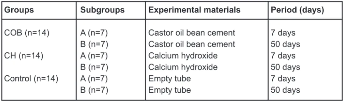
The sample comprised three experimental groups of 14 animals each, half of which were killed after 7 and half at 50 days. Each animal received a polyethylene tube containing COB cement or CH cement in the dorsum. In the control group, the animals received an empty polyethylene tube in the dorsum. Figure 1 shows the schematic design of the methodology.
For the surgical procedures, the animals were anesthetized by intramuscular administration of 38.5 mg/kg of ketamine HCL (Dopalen; Vetbrands do Brasil Ltda., Campinas, SP, Brazil), and 14.2 mg/kg of xylazine (Anasedan; Vetbrands do Brasil Ltda., Campinas, SP, Brazil). The back of the animal was shaved and cleaned with 1% iodine in ethanol. Incisions were made on the dorsum, and one subcutaneous pocket was carefully prepared by blunt dissection. The base of the pocket was located at 10 mm from the incision line. A tube containing cement was then placed into each pocket and the incision was closed with surgical gut sutures.
formalin.
3: #/&5J*(F&:0/&TR&.%& .#& *$$!#&4'$&,/01#$$#)& :0/&,'/':5(&#+"#))*(F2&B.#& !"#$&4#/#&/#+0<#)& )!/*(F& .*$&,/01#)!/#2&3&,'/':5(&"-01C&4'$&0/*#( #)&
in such a way that it was parallel to the long axis of the tube, and serial sections were cut to a 5-µm thickness. The sections were stained with hematoxylin and eosin. Histological qualitative
'()& ;!'( * ' *<#& '('-9$#$& 0:& .#& *(='++' 0/9&
response were performed on light microscope. Color images of stained sections were acquired with a high-resolution camera (Sony Cyber-Shot DSC-S85; Sony Com. Ind. Ltda., Tokyo, Japan) at
S88J&0/*F*('-&+'F(*51' *0(&:0/&.*$ 0+0/,.0+# /*1& '('-9$*$2&@(-9&P&<*#4&K0/*F*('-&+'F(*51' *0(&S88JN&
standardized total area analyzed. It was positioned in the exact center of end of tube in each one of the 5 semi-serial slides per rat.
A single investigator blinded to the groups examined all specimens. Inflammatory cells were counted with an automated image analysis software, a public domain image processing and analysis program (Image J, National Institutes of Health – US Department of Health and Human
Services, Bethesda, Maryland, USA), using the
point tool. The criteria of histological quantitative evaluation were based on microscopic aspects. Lymphocytes presented small, round, very darkly staining nuclei and little surrounding cytoplasm. Macrophages presented larger, paler, oval or bean shaped nuclei and a somewhat larger amount
0:& 19 0,-'$+2& ?#! /0,.*-$& 4#/#& #'$*-9& *)#( *5#)&
because of their polymorphic shape nucleus. Other
*(='++' 0/9&1#--$&4#/#&(0 &0"<*0!$&*(& .*$&$ !)92& W++' !/#& 5"/0"-'$ $& )*$,-'9*(F& -'/F#& 0<'-& (!1-#*& '()& +' !/#& 5"/0"-'$ $& 4* .& :!$*:0/+& (!1-#*& 4#/#& 0"$#/<#)&"! &4#/#&(0 &;!'( *5#)2&3&+#'(&(!+"#/& 0:&*(='++' 0/9&1#--$&4#/#&0" '*(#)&:0/&#'1.&'(*+'-2&
Data were subjected to descriptive and inferential
'('-9$*$2& B.#& +#'($& 0:& *(='++' 0/9& 1#--$& 4#/#&
tested by two-way ANOVA. The control, CH cement and COB cement groups were compared considering the following factors as variables: material and
,#/*0)&0:&$'1/*51#&K6&0/&78&)'9$&': #/&*+,-'( ' *0(N2& X.#(& .#& 3?@A3& $.04#)& $ ' *$ *1'--9& $*F(*51'( &
difference, the Tukey’s multiple-comparison test
4'$& !$#)2& B.#& -#<#-& 0:& $*F(*51'(1#& 4'$& 7E& :0/&
both tests.
RESULTS
!"#$%$&!'(%)*+(%!#(#!,-)(.(%/"!"
The groups were compared qualitatively and no difference was observed between the CH and COB cement groups. Figure 2 shows an overview of the tube end with each material and experimental period.
012)0-3-.#)45$+6)
In the sections obtained at the 7th day, a moderate to severe chronic inflammatory process was observed, except for one specimen that presented mild intensity. Inflammatory
*(5- /' #& 10+,0$#)& 0:& +0(0(!1-#'/& 1#--$%& +'*(-9&
lymphocytes, was present near the material. In some specimens, there were also plasma cells,
(#! /0,.*-$& '()& #0$*(0,.*-$& *(& .#& *(='++' 0/9& *(5- /' #2&@(-9&0(#&1'$#&#J.*"* #)&#J /'<'$' *0(&0:&
COB granules, which were surrounded by numerous
*(='++' 0/9&1#--$2&B.#&10((#1 *<#& *$$!#&*(&10( '1 &
with the material showed mild inflammatory reaction and a zone of necrosis was detected in few cases. The presence of multinuclear giant cells was not prominent in this experimental group. These cells appeared in a small number close to the
+' #/*'-&'()&,/#$#( #)& ./##& 0&5<#&(!1-#*2
In the sections obtained at the 50th day, a
+*-)& 0& +0)#/' #& *(='++' 0/9& 1#--& *(5- /' #& 4'$&
observed, mainly composed of lymphocytes and plasma cells. The presence of extravasated material in the subcutaneous tissue was associated with an
*(1/#'$#)& *(='++' 0/9& ,/01#$$2& W(& +0$ & 1'$#$%&
the tissue was organized in a capsular arrangement
4* .& ,'/'--#-& 10--'F#($& 5"#/$%& *( #/$,#/$#)& 4* .& 5"/0"-'$ $&'()&+' !/#&"-00)&<#$$#-$2&U!- *(!1-#'/& F*'( &1#--$&4* .& ./##& 0&5<#&(!1-#*&4#/#&:0!()&*(&
most specimens, although they appeared in a small number and were distributed near the material.
0 )0-3-.#)45$+6
In the sections obtained at the 7th day, moderate
0&$#<#/#&*(='++' 0/9&1#--&*(5- /' #&4'$&0"$#/<#)2&W &
Groups Subgroups Experimental materials Period (days)
COB (n=14) A (n=7) Castor oil bean cement 7 days
B (n=7) Castor oil bean cement 50 days
CH (n=14) A (n=7) Calcium hydroxide 7 days
B (n=7) Calcium hydroxide 50 days
Control (n=14) A (n=7) Empty tube 7 days
B (n=7) Empty tube 50 days
Figure 1- Schematic design of materials and methods
was increased around the extravasated CH granules in the subcutaneous tissue. Except for one specimen that presented numerous polymorphonuclear leukocytes, mainly neutrophils, the tissue reactions
$.04#)& '(& *(='++' 0/9& *(5- /' #& 10+,0$#)& 0:&
lymphocytes and macrophages. Few plasma cells were observed. The macrophages were distributed
'/0!()& .#& 0<#/=04& F/'(!-#$& 0/& )*::!$#-9& *(& .#&
granulation tissue. Sometimes, the cytoplasm of these macrophages presented some material particles. In some specimens, multinuclear giant cells with different amounts of nuclei and irregular cytoplasmic outline were found. The nuclei were distributed at random, characterizing a foreign body reaction. The connective tissue in contact with the material showed different degrees of reaction and a zone of necrosis was detected in many cases.
In sections obtained at the 50th day, moderate fibroblast and angioblastic proliferation was
0"$#/<#)2& B.#& 5"/0!$& '/#'& ,/#$#( #)& '& 1',$!-'/&
arrangement with moderate amount of collagen
5"#/$2&U0)#/' #& 0&$#<#/#&*(='++' 0/9&*(5- /' #& 4'$&<#/*5#)%&#J1#, &*(&$0+#&$,#1*+#($&*(&4.*1.&* & 4'$&0:&+*-)&*( #($* 92&B.#&*(='++' 0/9&1#--$&4#/#&
distributed around the extravasated CH granules. In this group, there was predominance of mononuclear cells, mainly lymphocytes and plasma cells, except for two cases exhibiting numerous neutrophils and eosinophils. Multinuclear giant cells and necrosis were not evident in this period.
Control Group
Seven days after implantation, granulation tissue was observed in contact with the tube, presenting
5"/0"-'$ $&'()&(#4&"-00)&<#$$#-$2&U0)#/' #& 0&+*-)& *(='++' 0/9&*(5- /' #&10+,0$#)&0:&+0(0(!1-#'/&
cells, mainly lymphocytes and plasma cells, was
observed. On day 50, a dense collagenous tissue was observed with scarce inflammatory cells,
1.'/'1 #/*Y*(F&'&5"/0!$&1',$!-#2&
!"#$3$567$3-#5!')(.(%/"!"
The analysis of the morphometric results shows
.' &*(& .#&I@L&F/0!,%& .#&(!+"#/&0:&*(='++' 0/9&
cells decreased from days 7 to 50.
I0+,'/*$0(&0:& .#&+#'(&(!+"#/&0:&*(='++' 0/9&
cells between the two experimental groups (CH and COB) and the control group by two-way ANOVA
/#<#'-#)&$*F(*51'( &)*::#/#(1#&K,O82888PN2&B.#/#& 4#/#& (0& $*F(*51'( & )*::#/#(1#$& "# 4##(& *$$!#& /#'1 *0(& 0& IM& KQRS& Z& PSQ2R& *(='++' 0/9& 1#--$N& '()& I@L& KQQ8& Z& P8[2\& *(='++' 0/9& 1#--$N& ': #/&
7 days. However, after 50 days, the CH group
,/#$#( #)&'&-'/F#/&+#'(&(!+"#/&0:&*(='++' 0/9&
cells than the COB group (404 ± 118.8 versus 176
Z&[82S&*(='++' 0/9&1#--$N2&B.#&+#'(&(!+"#/&0:& *(='++' 0/9&1#--$&*(& .#&10( /0-&F/0!,&' &6&'()&78&
days was 69.0 ± 35.1 and 50.0 ± 33.5, respectively.
DISCUSSION
Histomorphometric and/or quantitative analyses
1'(&"#&!$#)& 0&<#/*:9& .#&*(='++' 0/9&'()&/#,'*/&
phenomena and other reactions of dental materials in the subcutaneous tissue of rats. Some studies
have used quantitative analysis8,10,12,17,19 because it
is believed that, if made by calibrated examiners, it can determine reliable results. However, histomorphometric analysis based on counting of
.#& (!+"#/& 0:& *(='++' 0/9& 1#--$& ,/#$#( $& +0/#&
reliable results than qualitative analysis with scores,
for example15,23.
The Image J software is widely used in
quantitative experiments16. There are many ways
Figure 2- !"#$%"&'( )"**+%# $, +"(- .%$/) 0-+&"*$123'#4+$5'#6 $%'.'#"3 &".#'7("*'$# 891 &".#'7("*'$#:6 5-$;'#. ($#*"(*
to apply computerized tools in morphometry.
Semi-'! 0+' *1&10!( *(F&0:&*(='++' 0/9&1#--$&4'$&!$#)&
in this study to verify the intensity of the reaction caused by the implanted material. According to the obtained results, the simple qualitative analysis of
$,#1*+#($&)*)&(0 &)#+0($ /' #&'(9&*(='++' 0/9&
differences between CH and COB implants. On day 50, the histomorphometric analysis showed that
.#&I@L&1#+#( &,/#$#( #)&'&$*F(*51'( -9&$+'--#/& (!+"#/& 0:& *(='++' 0/9& 1#--$& .'(& IM%& '()& '-$0&
showed no difference from the control group. On the other hand, qualitative analysis showed an
*(='++' 0/9&/#'1 *0(&$*+*-'/& 0&"0 .&1#+#( $%&4* .&
mild to moderate intensity in the same evaluation period. Therefore, quantitative analysis was very important to show differences in tissue reaction to the tested cement.
IM& 1#+#( & ,/0)!1#)& +*(*+!+& *(='++' 0/9&
reactions in a previous study that determined
.#& =04& 1.'/'1 #/*$ *1$& '()& $!"1! '(#0!$& *$$!#&
reactions to CH and zinc oxide-eugenol endodontic
sealers15. Fourteen days after implantation,
the volume of tissue reaction was measured
.*$ 0+0/,.0+# /*1'--92& B.#& .*F.#$ & =04& <'-!#$& 4#/#&0" '*(#)&4* .&IM&1#+#( $%&"! & .#&=04&)*)&(0 & 10//#-' #&4* .& .#&)#F/##&0:&*(='++' 0/9&/#$,0($#2
Kolokouris, et al.19 (1998) evaluated the in vivo
biocompatibility of CH sealer in root canals. The intensity of reaction, initially severe, decreased on the 60th day, and this reduction continued progressively up to the 120th day.
W(& .#&,/#$#( &$ !)9%&': #/&78&)'9$%&* &4'$&<#/*5#)& .' & .#&IM&1#+#( &$ *--&$.04#)&'(&*(='++' 0/9&
process of moderate to severe intensity, mainly
1-0$#& 0&0<#/=04&IM&F/'(!-#$2&B.#&,/#$#(1#&0:& .*$&
material outside the tube increased not only the
#J #($*0(%&"! &'-$0& .#&*( #($* 9&0:& .#&*(='++' 0/9&
reaction. Therefore, before selecting a material for pulp capping procedures, it is important to know its mechanical properties. The ideal material
$.0!-)&,/#$#( &-04&=04&'()&$.0!-)&(0 &"#& 00&+!1.&
friable. Hydro C® CH cement (Dentsply, Petrópolis,
RJ, Brazil) exhibited the highest water sorption and solubility values after an evaluation of its
mechanical properties11.
Although CH capacity of inducing the formation of a hard tissue bridge is an important property of
pulp capping materials18,29, in the present study
the CH cement has less biocompatible than the COB cement. However, if we had used longer periods of evaluation, CH would be similar to COB
cement.A large number of dental materials present
cytotoxic effects when applied close or directly to the pulp, and the only material that seems to stimulate early pulp repair and dentin hard tissue barrier formation is CH. CH products are the best choice for conservative pulp treatments due to their therapeutic and biological potential, and the
property of stimulating the formation of sclerotic and reparative dentin with a consequent protection
of the pulp against thermal stimuli24.
Studies have persisted in the search for materials with high biocompatibility and good physicochemical properties, since the materials used in endodontic procedures can cause different reactions on pulp
tissue12. The COB cement has been used in Medicine
*(& .#&/#10($ /!1 *0(%&$!"$ * ! *0(&0/&5--*(F&0:&"0(#&
defects presenting good results14,21.
In Dentistry, Calixto, et al.3 (2001) and Carvalho,
et al.6 (1997) implanted a COB-derivative natural
resin in the extraction wounds in rats to add information about the biocompatibility of this material. Their results were similar to those of the present study because after 6 weeks there
4'$&(0&,#/$*$ #(1#&0:&*(='++' 0/9&/#'1 *0(%&9# &
a small number of giant cells were observed in the tissue in contact with the material. Also, there was no foreign-body reaction or persistence of the
*(='++' 0/9& /#'1 *0(& *(& .#& $ !)9& 0:& I'/<'-.0%&
et al.7 (1997). They evaluated histometrically the
bone healing around polyurethane resin implants
)#/*<#)&:/0+&1'$ 0/&"#'(&'()&<#/*5#)&,/0F/#$$*<#&
osteogenesis in conjunction with a decrease in the
5"/0!$&1',$!-#& .*1C(#$$2
Other authors have demonstrated that the incorporation of alkaline phosphatase to the Ricinus communitis polyurethane followed by synthetic
"0)9&=!*)&*(1!"' *0(&10!-)&"#&'&!$#:!-&'- #/(' *<#&
to improve the biological properties, as bone
formation, of this polyurethane2.
According to Costa, Marcantonio and Hebling8
(1997), the COB cement presented acceptable biocompatibility under microscopic analysis when tested in subcutaneous implants in rats. The biocompatibility of COB cement has also described
by Perassi, et al.28 (2004), who evaluated the tissue
/#$,0($#&0:&$!"1! '(#0!$&*+,-'( $&5--#)&4* .&I@L&
cement and other endodontic sealers. The COB cement showed less tissue response than any other sealer in both experimental periods (7 and 50 days), which can be explained by the structure of this material with high pureness, lack of solvent, debris, stabilizers and degradation products present in other polymers, which would lead to adverse
organic responses28.
Mastrantonio and Ramalho22 (2003) evaluated
the subcutaneous tissue reaction in rats after the implantation of COB cement with or without calcium carbonate and showed that both materials
*()!1#)&+*-)&*(='++' 0/9&/#$,0($#&': #/&6&)'9$2&
Barros, et al.1& KS88QN& <#/*5#)& .' & .#& '))* *0(&
of calcium phosphate or calcium carbonate to the Ricinus communis polyurethane improved its biocompatibility implanted in rabbit femurs.
These results are according with those of the
response and acceptable biocompatibility after 50 days of observation.
W(&$!++'/9%& .#&5()*(F$&0:& .#&,/#$#( &$ !)9&
indicate that the COB cement is a promising material. Furthermore, extracts of COB slightly induced cell proliferation and did not present genotoxicity without formation of micronuclei in
A6\&1#--$%&0/&+0)*51' *0(&0:& .#&(0/+'-&1#--&191-#&
in a previous in vitro study4 using primary human
pulp-derived cells. However, new complementary studies are necessary to evaluating this material over the pulp tissue.
CONCLUSION
These results demonstrate that the castor oil
"#'(& 1#+#( & KI@LN& *()!1#$& -#$$& *(='++' 0/9&
response within long periods.
REFERENCES
1- Barros VM, Rosa AL, Beloti MM, Chierice G. In vivo
biocompatibility of three different chemical compositions of
Riccinus communis polyurethane. J Biomed Mater Res A. 2003;67:235-9.
2- Beloti MM, Oliveira PT, Tagliani MM, Rosa AL. Bone cell responses to the composite of Ricinus communis polyurethane and alkaline phosphatase. J Biomed Mater Res A. 2008;84:435-41.
Q]&I'-*J 0&G^%&B#_5-0&`U%&L/#( #F'(*&ab%&I'/<'-.0&Ba2&W+,-'( ' *0(& 0:&='C#$&0:&1'$ 0/&0*-&/#$*(&*(&/' &)#( '-&'-<#0-!$2&c#$;&@)0( 0-&
Bras. 2001;15:257-62.
4- Camargo SE, Camargo CH, Hiller KA, Rode SM, Schweikl H, Schmalz G. Cytotoxicity and genotoxicity of pulp capping materials in two cell lines. Int Endod J. 2009;42:227-37.
5- Camp JH, Fuks AB. Pediatric Endodontics: endodontic treatment for the primary and young, permanent dentition. In: Cohen S, Hargreaves KM, editors. Pathways of the pulp. 9 ed. St. Louis: Mosby; 2006. p.822-82.
[]&I'/<'-.0&Ba%&3/'d>0&I3%&B#_5-0&`U%&L/#( #F'(*&ab2&M*$ 0-0F*1&
and histometric evaluation of rat alveolar wound healing around polyurethane resin implants. Int J Oral Maxillofac Surg. 1997;26:149-52.
6]&I'/<'-.0&Ba%&B#_5-0&`U%&3/'d>0&I3%&L/#( #F'(*&ab2&I./0(0-0F9&
of alveolar healing following immediate implantation of Ricinus communis polyurethane resin: histometric analysis in rats. J Biomed Mater Res A. 1997;37:449-52.
8- Costa CA, Marcantonio RA, Hebling J. Biocompatibility of oilbean polyurethane polymer in comparative study with zinc oxide eugneol cement: histologic evaluation of subcutaneous implants in rats. Odonto 2000. 1997;1:44-8.
9- Cox CF, Suzuki S. Re-evaluating pulp protection: calcium hydroxide liners vs. cohesive hybridization. J Am Dent Assoc. 1994;125:823-31.
10- Dominguez MS, Witherspoon DE, Gutmann JL, Opperman LA. Histological and scanning electron microscopy assessment of various vital pulp-therapy materials. J Endod. 2003;29:324-33. 11- Francisconi LF, Freitas AP, Scaffa PM, Mondelli RF, Francisconi PA. Water sorption and solubility of different calcium hydroxide cements. J Appl Oral Sci. 2009;17:427-31.
12- Holland R, Souza V, Nery MJ, Faraco IM Jr, Bernabé PF, Otoboni Filho JA, et al. Reaction of rat connective tissue to implanted
)#( *(& !"#$&5--#)&4* .&'&4.* #&+*(#/'-& /*0J*)#&'FF/#F' #2&L/'Y&
Dent J. 2002;13:23-6.
13- Hunter ML, Hunter B. Vital pulpotomy in the primary dentition: attitudes and practices of Specialists in Paediatric Dentistry practising in the United Kingdom. Int J Paediatr Dent. 2003;13:246-50.
14- Ignácio H, Mazzer N, Barbieri CH, Chierici G. Use of
,0-9!/# .'(#&)#/*<#)&:/0+&1'$ 0/&0*-& 0&5--&$#F+#( '-&)*',.9$#'-&
bone defects in radii: an experimental study on rabbits. Rev Bras Ortoped. 1997;32:815-21.
15- Kaplan AE, Ormaechea MF, Picca M, Canzobre MC, Ubios AM. Rheological properties and biocompatibility of endodontic sealers. Int Endod J. 2003;36:527-32.
16- Khurana R, Zhuang Z, Bhardwaj S, Murakami M, De Muinck E, Yla-Herttuala S, et al. Angiogenesis-dependent and independent phases of intimal hyperplasia. Circulation. 2004;110:2436-43. 17- Kim JS, Baek SH, Bae KS. In vivo study on the biocompatibility of newly developed calcium phosphate-based root canal sealers. J Endod. 2004;30:708-11.
18- Kitasako Y, Shibata S, Pereira PN, Tagami J. Short-term dentin bridging of mechanically-exposed pulps capped with adhesive resin systems. Oper Dent. 2000;25:155-62.
19- Kolokouris I, Economides N, Beltes P, Vlemmas I. In vivo
comparison of the biocompatibility of two root canal sealers implanted into the subcutaneous connective tissue of rats. J Endod. 1998;24:82-5.
20- Langeland K, Olsson B, Pascon EA. Biological evaluation of Hydron. J Endod. 1981;7:196-204.
21- Leite FR, Ramalho LT. Bone regeneration after demineralized bone matrix and castor oil (Ricinus communis) polyurethane implantation. J Appl Oral Sci. 2008;16:122-6.
22- Mastrantonio SS, Ramalho LT. Mouse connective tissue reaction to poliurethane derived from castor oil. Rev Odontol UNESP. 2003;32:31-7.
23- Modaresi J, Yavari SA, Dianat SO, Shahrabi S. A comparison of tissue reaction to MTA and an experimental root-end restorative material in rats. Austr Endod J. 2005;31:69-72.
24- Modena KC, Casas-Apayco LC, Atta MT, Costa CA, Hebling J, Sipert CR, et al. Cytotoxicity and biocompatibility of direct and indirect pulp capping materials. J Appl Oral Sci. 2009;17:544-54. 25- Murray PE, Litasako Y, Tagami J, Windsor LJ, Smith AJ. Hierarchy variables correlated to odontoblast-like cell numbers following pulp capping. J Dent. 2002;30:297-304.
26- Ozbas H, Yaltirik M, Bilgic B, Issever H. Reactions of connective
*$$!#& 0& 10+,0+#/$%& 10+,0$* #& '()& '+'-F'+& /00 ]#()& 5--*(F&
materials. Int Endod J. 2003;36:281-7.
27- Pascon EA, Sousa CJA, Langeland K. Biocompatibility of endodontic materials: cytotoxicity of a polyurethane resin derived from castor beam oil. Braz Endod J. 2001;5:5-12.
28- Perassi FT, Bonetti I Filho, Berbert FL, Carlos IZ, Toledo Leonardo R. Secretion of tumor necrosis factor-alpha by mouse peritoneal macrophages in the presence of dental sealers, sealapex and endomethasone. J Endod. 2004;30:534-7.