ABSTRACT
I N TROD UCTI ON
Sialocele is a subcut aneous cav it y cont aining saliva, usually r esult s fr om t raum a or infect ion t o t he par ot id gland par enchy m a, lacerat ion of t he par ot id duct or duct al st enosis w it h subsequent dilat at ion5. A post- t raum at ic sialocele is an acquired
lesion t hat ar ises fr om ext ravasat ion of saliva int o t he glandular or per iglandular t issues secondar y t o disr upt ion of t he par ot id duct or par enchym a1,7.
Traum at ic causes include sharp penet rat ing wounds in t he oral cavit y or in t he face6,17 and blunt t raum a,
su ch as zy gom at ic an d m an dible f r act u r es1 5 , 1 9.
Ext rinsic infect ions from m andibular t eet h3 m um ps,
act inom ycosis, t uber culosis and syphilis have been
recognized as causes of parotid istulae in the past14. Congenital istulae and the ones secondary
t o i n v a si v e m a l i g n a n t t u m o r s o f t h e p a r o t i d gland or t he oral cavit y can also be associat ed t o
S
ialocele is a subcut aneous cavit y cont aining saliva, caused by t raum a or infect ion in t he par ot id gland par enchym a, lacerat ion of t he par ot id duct or duct al st enosis w it h subsequent dilat at ion. I t is charact er ized by an asym pt om at ic soft and m obile sw elling on t he par ot id r egion. I m aging st udies ar e useful and help est ablishing t he diagnosis, such as sialography, ult rasonography, com put ed t om ography and m agnet ic r esonance im aging. This paper descr ibes a r ecur r ent case of a par ot id sialocele in a young fem ale pat ient . She pr esent ed a 6 cm x 5 cm sw elling on t he left par ot id r egion. The ult rasonographic scan of the area revealed a hypoechoic ovoid well deined image suggesting a cyst. A sialography of t he left par ot id show ed a cavit ar y sialect asia in a panoram ic and ant er opost er ior view. A conser vat ive m anagem ent was adopt ed by per cut aneous needle aspirat ion of t he sw elling, w hich was useful t o pr ovide m at er ial for analysis and helped healing. Dent ist s should be aware of t his pat hology and t he im port ance in adopt ing a conservat ive t reat m ent whenever it is possible.Ke y w or ds: Par ot id gland, diagnosis. Sialography. Sialocele.
Managem ent of a par ot id sialocelein a y oung
pat ient : case r epor t and lit erat ur e r eview
Melissa Rodrigues de ARAUJO1, Bruna Stuchi CENTURION2, Danielle Frota de ALBUQUERQUE3,
Luiz Henrique MARCHESANO4, José Humberto DAMANTE5
1- DDS, MSc, PhD. Department of Stomatology, Bauru Dental School, University of São Paulo, Bauru, SP, Brazil. 2- DDS, Department of Stomatology, Bauru Dental School, University of São Paulo, Bauru, SP, Brazil.
3- DDS, MSc. Department of Stomatology, Bauru Dental School, University of São Paulo, Bauru, SP, Brazil.
4- MSc, PhD. Clinical Analysis Laboratory, Craniofacial Anomalies Rehabilitation Hospital, University of São Paulo, Bauru, SP, Brazil. 5- DDS, MSc, PhD, Full Professor. Department of Stomatology, Bauru Dental School, University of São Paulo, Bauru, SP, Brazil.
Corresponding address: Melissa Rodrigues de Araujo - Rua Pedro Romildo Dall Stella, 100 - casa 5 - 82.115-470 - Phone: 41 3023 3357 or 8417 0800 - [email protected]
Received: March 13, 2009 - Modiication: September 5, 2009 - Accepted: October 9, 2009
sialocele1 5. Tem por om an dibu lar j oin t su r ger y1 3,
p ar ot id ect om y1 8, m ast oid ect om y1 2, m an d ib u lar
ost eot om ies11 and facial abscess drainage25 have
been m ent ioned as pot ent ial causes of sialocele, and in all t hese cases t he duct and/ or t he gland ar e dam aged. Pat ient ’s hist or y usually includes facial t raum a or sur ger y, days or w eeks befor e t he onset of t he sw elling5.
Sialocele is charact er ized by a sw elling on t he par ot id r egion. On palpat ion, t he lesion m ay be soft and m obile and unless secondar y infect ed, t he pat ient has no pain, fever, chills, or er yt hem a of t he skin7.
Th e d i a g n o si s i s co m p l e x a n d i n v o l v e s a co m b i n a t i o n o f t h o r o u g h h i st o r y a n d cl i n i ca l
assessment of the patient, ine needle aspiration
invest igat ion. I m aging st udies include sialography, ult rasonography, com put ed t om ography ( CT) and m agnet ic r esonance im aging5,7. The sialography is
a t echnique t hat m ay incr ease sialocele’s pr essur e
causing rupture and istulae, although this is not a
com m only obser ved com plicat ion18. CT m ay show
det ails of t he area, such as a single or m ult iloculat ed cyst ic lesion wit h regular m argins and lower densit y of t h e su r r ou n din g t issu es. Th e CT dif f er en t ial diagnosis would include ret ent ion cyst , sialodochit is, branchial cleft cyst and lym phoepit helial cyst7.
Many t reat m ent m odalit ies have been m ent ioned i n t h e l i t er a t u r e. Th ey b a si ca l l y co n si st o f a conservat ive or a surgical approach. A conservat ive m odalit y is based on r egu lar aspir at ion of t h e cont ent , com pr ession dr essing, and adm inist rat ion of an ant isialogogue8,18. Radiot herapy and t oxins
ar e ot her t r eat m ent m odalit ies. Adm inist rat ion of bot u lin u m t ox in cau ses t em por ar y ch em ical
denervation of the cholinergic nerve ibers and
has been used successfully. I t is a highly effect ive, s a f e a n d n o n i n v a s i v e m e t h o d2 1 , 2 2 , 2 8. D r u g s
act b y b lock in g acet h y lcolin e r elease, t h er eb y inhibit ing neur ot ransm ission at t he secr et om ot or p a r a s y m p a t h e t i c a u t o n o m i c n e r v e e n d i n g r esp on sib le f or saliv at ion9. Wh en con ser v at iv e
m an agem en t f ails, or w h en t h e ov er ly in g sk in becom e so t h in t h at t h er e is im m in en t r isk of r upt ur e, sur gical t r eat m ent is indicat ed11.Sur ger y
m ay be per for m ed by r epair or r econst r uct ion of
the duct, creation of a controlled internal istula, supericial or total parotidectomy, parasympathetic
dener vat ion, and duct al ligat ion. I f t he sialocele is
left untreated, it may develop into a signiicantly large facial swelling, istula formation and may drain
ext ra orally8,29
This paper r epor t s a case of a par ot id sialocele in a young pat ient m anaged w it h a conser vat ive appr oach.
CASE REPORT
A w hit e17- year- old fem ale pat ient pr esent ed w it h a 4- m ont h hist or y of sw elling over her left cheek ant er ior ly t o t he ear. She r efer r ed 3 pr evious episodes, t h e last on e h av in g st ar t ed 1 m on t h befor e t he appoint m ent . She denied t raum a t o t he r egion, had not have episodes of fever lat ely and her m edical hist ory wasnot cont ribut ory. There was no associat ed pain or alt erat ion of facial funct ion as
well as no motor or sensory deicits were observed.
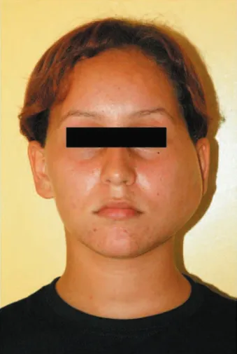
The swelling measured about 6 cm in length and 5
cm in width. On bimanual palpation an ill-deined and
resilient mass was noticed (Figure 1). This mass was evident extra orally with a considerable bulging of the skin in the left parotid region. The lesion was normal and no lymphadenopathy was detected. On intraoral examination, oral mucosa and teeth were healthy. The parotid duct in
the affected side was normal and salivary low had normal
physical aspects without debris or purulent discharge. The presumptive clinical diagnosis was an abscess associated
to mild inlammation, considering the patient’s history
and the previous episodes. Conventional panoramic radiography, ultrasonography and sialography were performed. The panoramic radiograph showed teeth and bones preserved without any evidence of abnormalities. The ultrasonographic scan revealed a hypoechoic
ovoid well deined image suggesting a cyst (Figure 2).
Figure 1- Anteroposterior facial view illustrating the 6 x 5
cm swelling on patient’s left cheek
Figure 2- The longitudinal section in the ultrasonographic
scan of the left parotid gland demonstrates a hypoechoic
Sialography of the left parotid was performed using Lipiodol® (Lipiodol; Guerbet, Jacarepaguá, RJ, Brazil) as
a substance of contrast. A partial illing of the gland was
enough to show cavitary sialectasia in a panoramic (Figure 3a) and anteroposterior view (Figure 3b), and the contrast was retained in the gland for at least 24 hours (Figure 3c).
A percut aneous needle aspirat ion of t he swelling was per for m ed 2 w eeks aft er t he sialography. I t
yielded 4 mL of a clear viscous luid. The material
w as su b m it t ed t o m icr ob iolog ical an aly sis an d show ed num er ous polym or phonuclear leukocyt es.
The luid did not show any bacterial growth and
Figure 3- A panoramic (A) and an anteroposterior (B) radiograph taken after sialography of the left parotid gland. A partial
illing of the gland was suficient to show cavitary sialectasia. The contrast was retained in the gland for at least 24 h, as
shown in the panoramic radiograph (C)
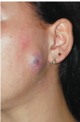
Figure 4- Erythema on the overlying skin after the
percutaneous needle aspiration
Figure 5- Complete recovery of the skin and remission of
pr esent ed high am ylase levels ( 7,810 unit s/ L) . The
aspiration procedure was suficient to empty the
cavit y. The pat ient was adver t ised t o com pr ession d r e ssi n g t w i ce a d a y. Se v e n d a y s a f t e r t h e per cut aneous aspirat ion a gr eat decr ease of t he sw elling was obser ved. The over lying skin show ed a discr eet er yt hem a on t he sur face of t he m ass, w it hout signs of infect ion, and t he pat ient did not r efer pain ( Figur e 4) . At t he 30- day follow- up visit , com plet e r ecover y was not iced, and t he r esidual sw elling had t ot ally disappear ed ( Figur e 5) . The pat ient was follow ed up during t w o years and a half and no r ecur r ence was det ect ed.
D I SCUSSI ON
Pr im ar y p ar ot id g lan d cy st s ar e v er y r ar e,
representing ive per cent of all parotid tumors10,23,25,. Mucoceles are round and well deined lesions that
cont ain m ucus, when t hey occur in t he parot id gland ar e called sialoceles. For pract ical pur poses t hey m ay be r egar ded as being of eit her ext ravasat ion or r et ent ion t ype. The t er m m ucous ext ravasat ion phenom enon ( cyst ) is used when m ucus is ext ruded int o t he connect ive t issues and is sur r ounded by a granulat ion t issue, w hile m ucous r et ent ion cyst is used t o descr ibe a cyst w it h r et ained m ucin t hat is lined w it h duct al epit helium w hich m ay hav e under gone squam ous or oncocyt ic m et aplasia26,27.
The fact or s t hat det er m ine a m ucocele ar e t he rat e of m ucus product ion and t he speed of phagocyt osis of t he ex t ravasat ed m ucus. The m aj or it y of t he m ucoceles pr eviously r epor t ed in t he par ot id gland ar e of r et ent ion t ype ( duct al cyst )26.
Par ot id sialoceles ar e lesions t hat occur aft er t raum a or inj ur y in t he face causing accum ulat ion of saliva in t he ar ea1,4,20. I t has not been descr ibed
yet a case w her e t he pat ient could not associat e a hist or y of t raum a, inj ur y or sur ger y. I t is possible t hat our pat ient suffer ed a t raum a of low int ensit y an d co u l d n o t r em em b er i t , b u t i t sh o u l d b e som et hing else t o explain t he 3 r ecur r ent episodes of t um efact ion.
Initially, our patient had a resilient, ill-deined mass, which was dificult to palpate, probably due to the position of the cystic diffuse inlammation, under
t he dense par ot id fascia, w hich m ak es phy sical exam inat ion unr eliable. Clinical assessm ent m ay be inaccurat e in t hese cases4,24. How ever, soft and
m obile lesions had also been descr ibed w hen t hey
are more supericial7.
Ou r p at ien t d id n ot p r esen t f ev er or ot h er com pr om ising signs in any episode of sw elling20.
The m anagem ent of a pat ient w it h a sw elling i n t h e p ar o t i d r eg i o n r eq u i r es car ef u l cl i n i cal ev alu at ion . Fin e- n eedle aspir at ion or biopsy is
necessary for a deinite diagnosis. Sialography,
com put ed t om ography and ult rasonographic scans
m ay be ver y helpful2. Sialography of t he par ot id
gland was m andat or y in r evealing t he sialect asia in our case and is also useful t o dist end t he duct w hen it is involved4. Lipiodol® is a solut ion cont aining
iodine t hat m ight have act ed as an ant ibact er ial ag en t an d h elp ed r ed u cin g t h e f acial sw ellin g com bined wit h t he aspirat ion. I n our case report t he sialography ser ved as a diagnost ic t ool and helped healing t he involved gland. Ther e was a clear r elief of t he signs aft er sialography.
Anot her key t o t he diagnosis and t reat m ent was
ine-needle aspiration, which provided material for
analy sis and helped em pt ing t he gland. Par ot id secret ion has a high am ylase cont ent t hat is usually ar ound 10,000 unit s/ L19. I n t he pr esent case, t he amylase content was 7,810 units/L, conirming the
diagnosis of par ot id saliva ext ravasat ion.
A var iet y of t r eat m ent s have been pr oposed f or p ar ot id sialoceles2 0. Th ese in clu d e m u lt ip le
aspirat ions and com pression dressings; lat e prim ary r epair or r econst r uct ion of t he duct ; cr eat ion of
a controlled internal istula; supericial or total
p a r o t i d ect o m y ; p a r a sy m p a t h et i c d en er v a t i o n ( se ct i o n i n g o f t h e a u r i cu l o t e m p o r a l n e r v e ) ; an t isialog og u es; r ad iat ion t h er ap y an d d u ct al ligat ion. Most of t hese pr ocedur es ar e invasiv e w it h r isks of inj ur y of t he facial ner ve, w it h var iable and oft en poor success rat es7. The ant icholiner gic
dr ugs have m any undesir ed side effect s such as xerost om ia, const ipat ion, phot ophobia, t achycardia and urinary ret ent ion16. At ropine and glycopyrrolat e
ar e ant isialagogue dr ugs t hat m ay be used t o t r eat sialoceles4. Th e p r esen t case r ep or t ed d id n ot
r equir e any invasive t r eat m ent or adm inist rat ion of a drug. A conservat ive t reat m ent such as suggest ed by Landau and St ewar t17 ( 1985) , t he inj ect ion of
an ant ibact er ial solut ion t o per for m a sialography, t he aspirat ion of t he cont ent and t he com pr ession dr essing w er e capable t o solve t he case.
Den t ist s sh ou ld b e aw ar e of t h is p at h olog y an d t h e im por t an ce in adopt in g a con ser vat iv e t r eat m ent w henever it is possible.
REFEREN CES
1 - Ak i n b am i BO. Tr au m at i c d i seases o f p ar o t i d g l an d an d sequalae. Review of lit erat ure and case report s. Niger J Clin Pract . 2009; 12( 2) : 212- 5.
2- Ant oniadis K, Karakasis D, Tzar ou V, Skor dalaki A. Benign cyst s of t he par ot id gland. I nt J Oral Maxillofac Sur g. 1990; 19( 3) : 139-40.
3- Bailey BM. A persistent parotid istula following the extraction of m andibular t eet h. J Lar yngol Ot ol. 1984; 98( 10) : 1051- 4. 4- Barron R, Margulis A, Icekson M, Zeltser R, Eldad A, Nahlieli O. I at r ogenic par ot id sialocele follow ing r hyt idect om y: diagnosis and t r eat m ent . Plast Reconst r Sur g. 2001; 108( 6) .
7- Canosa A, Cohen MA. Post - t raum at ic par ot id sialocele: r epor t of t w o cases. J Oral Maxillofac Sur g. 1999; 57( 6) : 742- 5. 8 - Ch olan k er il JV, Scioscia PA. Post - t r au m at ic sialoceles an d m ucoceles of t he salivary glands. Clin I m aging. 1993; 17( 1) : 41- 5. 9- Chow TL, Kw ok SP. Use of bot ulinum t oxin t ype A in a case of per sist ent par ot id sialocele. Hong Kong Med J. 2003; 9( 4) : 293- 4. 10- Cohen MN, Rao U, Shedd DP. Benign cyst s of t he parot id gland. J Sur g Oncol. 1984; 27( 2) : 85- 8.
1 1 - Dem et r iad es D, Rab in ow it z B. Man ag em en t of p ar ot id sialoceles: a sim ple surgical t echnique. Br J Surg. 1987; 74( 4) : 309. 12- Dierks EJ, Granite EL. Parotid sialocele and istula after m andibular ost eot om y. J Oral Sur g. 1977; 35( 4) : 299- 300 13- Dolw ick MF, Kr et zschm ar DP. Mor bidit y associat ed w it h t he preauricular and perim eat al approaches t o t he t em porom andibular j oint . J Oral Maxillofac Sur g. 1982; 40( 11) : 699- 700.
14- Hemenway WG, Bergstrom L. Parotid duct istula: a review. Sout h Med J. 1971; 64( 8) : 912- 8.
1 5 - Hu t ch i so n I L, Ry an D. A p ar o t i d f i st u l a an d si al o cel e com plicat ing t em porom andibular j oint surgery. Br J Oral Maxillofac Sur g. 1989; 27( 3) : 203- 8.
1 6 - Kr au sen AS, Og u r a JH. Si al o cel es: m ed i cal t r eat m en t irst. Trans Sect Otolaryngol Am Acad Ophtalmol Otolaryngol. 1977; 84( 5) : ORL890- 5.
17 Landau R, St ewar t M. Conser vat ive m anagem ent of post -traumatic parotid istulae and sialoceles: a prospective study. Br J Sur g. 1985; 72( 1) : 42- 4.
1 8 - Lan g d on JD. Com p licat ion s of p ar ot id g lan d su r g er y. J Maxillofac Sur g. 1984; 12( 5) : 225- 9.
1 9 - Mey er RA, Gor d on RC. Met h od f or r ep air of t r au m at ic p se u d o cy st o f p a r o t i d d u ct : r e p o r t o f ca se . J Or a l Su r g . 1969; 27( 4) : 281- 3.
20- Monfared A, Ort iz J, Roller C. Dist al parot id duct pseudocyst as a result of blunt facial t raum a. Ear Nose Throat J. 2009; 88( 8) : 15- 7. 21- Par ek h D, Glezer son G, St ew ar t M, Esser J, Law son HH. Post-traumatic parotid istulae and sialoceles. A prospective st u d y of con ser v at iv e m an ag em en t in 5 1 cases. An n Su r g . 1989; 209( 1) : 105- 11.
22- Per eira KD, Sm it h SL, Mit chell RB. Par ot id sialocele in a 10- year- old gir l. Ear Nose Thr oat J. 2007; 86( 1) : 27- 8.
23- Piet er se AS, Seym our AE. Par ot id cyst s. An analysis of 16 cases and suggested classiication. Pathology. 1981;13(2):225-34. 24- Rej ali D, Sim o R, Sm all M. Mucocele m im icking a War t hin's t um our r ecur r ence. J Lar yngol Ot ol. 1998; 112( 11) : 1092- 4. 25- Richar dson GS, Clair m ont AA, Er ick son ER. Cy st ic lesions of t he par ot id gland. Plast Reconst r Sur g. 1978; 61( 3) : 364- 70. 26- Seifer t , G., Miehlke, A., Haubr ich, J., Chilla, R. Diseases of t he salivar y gland. St ut t gar t : Geor g Thiem e Ver lag; 1986. p. 171- 80. 27- Tal H, Alt ini M, Lem m er J. Mult iple m ucous r et ent ion cyst s of t he oral m ucosa. Oral Surg Oral Med Oral Pat hol. 1984; 58( 6) : 692-5.