Yoğun Persistan Pupiller Membran / Dense Persistent Pupillary Membrane
Dense Persistent Pupillary Membrane in an Adult Patient
Yetişkin Bir Hastada Yoğun Persistan Pupiller Membran
DOI: 10.4328/JCAM.3204 Received: 03.01.2015 Accepted: 12.02.2015 Printed: 01.06.2014 J Clin Anal Med 2014;5(suppl 3): 397-9 Corresponding Author: Yeşim Altay, 48. Cadde Yonca Apt. No: 33/31, Cukurambar, Ankara, Turkey.
GSM: +905324902298 E-Mail: [email protected]
Özet
Persistan pupiller membran (PPM) tunika vaskuloza lentis’in tam olmayan gerile-mesi sonucunda ortaya çıkan ve yetişkinlerde nadir rastlanan bir konjenital anor-malidir. Otuz yaşındaki erkek hasta sol gözde ilerleyici görme bulanıklığı ve her iki gözde farklı görünen pupil varlığı yakınmalarıyla başvurdu. Muayenede, düzeltil-memiş görme (Snellen) sağ gözde 20/100, sol gözde 20/640 idi ve solda amblyopi mevcut idi. Biyomikroskopik incelemede, bilateral dens PPM ve sol gözde kataraktı vardı. Sağ ve sol gözün görme alanı incelemesinde, görme alanlarında belirgin da-ralma saptandı. Biz bu olguyu PPM’lı hastalarda tedaviyi planlarken, görme alanı incelemesinin de en az santral görme kadar önemli olduğunu vurgulamak amacıy-la sunduk. PPM’lı hastaamacıy-ların tedavisini pamacıy-lanamacıy-larken görme bozukluğu, pupiller açıklı-ğın boyutu ve görme alanının muayenesi dikkate alınmalıdır.
Anahtar Kelimeler
Persistan Pupiller Membran; Katarakt; Ambliyopi; Görme Alanı; Konjenital Okü-ler AnomaliOkü-ler
Abstract
Persistent pupillary membranes (PPM) are congenital abnormalities which re-sults from an incomplete involution of tunica vasculosa lentis and are rarely seen in adults. A thirty-year old man applied to the hospital with the complaint of uncommon-looking pupils and progressive blurring of vision in the let eye. On examination, uncorrected visual acuity (Snellen ) were 20/100 in the right eye and 20/640 in the let eye with amblyopia. On biomicroscopic examination, there were bilateral dense PPM and cataract in the let eye. Visual ield analysis of right and let eyes showed great narrowing of visual ields. We present our case in order to emphasize that analysis of visual ield of patients with PPM is as important as central vision when planning its treatment. For planning treatment of patients with PPM, visual impairment, size of pupillary opening, and visual ield analysis should be considered.
Keywords
Persistent Pupillary Membrane; Cataract; Amblyopia; Visual Field; Congenital Ocu-lar Anomalies
Yeşim Altay, Özgür Balta, Ayşe Burcu Department of Ophthalmology, Ankara Training and Research Hospital, Ankara, Turkey
| Journal of Clinical and Analytical Medicine
Yoğun Persistan Pupiller Membran / Dense Persistent Pupillary Membrane
2
Introduction
Persistent pupillary membrane (PPM) is an ocular congenital
anomaly which results from an incomplete involution of tunica
vasculosa lentis. Although familial forms have been reported,
most cases are sporadic in nature [1].
The incidence of PPM ranges from 30% to 95% in normal
new-borns, and majority of them regress within the irst year of life
[2]. Dense PPMs are rare in adult eyes.
We report a case of 30 years old man diagnosed with bilateral
dense persistent pupillary membrane and unilateral cataract.
Case Report
30 year old man presented to the Ophthalmology Clinic of our
hospital with the complaint of uncommon-looking pupils and
progressive blurring of vision in the let eye. The patient claimed
that his vision of both eyes was inadequate from childhood and
there were no documented systemic or ocular illnesses.
On examination, uncorrected visual acuity (Snellen) were
20/100 in the right eye and 20/640 in the let eye with
amblyo-pia. Best corrected visual acuity were 20/32 in the right eye and
20/100 in the let eye with myopic correction.
On biomicroscopic examination, both eyes had clear corneas
and normal deep anterior chambers. Examination revealed the
pupils were covered with a network of pigmented tissue,
run-ning from the iris surface spreading over the pupils. (Figure 1)
In the let eye this tissue was sticky to the lens and there was a
cataractous change (Figure 2). Gonioscopy results were normal
in both eyes. Intraocular pressure were 20 mmHg in the right
eye and 22 mmHg in the let eye. Axial length of right and let
eye 23,05 mm and 23,86 mm respectively.
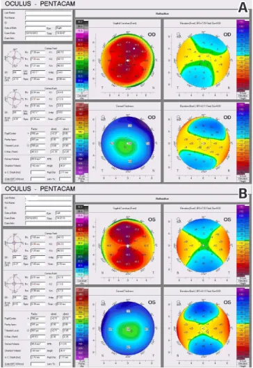
Evaluation with pentacam corneal topography revealed that
central corneal thickness was 590 µm in the right eye and 596
µm in the let eye and there were bilateral bowtie corneal
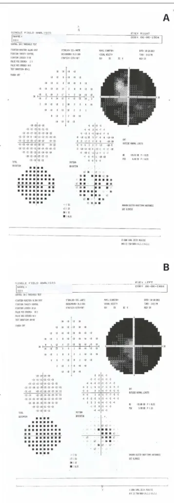
astig-matism (Figure 3 a-b). Vision analysis of right and let eyes
showed great narrowing of visual ield. (Figure 4 a-b) Ater the
pupils were dilated both fundi were within normal limits.
We planned surgical excision of membranes and cataract
sur-gery for the let eye. But the patient refused the treatment.
Discussion
During intrauterine life, the lens receives nourishment from the
posterior and anterior tunica vasculosa lentis [2]. The persistent
pupillary membrane (PPM) represents the congenital remnant
of the anterior tunica vasculosa lentis. The pupillary membrane
itself begins to regress in the sixth month and dissappears
completely by the eight month of gestation, if an arrest occurs
in this process it will result in PPM [3]. PPM may be seen as
iso-lated or in association with other ocular pathologies like
mic-rocornea, posterior keratoconus and macular hypoplasia [4,5].
PPM oten spans the pupil from its insertion in the iris collarate,
and attachment of it to the lens can cause cataract and visual
deprivation as in our case. PPM may not afect central vision
Figure 1. Persistent pupillary membrane of the right eye.Figure 2. Persistent pupillary membrane with cataract of the let eye.
Figure 3. Pentacam corneal topography of the right eye(A). Pentacam corneal topography of the let eye(B).
A
B
| Journal of Clinical and Analytical Medicine
398
| Journal of Clinical and Analytical Medicine
Yoğun Persistan Pupiller Membran / Dense Persistent Pupillary Membrane
3
unless the pupillary opening is less than 1,5 mm in size. Thus
it seems that most cases are not signiicant enough to have
visual impairment and so may go undetected [6]. However, as in
this patient’s right eye, although visual acuity was good, visual
ield analysis might show impairment despite enough pupillary
opening. Besides PPM, narrowing of the visual ield of the let
eye might be afected by cataract as well.
Primary indications for the treatment of PPM are restoration of
vision and prevention of amblyopia in children [6].
First-line treatment in patients with visual impairment
gener-ally consist of medical therapy with mydriatic agents. If
medi-cal therapy is inefective, surgimedi-cal removal is an alternative
management. Treatment options include Nd-YAG or Argon laser
therapy to the central portion of the membrane and surgical
excision of the membranes [7]. Argon laser treatment reported
as causing less hyphema than Nd-YAG laser [7].
Since surgical excision may be associated with complications
such as cataract, hyphema and infection, some authors
recom-mend a conservative approach if a patient have a good vision,
even in the case of dense PPMs [8].
In conclusion, bilateral dense persistent pupillary membranes
are rare congenital abnormalities in adults. Although patients
with PPM have adequate central vision due to pinhole efect,
PPM may cause signiicant decrease in visual ield as in our
patient. We presented this case in order to emphasize that
analysis of visual ield of patients with PPM is as important as
central vision when planning its treatment. We have not found
any reported case or case series in literature evaluating visual
ield of patients with PPM. For planning treatment of patients
with PPM, visual impairment, size of pupillary opening, and
vi-sual ield analysis should be considered.
Competing interests
The authors declare that they have no competing interests.
References
1. Norris HJ, Backhouse OC. The congenital pinhole:a persistant pupillary mem-brane. Clin Exp Optom 2010;93(2):100-1.
2. Forrester JV, Dick AD, McMenamin P, Lee WR. Embryology and early devel-opment of the eye and adnexa. In: Forrester JV, Dick AD, McMenamin P, Lee WR, editors.The Eye Basic Sciences in Practice.2nd edition. Edinburgh: WB Saunders;2002.p.99-129.
3. Wright KW, Speigel PH, Buckley EG. Embryology. In: Wright KW, Speigel PH,editors.Pediatric Ophthalmology and Strabismus.2nd edition. New York: Springer-Verlag; 2003.p.17.
4. Viswanathan D, Padmanabhan P, Johri A. Hyperplastic persistant pupil-lary membranes with congenital corneal anomalies. J Cataract Refract Surg 2007;33(6):1123-6.
5. Altun A, Kurna SA, Bozkurt E, Erdoğan G, Altun G, Olcaysu OO, et al. Bilat-eral persistant pupillary membrane with tetralogy of fallot: A case report and review of the literature. Case Reports in Ophthalmological Medicine 2014; doi:10.1155/2014/581273
6. Lee SM, Yu YS. Outcome of hyperplastic persistant pupillary membrane. J Pedi-atr Ophthalmol Strabismus 2004;41:163-71.
7. Bucan K, Znaor L, Jurasin K, Buban D, Galetovic D, Lesin M. Laser manage-ment of persistant pupillary membrane prior to cataract surgery. Acta Clin Croat 2008;47(Suppl. 1):7-9.
8. Meyer-Rüsenberg B, Thill M, Vujancevic S, Meyer-Rüsenberg HW. Conservative management of bilateral persistant pupillary membranes with 18 years of follow up. Graefes Arch Clin Exp Ophthalmol 2010;248:1053-4.
How to cite this article:
Altay Y, Balta Ö, Burcu A. Dense Persistent Pupillary Membrane in an Adult Patient. J Clin Anal Med 2014;5(suppl 3): 397-9.
Figure 4. Visual ield analysis of the right eye (A). Visual ield analysis of the let eye (B).