Virtual orthodontic setup in orthodontic camouflage
planning for skeletal Class III malocclusion
Felipe Augusto M. Barreto1, João Roberto R. da Costa Santos2
The purpose of this paper was to emphasize the importance of the orthodontic setup in treatment planning for skeletal Class III malocclusion correction in an adult patient with moderate lower anterior crowding and anterior crossbite associated with two supernumerary lower incisors.
Keywords: Orthodontics, corrective. Crossbite. Tooth, supernumerary. Incisor. Tooth extraction.
How to cite: Barreto FAM, Santos JRRC. Virtual orthodontic setup in ortho-dontic camouflage planning for skeletal Class III malocclusion. Dental Press J Or-thod. 2018 Mar-Apr;23(2):75-86.
DOI: https://doi.org/10.1590/2177-6709.23.2.075-086.bbo » The author reports no commercial, proprietary or financial interest in the products
or companies described in this article.
» Patients displayed in this article previously approved the use of their facial and in-traoral photographs.
DOI: https://doi.org/10.1590/2177-6709.23.2.075-086.bbo
INTRODUCTION
The treatment of skeletal Class III malocclusion in adult patients is a challenge for the orthodontist, main-ly in the choice between compensatory orthodontic treatment or orthodontic preparation for orthognathic surgery.1-7
In many situations, the fear of the surgical proce-dure associated with the patient’s satisfaction with their facial appearance makes orthodontic camouflage the patient’s treatment of choice.1,4,6,8 Thus, orthodontic treatment associated with tooth extractions is an ap-proach for orthodontic compensation in patients with mild or moderate skeletal Class III malocclusion.6,8,9,10
Traditionally, premolars are the most indicated teeth for extraction with camouflage purposes; however, other extraction alternatives have been used, such as extrac-tion of a lower incisor.3,5,6,9,11
The extraction of a lower incisor in Class III treat-ment is an option primarily used in patients with Bolton’s tooth-size discrepancy.5 The presence of this discrepancy significantly influences orthodontic plan-ning, requiring reduction of dental mass, either by in-terproximal stripping or by tooth extraction.12,13
The presence of supernumerary teeth promotes upper and lower dental disproportion. This dis-harmony may lead to malocclusions and
difficul-O objetivo desse artigo é enfatizar a importância do setup ortodôntico no planejamento do tratamento da má oclusão de Clas-se III esquelética de uma paciente adulta com apinhamento anteroinferior moderado e mordida cruzada anterior associada à presença de dois incisivos inferiores supranumerários.
for the good orthodontist. Thus, an orthodontic setup is necessary to allow visualization of the final occlusion and to determine how many and which teeth should be extracted.5,10,18,19
Thus, the present article describes the treatment of an adult patient presenting skeletal Class III malocclu-sion with moderate lower anterior crowding and ante-rior crossbite associated with the presence of two super-numerary lower incisors.
CLINICAL CASE REPORT
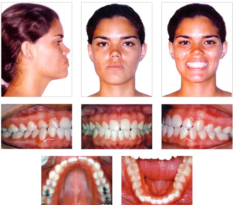
Brown-skinned patient, 20 years old, female, pre-sented with good general health and excellent oral health. Her main complaint concerned the projection of the lower incisors out of the mouth. She presented a Class III skeletal pattern, aggravated by the lack of space for correct alignment of the lower arch, due to the pres-ence of two supernumerary lower incisors.
Frontal facial examination revealed mandibular asym-metry to the right side. In sagittal view, the lower facial
ited anterior contraction on the right side (Fig 1).
Bolton’s analysis revealed inferior excess of 7.5 mm, considering the proportion between the sum of the me-siodistal widths of the fourteen lower and twelve upper teeth; and inferior excess of 9.2 mm, considering the proportion between the anterior lower teeth with the anterior upper teeth.
The periapical and panoramic radiographs revealed intact roots, absence of the upper and lower third molars on the right side, presence of two fully erupted super-numerary incisors, as well as light horizontal bone loss in the lower arch (Figs 2 and 3).
Figure 2 - Initial panoramic radiograph.
Figure 3 - Initial periapical radiographs.
Figure 4 - Initial lateral cephalometric radio-graph (A) and cephalometric tracing (B). B
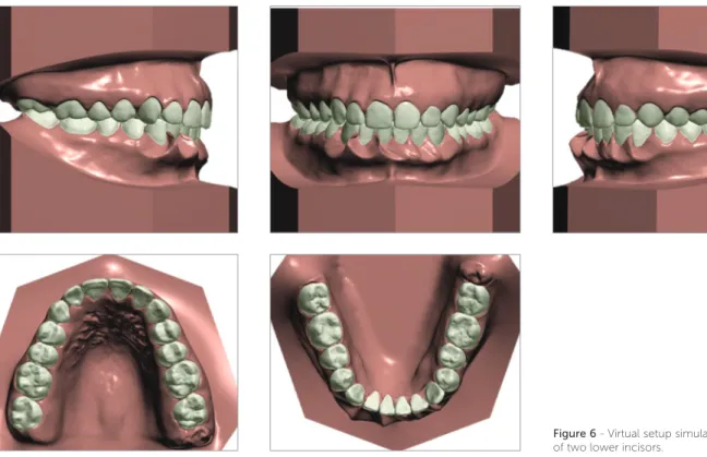
Figure 5 - Virtual setup with excessive overjet quantification.
TREATMENT PLAN AND MECHANICS EMPLOYED
Considering that the patient discarded the possibil-ity of orthodontic-surgical treatment, the orthodontic planning consisted in correcting Bolton’s tooth-size discrepancy at the anterior region to obtain a harmoni-ous relationship between the dental arches. The virtual orthodontic setup was performed with extraction of two lower incisors, which revealed an unsatisfactory occlusal result, with excessive overjet of 3.92 mm (Figs 5 and 6). Therefore, it was decided to extract only one supernu-merary incisor, adjacent to the lower right canine, asso-ciated with interproximal 3-mm stripping, distributed among the five remaining incisors. It was verified on the orthodontic setup that this strategy would be suffi-cient to balance the tooth-size discrepancy between the upper and lower arches, to dissolve the lower anterior crowding and to correct the anterior crossbite.
Treatment was started by bonding Edgewise brack-ets, 0.022 x 0.028-in slot, using 0.015-in Twist Flex arches for alignment and leveling and 0.014-in, 0.016-in, 0.018-in stainless steel arches for projection of upper incisors and retraction of lower incisors, to aid the correction of the negative overjet. After
extrac-tion of the supernumerary incisor, lingual tracextrac-tion of the right lower canine was started, with a chain elastic anchored at a button bonded to its lingual surface and connected to a hook welded in the previously placed Nance lingual arch. In the posterior region, occlusal stops made of self-curing resin (Triad VLC, Dentsply GAC) were added bilaterally, causing anterior disoc-clusion to aid uncrossing the bite. After closing the ex-traction space and removing the occlusal stops, it was verified that the incisors presented an edge-to-edge relationship. The planned interproximal stripping was performed and the spaces were closed with active tie-backs in the 0.018-in arch with lingual traction, since new hooks were welded to the lingual arch and but-tons were bonded to the lingual surface of the lower incisors. Class III intermaxillary elastics (5/16-in) were used under protocol of continuous use and daily ex-changes, thus obtaining positive overjet. Orthodontic finalization was performed using round arches, with-out the need of incorporating torques into the teeth.
RESULTS
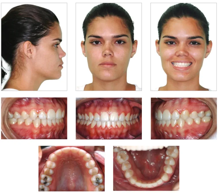
At treatment completion, the facial aesthetics was improved and, although five lower incisors were main-tained, normal overbite and overjet were obmain-tained, lead-ing canines and molars to Angle Class I, upper midline coinciding with the center of the central lower incisors, resulting in anterior guidance and right and left func-tional laterality, providing masticatory efficiency and TMJ integrity (Fig 7).
The most marked effects were observed in the lower arch, in which extraction of only one supernumerary incisor and interproximal stripping corrected the ante-rior crossbite and crowding, with better positioning of teeth in the bone base.
Bolton’s analysis of the final models revealed cor-rection of the discrepancy between the sum of mesio-distal diameters of the thirteen lower teeth and twelve upper teeth, with reduction of the anterior discrep-ancy to 1.0 mm.
The final panoramic radiograph shows preserva-tion of root lengths and integrity of the alveolar bone crests — except for the mandibular incisor that fin-ished centralized with the upper midline, good root parallelism was observed (Fig 8).
Since the treatment comprised dental compensa-tion, there were no changes in maxillary and mandibu-lar positioning. The patient continued with a Class III skeletal pattern, ANB equal to -2o (SNA = 85o and SN.GoGn = 87o), and maintained the horizontal pat-tern (SN.GoGn = 22o and FMA = 13o). However, the patient’s facial profile became more harmonious due to the better relation between the upper and lower lips (Upper lip - S line = 2mm and Lower lip - S line = 3mm). In addition, there was evident decrease in the inclina-tion of mandibular incisors and a small increase in the labial inclination of upper incisors, which allowed cor-rection of the anterior crossbite (1.NA from 29o to 30o and 1.NB from 37o to 25o) (Fig 9).
Figure 9 - Final lateral cephalometric radio-graph (A) and cephalometric tracing (B). A
DISCUSSION
The orthodontic compensatory treatment of a skel-etal malocclusion is one of the possible alternatives for adult patients who choose not to undergo surgical procedures1,4,8. In the case reported, orthognathic sur-gery was discarded in response to the patient’s desire, since there were no complaints regarding facial esthet-ics and the skeletal discrepancy was small. The patient had hyperdontia, an anomaly related to the presence of supernumerary teeth, whose etiology may be related to heredity.17,20,21,22 However, in the present case there were no reports of supernumerary teeth in the family. Hyperdontia affects more males, at a 2:1 ratio,17,20,21,22 presenting more frequently as a single supernumerary tooth, with strong predilection for the maxillary ante-rior region.16,17,20,22 In contrast, in the case reported, the patient is female and had two supernumerary teeth in the mandibular anterior region.
Although the presence of supernumerary teeth pre-cludes the achievement of occlusion with normal overjet and overbite,12-15 the patient always had molars and canines in Class I relation, since the tooth-size excess was located at the incisors region.
The extraction of a lower incisor is a therapeutic alter-native for adult patients with mild to moderate Class III malocclusion, which does not fit in the conventional op-tions of extracop-tions.18,19 In the present case, the decision to extract only one supernumerary lower incisor was
deter-mined by the virtual orthodontic setup, which evidenced that it was not necessary to extract two supernumerary incisors to solve the Bolton’s tooth-size discrepancy be-tween the upper and lower arches. With extraction of both supernumerary incisors, Bolton’s analysis showed a superior excess of 3.8 mm; and 2.5 mm considering only the six anterior teeth.
The extraction of the supernumerary tooth adjacent to the right lower canine was indicated because this tooth was closer to the malocclusion region. Reduction of the mesiodistal diameter by interproximal stripping of the five lower incisors provided balance between the dental masses, allowing the overjet correction. Determination of the required amount of interproximal stripping was also based on the Bolton’s analysis of the initial models, which pointed to a tooth size excess of 3 mm in the lower ante-rior region, even after extracting the supernumerary tooth adjacent to the right lower canine.
Cases treated with extraction of a lower incisor pres-ent a lower rate of post-treatmpres-ent crowding relapse, due to maintenance or reduction of intercanine distance.18,19 This stability can be clinically and radiographically dem-onstrated after two years of treatment completion in the present case, as shown in Figures 10, 11 and 12.
Tracings superimposition shows decrease of the profile’s concavity because the upper lip acquired an anterior position, improving its relationship with the lower lip (Fig 13).
Figure 12 - Lateral cephalometric radiograph (A) and cephalometric tracing (B) two years after treatment completion.
Figure 13 - Total (A) and partial (B) superimpo-sitions of cephalometric tracings obtained at treatment onset, completion and two years after treatment completion.
A
Figure 11 - Panoramic radiography two years af-ter treatment completion.
B A
Table 1 - Cephalometric values: A) initial, B) final and C) two years after the end of orthodontic treatment.
Measurements Normal A B Dif. A/B C
Skeletal pattern
SNA (Steiner) 82o 85o 85o 0 86o
SNB (Steiner) 80o 88o 87o 1 88o
ANB (Steiner) 2o -3o -2o 1 -2o
Angle of convexity (Downs) 0o -6o -5o 1 -4o
Y-axis (Downs) 59o 52o 52o 0 52o
Facial angle (Downs) 87o 98o 97o 1 96o
SN-GoGn (Steiner) 32o 22o 22o 0 22o
FMA (Tweed) 25o 12o 13o 1 14o
Dental pattern
IMPA (Tweed) 90o 107o 97o 10 95o
1.NA (degrees) (Steiner) 22o 29o 30o 1 29o
1-NA (mm) (Steiner) 4 mm 14mm 12mm 2 11mm
1.NB (degrees) (Steiner) 25o 37o 25o 12 25o
1-NB (mm) (Steiner) 4 mm 8mm 8mm 0 6mm
1
1- Interincisal angle (Downs) 130o 119o 128o 9 128o
1-APo (Ricketts) 1 mm 1mm 2mm 1 1mm
Profile
Upper lip — S-line (Steiner) 0 mm 0mm 2mm 2 1mm
Lower lip — S-line (Steiner) 0 mm 4mm 3mm 1 1mm
CONCLUSIONS
Bolton’s discrepancy analysis and virtual orthodontic set-up are important diagnostic tools that assist in the planning of atypical cases, such as the present one. Compensatory
5. Araújo EA, Araújo CV. Abordagem clínica não cirúrgica no tratamento da má oclusão de Classe III. Rev Dental Press Ortod Ortop Facial. 2008 Nov-Dez;13(6):128-57.
6. Almeida KCM, Paulin RF, Raveli TB, Raveli DB, Santos-Pinto A. Two-Step extraction of the lower first molar for Class III treatment in adult patient. Case Rep Den. 2016;2016:1580313.
7. Janson G, Souza JE, Alves FA, Andrade P Jr, Nakamura A, Freitas MR, et al. Extreme dentoalveolar compensation in treatment of Class III malocclusion. Am J Orthod Dentofacial Orthop. 2005 Dec;128(6):787-94. 8. Lin J, Gu Y. Lower second molar extraction in correction of severe skeletal
Class III malocclusion. Angle Orthod. 2006 Mar;76(2):217-25. 9. Ruellas ACO, Baratieri C, Roma MB, Izquierdo AM, Boaventura L,
Rodrigues CS, et al. Angle Class III malocclusion treated with mandibular first molar extractions. Am J Orthod Dentofacial Orthop. 2012 Sept;142(3):384-92. 10. Daher W, Caron J, Wechsler MH. Nonsurgical treatment of an adult
with a Class III malocclusion. Am J Orthod Dentofacial Orthop. 2007 Aug;132(2):243-51.
11. Moura ROL, Cruz KS. Tratamento ortodôntico compensatório da má oclusão de Classe III esquelética. Orthod Sci Pract. 2015;8(29):80-8. 12. Cançado RH, Gonçalves Júnior W, Valarelli FP, Freitas KMS, Crêspo JAL.
Association between Bolton discrepancy and Angle malocclusion. Braz Oral Res. 2015;29(1):1-6.
13. Gaddam R, Arya S, Shetty KS. Incidence of tooth size discrepancy in different groups of malocclusion and its relation to extraction. J Int Oral Health. 2015;7(Suppl 1):48-53.
UNESP. 2015 Set-Out;44(5):280-4.
17. Matsumoto MAN, Romano FL, Ferreira JTL, Tanaka S, Morizono EM. Lower incisor extraction: An orthodontic treatment option. Dental Press J Orthod. 2010 Nov-Dec;15(6):143-61.
18. Lima CMF, Lacet E, Marques CR. Extração de incisivo inferior: uma opção terapêutica. Rev Dental Press Ortod Ortop Facial. 2005 Jul-Ago;10(4):47-59.
19. Lo Giudice G, Nigrone V, Longo A, Cicciù M. Supernumerary and supplemental teeth: case report. E Eur J Paediatr Dent. 2008 June;9(2):97-101.
20. Moura WL, Cravinhos JCP, Moura CDVS, Freire SASR, Monteiro AMO, Pinheiro SDA, et al. Prevalência de dentes supranumerários em pacientes atendidos no Hospital Universitário da UFPI: um estudo retrospectivo de cinco anos. Rev Odontol UNESP. 2013 Maio-Jun;42(3):167-71.
21. Simões FXPC, Crusoé-Rebello I, Neves FS, Oliveira-Santos C, Ciamponi AL, Silva Filho OM. Prevalence of supernumerary teeth in orthodontic patients from Southwestern Brazil. Int J Odontostomat. 2011 Aug;5(2):199-202. 22. Riedel RA, Little RM, Bui TD. Mandibular Incisor extraction:


