333
GENETIC DIVERSITY AMONG
FUSARIUM GRAMINEARUM
AND
F. CULMORUM
ISOLATES BASED ON ISSR MARKERS
Gülruh Albayrak1,*, Emre Yörük2, Aylin Gazdağli2 and Bahram Sharifnabi3
1Istanbul University, Faculty of Science, Department of Molecular Biology and Genetics, Istanbul, Turkey
2Istanbul University, Institute of Science, Program of Molecular Biology and Genetics, Istanbul, Turkey
3Department of Plant Protection, College of Agriculture, Isfahan University of Technology, Isfahan, Iran
*Corresponding author: [email protected]
Received: June 30, 2015; Revised: September 1, 2015; Accepted: October 6, 2015; Published online: March 21, 2016
Abstract: To characterize the isolates of F. graminearum and F. culmorum fungi from Turkey and Iran, we performed ISSR analysis with 18 non-anchored and 23 anchored (including ten novel) primers. Amplification product sizes were 0.2-3.5 kb. In total, 405 bands were scored, 24 of which (5.92%) were polymorphic. The similarities among F. graminearum isolates were calculated as 62.3-99%, and among F. culmorum as 65.7-94.3%. Moderate genetic variation at intra- and inter-specific levels was determined, and the average intraspecific genetic diversity values were 80.65% for F. graminearum,and 80% for F. culmorum. Cluster analysis separated the isolates into two main clades. Group I consisted of F. culmorum isolates from Turkey that produced DON mycotoxin. Group II contained all F. graminearum isolates that were deoxynivalenol (DON) and nivalenol (NIV) chemotypes from Turkey and Iran. Both groups I and II were divided into two subgroups including their divisions. Phenons in group II included isolates distributed in the same geographic region. ISSR markers clustered isolates within a definite order according to their species. Isolates from the same agro-ecological locations were also kept together in subdivisions. The novel ISSR markers developed for the first time in this study contribute to differentiating between Fusarium isolates according to their species and geographic regions.
Key words: Fusarium graminearum; Fusarium culmorum; chemotype; inter simple sequence repeats (ISSRs); mating type
INTRODUCTION
Fusarium genus has more than ten phytopathogenic species, including F. graminearum and F. culmorum, and infect several plant species. Fusarium gramine-arum and F. culmorum cause diseases such as Fusari-um head blight (FHB) and root rot in cereals, resulting in severe yield loses, reduction in crop quality and quantity [1,2]. Fusarium graminearum is a predomi-nating species all over the world [3-5]. According to molecular studies, the pathogen is a species complex consisting of at least nine phylogenetically distinct and biogeographically structured species [6-10]. The pre-dominant F. graminearum complex in Turkey, Europe and temperate regions of Asia is F. graminearum sensu stricto (lineage 7) [11]. However, F. graminearum sensu lato (lineage 7) has been reported as the dominant
FHB species in northern and northwestern provinces of Iran [12]. Outbreaks caused by F. culmorum have increased worldwide [3,4]. Contamination of plants with these two species results in the accumulation of mycotoxins, including deoxynivalenol (DON), niva-lenol (NIV) and zearalenone (ZEA), which threaten human health [13, 14].
mating types of these species were identified by the amplification of specific genes (MAT-1 and MAT-2). Besides, transcription of both of the genes was inves-tigated in F. culmorum [25], but detailed information is still lacking. Furthermore, sexual reproduction is not reported in F. culmorum as yet [26]. Therefore, reliable, reproducible and fast approaches are needed to determine the genetic structure and diversity of these species.
In Turkey, more than ten different Fusarium species were reported to cause FHB and root rot on cereals including wheat, barley and maize, and F. graminearum and F. culmorum were the most com-monly isolated Fusarium species [27], whereas F. graminearum species caused FHB in Iran [28]. Stud-ies of F. graminearum and F. culmorum isolates from Turkey and Iran, including chemotype and genotype analyses, have increased [29-31].
Molecular marker techniques based on polymer-ase chain reaction (PCR) are currently used in deter-mining the genetic variability of Fusarium species. Some of these techniques, such as universally primed PCR (UP-PCR) [32], random amplified polymorphic DNA (RAPD) [33], microsatellite PCR [34], restric-tion fragment length polymorphism (RFLP) [35] and amplified fragment length polymorphism (AFLP) [36], detect genetic variation at the genomic level. In addition, the internal transcribed (ITS) region [37], restriction polymorphism of the intergenic spacer (IGS) region [38], rRNA coding regions of nuclear DNA [39] and mitochondrial DNA [40] were also analyzed in the genotyping of Fusarium species.
Determination of inter simple sequence repeats (ISSR) is widely used in genotyping Fusarium species as well as other fungal genera [17,18,41,42]. ISSR anal-ysis is a PCR-based technique. ISSR primers are gener-ated from simple sequence repeat (SSR) loci. They can be anchored, consisting of repeated motifs fallowed by one or a few additional nucleotides, or non-anchored composed of repeated nucleotides. ISSR primers pro-duce dominant molecular markers. ISSR fingerprint-ing shows higher levels of polymorphisms than some of the other PCR-based techniques. The method does
not require previous knowledge of the nucleotide se-quences in a target region. Since ISSR fingerprinting is reliable and reproducible, it is preferred over other dominant molecular marker techniques [32,43,44].
Knowledge from the genetic characterization of these phytopathogenic species provides valuable re-sources for the development of disease control strate-gies. Therefore, in this study, our focus was on genetic characterization, and intra- and interspecific genetic variation among F. graminearum and F. culmorum iso-lates from different hosts in different agro-ecological locations in Turkey and Iran. The determination of genetic diversity among Fusarium isolates based on ISSR analysis will contribute to integrated pest man-agement (IPM) programs against head blight disease and may also help to develop new control strategies of the disease.
MATERIALS AND METHODS
Fungal isolates and DNA extraction
Twenty F. culmorum and 43 F. graminearum single-spore isolates from scabby kernels were kindly pro-vided by Prof. Dr. Berna Tunali, Department of Plant Protection, Agricultural Faculty, Samsun Ondokuz Mayis University and Prof. Dr. Bahram Sharifnabi, Department of Plant Protection, College of Agricul-ture, Isfahan University of Technology. Collection places, hosts and codes of fungal isolates used in this study are listed in Table 1. Fusarium isolates were grown on potato dextrose agar (PDA) plates for six days at 25ºC. Genomic DNA was isolated from 50 mg of fresh mycelia grown on PDA using a genomic DNA isolation kit (Macherey-Nagel, Germany), which was developed on the basis of the established cetyltri-methylammonium bromide (CTAB) method.
Sequence characterized amplified region (SCAR) assay
Table 1.Fusarium spp. isolated from different hosts in different agro-ecological locations, their chemotypes and mating types.
Code Species Host Location Chemotype Mating type DON NIV MAT-1 MAT2
Code Species Host Location Chemotype Mating type DON NIV MAT-1 MAT2
18 F. graminearum Wheat Moghon/Iran - + + + sh4 F. graminearum Wheat Mazandaran/Iran - + + + sh14 F. graminearum Wheat Unknown/Iran + - + + sh15 F. graminearum Wheat Unknown/Iran + - + + sh7 F. graminearum Wheat Mazandaran/Iran + - + + sh1 F. graminearum Wheat Mazandaran/Iran + - + + sh13 F. graminearum Wheat Unknown/Iran - + + + M5 F. graminearum Wheat Mazandaran/Iran - + + + sh5 F. graminearum Wheat Mazandaran/Iran - + + + F49 F. graminearum Wheat Moghon/Iran + - + + Fg4 F. graminearum Wheat Mazandaran/Iran - + + + T3 F. graminearum Wheat Mazandaran/Iran - + + + M3 F. graminearum Wheat Mazandaran/Iran - + + + M9 F. graminearum Wheat Mazandaran/Iran - + + + 174 F. graminearum Wheat Gorgan/Iran - + + + Fg5 F. graminearum Wheat Sari/Iran - + + + sh10 F. graminearum Wheat Mazandaran/Iran - + + + M7 F. graminearum Wheat Mazandaran/Iran - + + +
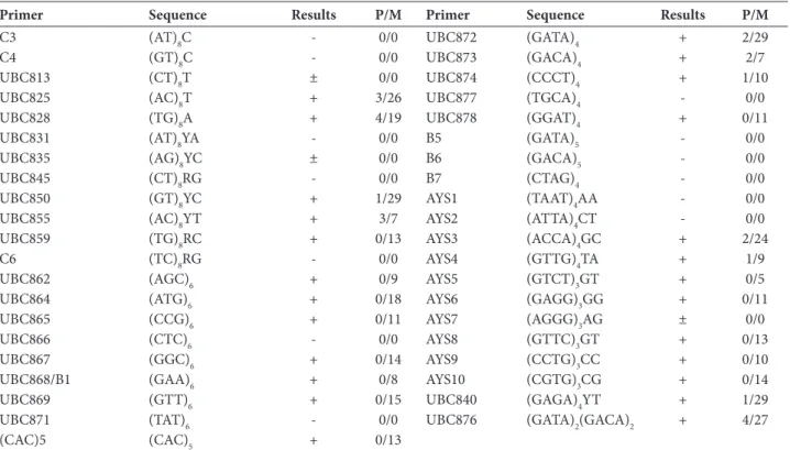
Table 2. ISSR primers used in this study
Primer Sequence Results P/M Primer Sequence Results P/M
C3 (AT)8C - 0/0 UBC872 (GATA)4 + 2/29
C4 (GT)8C - 0/0 UBC873 (GACA)4 + 2/7
UBC813 (CT)8T ± 0/0 UBC874 (CCCT)4 + 1/10
UBC825 (AC)8T + 3/26 UBC877 (TGCA)4 - 0/0
UBC828 (TG)8A + 4/19 UBC878 (GGAT)4 + 0/11
UBC831 (AT)8YA - 0/0 B5 (GATA)5 - 0/0
UBC835 (AG)8YC ± 0/0 B6 (GACA)5 - 0/0
UBC845 (CT)8RG - 0/0 B7 (CTAG)4 - 0/0
UBC850 (GT)8YC + 1/29 AYS1 (TAAT)4AA - 0/0
UBC855 (AC)8YT + 3/7 AYS2 (ATTA)4CT - 0/0
UBC859 (TG)8RC + 0/13 AYS3 (ACCA)4GC + 2/24
C6 (TC)8RG - 0/0 AYS4 (GTTG)4TA + 1/9
UBC862 (AGC)6 + 0/9 AYS5 (GTCT)3GT + 0/5
UBC864 (ATG)6 + 0/18 AYS6 (GAGG)3GG + 0/11
UBC865 (CCG)6 + 0/11 AYS7 (AGGG)3AG ± 0/0
UBC866 (CTC)6 - 0/0 AYS8 (GTTC)3GT + 0/13
UBC867 (GGC)6 + 0/14 AYS9 (CCTG)3CC + 0/10
UBC868/B1 (GAA)6 + 0/8 AYS10 (CGTG)3CG + 0/14
UBC869 (GTT)6 + 0/15 UBC840 (GAGA)4YT + 1/29
UBC871 (TAT)6 - 0/0 UBC876 (GATA)2(GACA)2 + 4/27
(CAC)5 (CAC)5 + 0/13
+ − amplification in all isolates; ± − amplification in some isolates; - − no amplification products; P − the number of polymorphic markers; M − mono-morphic markers.
[45] and adjusted as described previously [30]. Fg16F/ Fg16R (5ʹ-CTCCGGATATGTTGCGTCAA-3ʹ/5ʹ-GGTAGGTATCCGACATGGCAA-3ʹ) and Fc01F/ Fc01R (5ʹ-ATGGTGAACTCGTCGTGGC-3ʹ/5ʹ-CCCTTCTTACGCCAATCTCG-3ʹ) primer sets were used in F. graminearum- and F. culmorum-specific PCR assays.
Anchored primer design and ISSR assays
Forty-one ISSR primers were used in this study (Ta-ble 2), of which 31 belonged to the Universal ISSR primer set (NAPS-UBC, Canada) and the remaining primers were newly designed according to the nu-cleotide sequence information about F. graminearum [46]. SSR motifs in the Fusarium genus that had not been studied before were selected via “blasting” of four chromosomes of the F. graminearum PH-1 strain. All contigs and supercontigs were separately screened for microsatellite regions by the online microsatellite re-peats finder. After selection of SSR regions, all primers were designed with 2-mer anchors.
Amplification of ISSR loci by PCR was conducted in a volume of 25 µL reaction mixture containing 1 x buffer, 25 ng genomic DNA, 0.5 µM primer, 0.2 mM of dNTPs, 2.5 mM MgCl2 and 1 U of Taq polymerase. PCR was performed under the following program: 35 cycles at 94°C for 45 s, 48-55°C for 45 s and 72°C for 2 min. The first cycle had an extra 5 min at 94°C and the final extension an extra 10 min at 94°C. Amplicons were separated by electrophoresis in 1.5% agarose gels and stained with ethidium bromide. Images were pho-tographed with a gel visualization system (Avegene, Taiwan). Each experiment set was replicated at least three times.
Mating type assays
MAT-1 and MAT-2 specific primer pairs fusALPHA-for/rev (5’-CGCCCTCTKAAYGSCTTCATG-3’/5’-GGARTARACYTTAGCAATYAGGGC-3’) and fusH-MGfor/rev (5’-CGACCTCCCAAYGCYTACAT-3’/5’-TGGGCGGTACTGGTARTCRGG-3’) were used in mating type determination according to Kerényi et al.
[25]. PCRs were carried out in a reaction volume of 25 µl containing 1 × buffer, 50 ng of genomic DNA, 0.5 µM of primer, 0.2 mM of dNTPs, 2.5 mM MgCl2 and 1 U of Taq polymerase. PCRs were performed under the following program: 35 cycles at 94°C for 1 min, 53°C for 1 min and 72°C for 1 min. The first cycle had an extra 3 min at 94°C and the final extension had an extra 3 min at 94°C. Amplicons were analyzed as mentioned before. MAT-1 and MAT-2 experiment sets were replicated three times.
Statistical analysis
ISSR markers were visually scored as presence (1) or absence (0) of a band. A similarity matrix was gener-ated from the data using Nei-Li’s coefficient [47]. The dendrogram was created according to cluster analysis of ISSR markers using UPGMA (unweighted pair-group method with arithmetic average) algorithm via MVSP 3.1 software.
RESULTS
Isolates used in this study were completely identified at species level by their morphological characteristics [27,29]. In addition, verification of fungal isolates at species level was carried out via amplification of SCAR markers. A UBC85F/UBC85R primer set yielded 332 bp of PCR product in 43 F. graminearum isolates. The primer pair OPT18F/OPT18R produced a common amplicon of 472 bp in all F. culmorum isolates (data not shown). All isolates characterized at species level were incorporated into further ISSR fingerprinting analysis.
0.2 to 3.5 kb. Maximum and minimum number of ISSR bands were obtained from primers UBC872 and UBC876 (31 amplicons) and IF5 (five amplicons). The calculated similarity coefficient among F. gramine-arum isolates ranged from 62.3 to 99% (data not shown). The mean value of similarity was calculated as 0.809±0.002 (SE). Genetically, the most similar (99%) isolates were sh10 and M7. However, the maximum variation level was 62.3% between M3 and sh7. These four isolates belonged to the same geographic location (Neka). The similarity coefficients among F. culmorum isolates were 65.7-94.3% (Table 3). The mean value of
similarity was 0.798±0.004 (SE). Genetically, the clos-est (94.3%) isolates were 10F and F12. Also, the most dissimilar (65.7%) were F10 and 11F from Bilecik and Corum, respectively (Table 1). Interspecific similarity between two species ranged from 42.5 to 59.2%. The mean value of similarity belonging to two species was 0.525±0.001 (SE). The most closely related isolate of F. culmorum F10 from Bilecik was sh1 of F. gramine-arum from Mazandaran. Similarly, the most distinct isolates were F. culmorum 12F from Amasya and F. graminearum M9 from Mazandaran.
Table 3. Similarity matrix of F. culmorum isolates used in this study. Decimal places were accepted as “2”.
F1 F2 F3 F4 F10 F12 F14 F15 F16 F17 F19 F20 F21 F24 8F 9F 10F 11F 12F 13F F1 1
F2 0.86 1
F3 0.87 0.9 1
F4 0.81 0.87 0.91 1
F10 0.8 0.83 0.85 0.86 1
F12 0.88 0.84 0.88 0.86 0.87 1
F14 0.86 0.85 0.92 0.89 0.87 0.91 1
F15 0.81 0.77 0.83 0.82 0.81 0.87 0.87 1
F16 0.82 0.81 0.86 0.81 0.81 0.84 0.87 0.86 1
F17 0.84 0.83 0.85 0.83 0.83 0.87 0.84 0.83 0.88 1
F19 0.81 0.79 0.81 0.79 0.78 0.85 0.83 0.81 0.85 0.88 1
F20 0.85 0.81 0.85 0.8 0.81 0.86 0.86 0.84 0.88 0.9 0.92 1
F21 0.8 0.78 0.83 0.82 0.78 0.82 0.85 0.78 0.79 0.81 0.86 0.86 1
F24 0.78 0.75 0.8 0.77 0.8 0.81 0.82 0.79 0.81 0.84 0.82 0.88 0.85 1
8F 0.71 0.68 0.73 0.68 0.69 0.72 0.75 0.71 0.77 0.71 0.75 0.78 0.78 0.79 1
9F 0.73 0.7 0.74 0.7 0.7 0.74 0.76 0.75 0.77 0.74 0.76 0.8 0.77 0.81 0.91 1
10F 0.75 0.72 0.77 0.73 0.68 0.76 0.77 0.75 0.78 0.75 0.78 0.82 0.8 0.78 0.86 0.85 1
11F 0.73 0.69 0.74 0.71 0.66 0.73 0.75 0.71 0.74 0.72 0.73 0.76 0.75 0.73 0.84 0.8 0.91 1
12F 0.75 0.71 0.76 0.72 0.67 0.75 0.76 0.75 0.78 0.72 0.76 0.79 0.77 0.75 0.87 0.86 0.94 0.92 1
13F 0.73 0.74 0.78 0.74 0.7 0.75 0.79 0.73 0.75 0.72 0.75 0.78 0.8 0.75 0.84 0.83 0.94 0.9 0.94 1
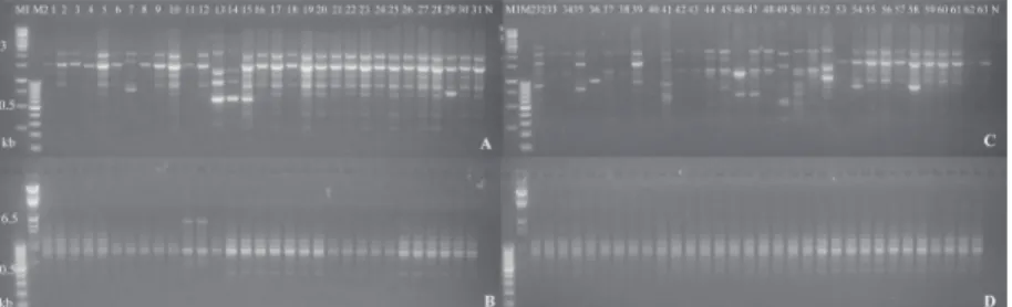
Fig. 1. ISSR profiles obtained from F. graminearum (A, B) and F. culmorum isolates (C, D) with primers AYS8 (A, C) and AYS6 (B, D) primers. M1: 1 kb DNA ladder (Thermo, SM0313), M2: λ/HindIII DNA marker (Thermo, SM0101), 1-31 and 52-63:
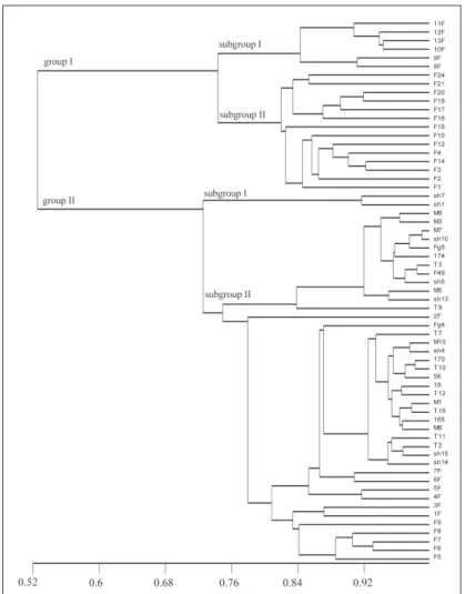
Cluster analysis showed that all isolates were divided into two main clades: group I and group II (Fig. 2). As illustrated in the dendrogram generated by UPGMA, group I included 20 isolates belonging to F. culmorum species and was smaller than group II, which consisted of 43 F. graminearum isolates. Group I consisted of F. culmorum isolates from Turkey that produced DON mycotoxin. Group II had only F. graminearum isolates from both Turkey and Iran. Their chemotypes showed heterogeneity, and both have DON and NIV chemotypes. Phenons in group II included isolates distributed in same geographic region.
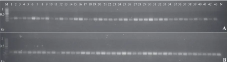
According to mating type analysis, all F. gramine-arum and three F. culmorum isolates (13F, 9F, F16) yielded specific amplicons of both MAT-1 and MAT-2 genes of 210 and 260 bp, respectively. The 210 bp of DNA fragment corresponding to MAT-1 was ampli-fied from all F. graminearum and nine F. culmorum isolates (Fig. 3). Similarly, all F. graminearum and 14 F. culmorum isolates produced a common band of 260 bp belonging to MAT-2. Findings obtained from this analysis showed that all F. graminearum isolates have sexual reproduction, while three of F. culmorum (13F, 9F, F16) isolates have potential of a sexual stage.
Fig. 2. UPGMA dendrogram generated via ISSR markers amplified in Fusarium
DISCUSSION
More than ten Fusarium species are responsible for FHB and root rot diseases worldwide. Among them, F. graminearum and F. culmorum are the main causal agents of the diseases. However, predominating spe-cies in one location can vary according to climatic conditions, their hosts and other environmental fac-tors [1,14,48,49]. Fusarium species possess a high level of phenotypic and genotypic diversity. Genotyping of F. graminearum and F. culmorum has a great im-portance to the genetic characterization of these phy-topathogenic species; however, comprehensive and detailed studies are required.
Mating type analysis provides valuable knowledge about the reproduction of fungal pathogens. There-fore, the identification of mating type is important for the characterization of a fungus. In this study, all isolates belonging to F. graminearum yielded both mating type genes, i.e. a sexual stage is present in the species. In F. culmorum isolates, no sexual stage was determined in 17 isolates according to either MAT-1 or MAT-2 amplification profiles. The presence both of two mating type genes in three F. culmorum isolates (13F, 9F and F16) is an interesting finding of asexual reproduction. Çepni et al. [50] used the same F. cul-morum isolates from Turkey in their study includ-ing genetic diversity and matinclud-ing type analysis. They reported that two F. culmorum isolates (F4 and F15) contained both MAT-1 and MAT-2 loci. Also, they stated that no amplification product was obtained from the F21 isolate. In this study, mating type anal-ysis of F. culmorum and F. graminearum isolates was performed. In contrast to these authors’ findings, we
observed that the F21 isolate possesses only the MAT-1 gene. In addition, while F4 carries only MAT-2, FMAT-15 carries only MAT-1. There are incompatible findings between two studies. Nevertheless, the presence of both mating types in F. culmorum could indicate a parasexual recombination as previously reported [22]. In the wild, hyphal anastomosis can arise when geneti-cally different hyphae of the same species come into contact. After the fusion of two cells, different types of nuclei come together in the heterokaryon, giving a somatic diploid nucleus. During the division of such nuclei, mitotic crossing-over may occur, resulting in parasexual recombination. This is the reason why both MAT-1 and MAT-2 idiomorphs can be amplified in the 13F, 9F and F16 isolates of F. culmorum [51].
A wide range of molecular marker techniques based on PCR was employed in genotyping Fusarium isolates. Many of them involve the amplification of genomic DNA. RAPD [24], AFLP [36] and microsat-ellite-PCR [52] are just some of them and they target chromosomal DNA. Moreover, mitochondrial DNA analysis is also used for genotyping [40]. The ISSR technique is one of the most widely used and versatile tools in genetic characterization in fungi. ISSR mark-ers obtained from PCR amplification represent dif-ferentiation between SSR regions [42]. In ISSR analy-sis, no prior genomic information is required. Both anchored and non-anchored primers are effectively used in PCR amplification. Although non-anchored primers in particular are a good choice for DNA fin-gerprinting, stable and reproducible fragments are also obtained from anchored ones [43]. As a result, only a limited number of polymorphic markers (5.92%) could be produced in the current study because (i)
anchored primers were used for amplification and (ii) more than half of the primers contained four nucle-otide repeats. Nevertheless, a moderate genetic varia-tion at intra- and interspecific levels was determined in Fusarium isolates. Mishra et al. [18,19] reported that there are high levels of genetic diversity using ISSR marker amplification in F. graminearum and F. culmo-rum isolates from different countries. They found 81% of ISSR amplicons to be polymorphic. Similarly, from ISSR analysis among F. poae isolates from England and Argentina, Dinolfo et al. [42] showed that there was a high level of genetic variation. They reported that 89% of the produced ISSR bands were polymorphic. Moreover, Li et al. [53] and Mishra et al. [54] reported a high level of polymorphic bands (82.3% and 98.3%, respectively) obtained from in their ISSR analysis. Both Mishra et al. [18] and Dinolfo et al. [42] used 4 and 25 oligonucleotide primers, respectively, most of which were non-anchored. However, in the current study, 41 primers were examined for ISSR fingerprinting, of which 23 were anchored and 18 non-anchored. Among them, 10 were novel and were developed and used for the first time in this study. Of the 24 primers (60%), 7 were novel amplified scorable ISSR markers. As a result, variations at the intra- and interspecific level were determined via these primers. Anchored prim-ers have the potential to produce more monomorphic markers. A high number of anchored primers could result with a stable and reproducible but low number of polymorphic bands. The mechanisms accounting for variations in ISSR markers are replication slippage, structural rearrangements of chromosomes and inser-tion/deletion SNPs occurring in amplified regions [18]. ISSR analysis is not time consuming and laborious, and it also provides reproducible and reliable results in fungal genetics [19,42-44].
Many genotyping studies of Fusarium species re-sulted in no clear correlation between genetic variabil-ity and the host and/or geographic origin of isolates [18,19,32]. Isolates either from a single population and/ or a few populations, or from limited agro-ecological regions have been used in ISSR studies to date. In this study, the 63 isolates used in this study belonged to two different species, from 3 host plants in 13 differ-ent agro-ecological regions of two neighboring
coun-tries. Wheat is the prevalent host and therefore, there was no correlation between genetic diversity and host plant. However, isolates were found in subdivisions, especially phenons in group II clustered together at the same agro-ecological locations. F. graminearum and F. culmorum isolates were definitely separated into clades. Group I consisted of only F. culmorum isolates producing DON mycotoxin from Turkey and group II involved only F. graminearum isolates from both Turkey and Iran. Their chemotypes and mating types showed heterogeneity, with both having DON and NIV chemotypes and MAT-1 and MAT-2 mating types.
The fingerprinting results obtained from this study showed that ISSR fingerprinting is a powerful tool for the discrimination of phytopathogenic species at the species level. Moreover, the technique provides reliable and reproducible results for the correlation between genotype and geographic origins. The novel ISSR markers developed for the first time in this study, should contribute to the differentiating of Fusarium isolates according to their species and geographic re-gion. Genotyping via molecular marker techniques is currently a promising and reliable tool for fungal genetic characterization.
Acknowledgements: This work was supported by the Scientif-ic Research Projects Coordination Unit of Istanbul University, Project No. 23672. The authors thank Prof. Dr. Berna Tunali from the Department of Plant Protection, Agricultural Faculty, Samsun Ondokuz Mayis University, for providing fungal material.
Conflict of interest disclosure: The authors have no conflict of interest.
REFERENCES
1. Parry DW, Jenkinson P, McLeod I. Fusarium ear blight (scab) in small grain cereals-a review. Plant Pathol. 1995; 44:207-238.
2. Foroud NA, Eudes F. Trichothecenes in cereal grains. Int J of Mol Sci. 2009; 10:147-173.
3. Miedaner T, Cumagun CJR, Chakraborty S. Population genetics of three important head blight pathogens Fusarium graminearum, F. pseudograminearum and F. culmorum. Plant Pathol J.2008;156: 129-139.
Europe as compared to those in Southern Europe. Microor-ganisms. 2013; 1: 162-174.
5. Przemieniecki SW, Kurowski TP., Korzekwa, K. Chemotypes and geographic distribution of the Fusarium graminearum
species complex. Environ Biotech. 2014; 10(2):45-54. 6. O’Donnell K, Kistler HC, Tacke BK, Casper, HH. Gene
gene-alogies reveal global phylogeographic structure and repro-ductive isolation among lineages of Fusarium graminearum, the fungus causing wheat scab. Proc Natl Acad Sci 2000; 97(14): 7905-7910.
7. O’Donnell K, Ward TJ, Geiser DM, Kistler HC, Aoki T. Genealogical concordance between the mating type locus and seven other nuclear genes supports formal recognition of nine phylogenetically distinct species within the Fusarium graminearum clade. Fungal Genet. Biol. 2004; 41:600-623. 8. Starkey DE, Ward TJ, Aoki T, Gale LR, Kistler HC, Geiser
DM, Suga H, Toth B, Varga J, O’Donnell K. Global molecular surveillance reveals novel Fusarium head blight species and trichothecene toxin diversity. Fungal Genet Biol. 2007; 44: 1191-1204.
9. Yli-Mattila T, Gagkaeva T, Ward TJ, Aoki, T, Kistler HC, O’Donnell K. A novel Asian clade within the Fusarium graminearum species complex includes a newly discovered cereal head blight pathogen from the Russian far east. Myco-logia. 2009; 101: 841-852.
10. Sarver BAJ, Ward TJ, Gale LR, Broz K, Kistler HC, Aoki T, Nicholson P, Carter J, O’Donnell K. Novel Fusarium Head Blight pathogens from Nepal and Louisiana revealed by mul-tilocus genealogical concordance. Fungal Genet Biol. 2011; 48:1096-1107.
11. Gale LR, Ward TJ, Balmas V, Kistler HC. Population Subdi-vision of Fusarium graminearum sensu stricto in the Upper Midwestern United States. Phytopathol. 2007; 97(11):1434-1439.
12. Davari M, Safaie N, Darvishnia M, Taleshmikaeel RD. Occurrence of deoxynivalenol producing isolates of Fusar-ium graminearum species complex associated with head blight of wheat in Moghan area. J Crop Prot. 2014; 3(2): 113-123.
13. Champail A, Dore T, Fourbet JF. Fusarium head blight: epi-demiological origin of the effects of cultural practices on head blight attacks and the production of mycotoxins by
Fusarium in wheat grains. Plant S ci. 2004; 166:1389-1415. 14. Nicholson P, Simpson DR, Wilson AH, Chandler E,
Thom-sett M. Detection and differentiation of trichothecene and enniatin-producing Fusarium species on small-grain cereals. Eur J Plant Pathol. 2004;110:503-514.
15. Puhalla JE. Genetic considerations of the genus Fusarium. In: Nelson PE, Toussoun TA, Cooki RJ, editors. Fusarium Dis-eases, Biology, and Taxonomy. Pennsylvania:Pennsylvania State University Press; 1981. p. 291-305.
16. Carter JP, Rezanoor HN, Holden D, Desjardins AE, Plattner RD, Nicholson P. Variation in pathogenicity associated with the genetic diversity of Fusarium graminearum. Eur J Plant Pathol. 2002; 108:573-583.
17. Yli-Mattila T, Paavanen-Huhtala S, Parikka P, Konstanti-nova P, Gagkaeva T. Molecular and morphological diversity of Fusarium fungi in Finland and north-western Russia. Eur J Plant Pathol. 2004; 110:537-585.
18. Mishra PK, Fox RTV, Culham A. Inter simple sequence repeat and aggressiveness analysis revealed high genetic diversity, recombination and long-range dispersal in Fusar-ium culmorum. Ann Appl Biol. 2003a; 143:291-301. 19. Mishra PK, Fox RTV, Culham A. Development of a
PCR-based assay for rapid and reliable identification of pathogenic
Fusaria. FEMS Microbiol Lett. 2003b; 218:329-332. 20. Gagkaeva TY, Mattila, TY. Genetic diversity of Fusarium
graminearum in Europa and Asia. Eur J Plant Pathol. 2004; 110:551-562.
21. Miedaner T, Schilling AG, Geiger HH. Competition effects among isolates of Fusarium culmorum differing in aggres-siveness and mycotoxin production on heads of winter rye. Eur J Plant Pathol. 2004; 110:63-70.
22. Miedaner T, Schilling AG, Geiger HH. Molecular genetic diversity and variation for aggressiveness in populations of
Fusarium graminearum and Fusarium culmorum sampled from wheat fields in different countries. J Phytopathol. 2001; 149:641-8.
23. Gürel F, Albayrak G, Diken O, Cepni E, Tunali B. Use of REP-PCR for genetic diversity analysis in Fusarium culmo-rum. J Phytopathol. 2010; 158:387-9.
24. Yörük E, Albayrak G. Genetic characterization of Fusarium graminearum and F. culmorum isolates from Turkey by using random-amplified polymorphic DNA. Genet Mol Res. 2013; 12(2):1360-72.
25. Kerényi Z, Moretti A, Waalwijk C, Olah B, Hornok L. Mating type sequences in asexually reproducing Fusarium species. Appl Environ Microb. 2004; 70:4419-4423.
26. Obanor F, Erginbas-Orakci G, Tunali B, Nicol JM, Chakraborty S. Fusarium culmorum is a single phylogenetic species based on multilocus sequence analysis. Fungal Biol. 2010; 114:753–765.
27. Tunali B, Özseven İ, Büyük O, Erdurmuş D, Demirci A. Fusarium head blight and deoxynivalenol accumulation of wheat in Marmara region and reactions of wheat cultivars and lines to F. graminearum and Fusarium culmorum. Plant Pathol J. 2006; 5(2):150-6.
28. Tizaki MA, Sabbagh SK. Detection of 3-Acetyldeoxynivale-nol, 15-Acetyldeoxynivalenol and Nivalenol-Chemotypes of
Fusarium graminearum from Iran using specific PCR assays. Plant Knowledge Journal. 2013; 2(1):38-42.
29. Haratian M, Sharifnabi B, Alizadeh A, Safaie N. PCR analy-sis of the tri13 gene to determine the genetic potential of
Fusarium graminearum isolates from Iran to produce nivale-nol and deoxynivalenivale-nol. Mycopathologia. 2008; 166:109-116.
30. Yörük E, Albayrak G. Chemotyping of Fusarium
31. Mert-Türk F, Gencer G. Distribution of the 3-AcDON, 15-AcDON, and NIV chemotypes of Fusarium culmorum in the North-West of Turkey. Plant Prot Sci. 2013; 49(2):57-64. 32. Yli-Mattila T, Mironenko NV, Alekhina IA, Hannukkale
A, Bulat SA, Universally primed polymerase chain reac-tion analysis of Fusarium avencceum, F. arthrosporioides, F. tricinctum species complex – a polyphasic approach. Mycol Res. 1997; 106:655-669.
33. Saharan MS, Naef A, Kumar J, Tiwari R. Characterization of variability among isolates of Fusarium graminearum associ-ated with head scab of wheat using DNA markers. Curr Sci. 2007; 92:230-5.
34. Vogelgsang S, Widmer F, Jenny E, Enkerli J. Characterisa-tion of novel Fusarium graminearum microsatellite markers in different Fusarium species from various countries. Eur J Plant Pathol. 2009; 123:477-482.
35. Llorens A, Hinojo MJ, Mateo R, Medina A, Valle-Algarre FM, Gonzalez-Jaen MT, et al. Variability and characteriza-tion of mycotoxin producing Fusarium spp. isolates by PCR-RFLP analysis of the IGS-rDNA region. Anton Leeuw. 2006; 89(3-4):465-478.
36. Monds RD, Cromey MG, Lauren DR, Di Menna M, Marshall J. Fusarium graminearum, F. cortaderiae and F. pseudogra-minearum in New Zealand: molecular phylogenetic analysis, mycotoxin chemotypes and co-existence of species. Mycol Res. 2005; 109(4):410-420.
37. Chung WH, Ishii H, Nishimura K, Ohshima M, Iwama T, Yoshimatsu H. Genetic analysis and PCR-based identifica-tion of major Fusarium species causing head blight on wheat in Japan. J Gen Plant Pathol. 2008; 10:110-8.
38. Konstantinova P, Yli-Mattila T. IGS-RFLP analysis and devel-opment of molecular markers for identification of F. poae, F. pulverosum, F. sporotrichioides and F. kyushuemse. Int J Food Microbiol. 2004; 95(3):321-331.
39. To´th B, Mesterha´zy A, Horva´th Z, Barto´k T, Varga M, Varga J. Genetic variability of central European isolates of the
Fusarium graminearum species complex. Eur J Plant Pathol. 2005; 113:35–45.
40. Laday M, Juhasz A, Mule G, Moretti A, Szecsi A, Logrieco A. Mitochondrial DNA diversity and lineage determination of Europan isolates of Fusarium graminearum (Gibberella zeae). Eur J Plant Pathol. 2004; 110:545-550.
41. Baysal Ö, Siragusa M, Gümrükcü E, Zengin S, Carimi F., Sajeva M, et al. Molecular characterization of Fusarium oxy-sporum f. melongenae by ISSR and RAPD markers on egg-plant. Biochem Genet. 2010; 48:524-537.
42. Dinolfo MI, Stenglein SA, Moreno MV, Nicholson P, Jen-nings P, Salerno GL. ISSR markers detect high genetic varia-tion among Fusarium poae isolates from Argentina and Eng-land. Eur J Plant Pathol. 2010; 127:483-491.
43. Bornet B, Branchard M. Nonanchored inter simple sequence repeat (ISSR) markers: Reproducible and specific tools for genome fingerprinting. Plant Mol Biol Rep. 2001; 19:209-215. 44. Bayraktar H, Dolar FS, Maden S. Use of RAPD and ISSR
markers in detection of genetic variation and population structure among Fusarium oxysporum f. sp. ciceris isolates on chickpea in Turkey. J Phytopathol. 2008; 156:146-154. 45. Nicholson P, Simpson DR, Weston G, Rezanoor HN, Lees
AK. Detection and quantification of Fusarium culmorum and
Fusarium graminearum in cereals using PCR assays. Physiol Mol Plant Pathol. 1998;53:17-37.
46. Broad Institute: Genome Index: Broad Institute; 2007 [ updated 2010 March 18; cited 2015 January 2]. Avaible from www.broadinstitute.org/annotation/genome/fusar-ium_group/GenomesIndex/
47. Nei M, Li WH. Mathematical model for studying genetic variation in terms of restriction endonucleases. Proc Natl Acad Sci USA. 1979; 76:5269-5273.
48. Bottalico A, Perrone G. Toxigenic Fusarium species and mycotoxins associated with head blight in small-grain cere-als in Europe. Eur J Plant Pathol. 2002; 108:611-624. 49. Saharan MS, Kumar J, Nagarajan S. Fusarium head blight
(FHB) or head scab of wheat- a review. Proc Natl Acad Sci India 2004; 3:255-268.
50. Çepni E, Tunalı B, Gürel F. Genetic diversity and mating types of Fusarium culmorum and Fusarium graminearum
originating from different agro-ecological regions in Turkey. 2013; J Basic Microb; 53:686-694.
51. Carlile MJ, Watkinson SC, Gooday GW. The Fungi: Genetic variation and evolution. 2nd ed. Amsterdam: Academic Press; 2001.
52. Saharan MS, Naef A. Detection of genetic variation among Indian wheat head scab pathogens (Fusarium spp./isolates) with microsatellite markers. Crop Prot. 2008; 27: 1148-1154. 53. Li RQ, He R, Zhang YB, Xu YM, Wang JM. Establishment of ISSR reaction system of Fusarium and its analysis of genetic diversity. Agric Sinica. 2009; 42(9):3139-3146.