CASE REPORT
Association of myelodysplastic syndrome with CD5
+
,
CD23
+
monoclonal B-cell lymphocytosis
Alex F. Sandes,IMaria de Lourdes L. F. Chauffaille,IAlberto Orfao,II Graziella C. Siufi,I Maria Regina R. Silva,III Mihoko YamamotoI
IUniversidade Federal de Sa˜o Paulo (UNIFESP), Disciplina de Hematologia e Hemoterapia, Sa˜o Paulo/SP, Brazil.IIUniversity of Salamanca, Servicio General
de Citometria, Department of Medicine and Cancer Research Center (IBMCC-USAL/CSIC), Salamanca, Spain.IIIUniversidade Federal de Sa˜o Paulo
(UNIFESP), Pathology Department, Sa˜o Paulo/SP, Brazil.
Email: alex.sandes@bol.com.br / yamamoto@unifesp.br Tel.: 55 11 5576-4240
INTRODUCTION
Myelodysplastic syndromes (MDS) are clonal disorders characterized by bone marrow (BM) failure and an increased risk of transformation into acute myeloid leukemia (AML). Typically, MDS patients are elderly and are already anemic, leucopenic and/or thrombocytopenic upon presentation. Despite their myeloid origin, many MDS cases show abnormalities in B-cells, usually related to a decrease in the B-cell production and/or an increase in apoptosis (1,2). Such alterations may be due to an increased production demand for essential hematopoietic cells, such as red cells and neutrophils. Alternatively, a blockade on B-cell maturation might occur (2). A low level of monoclonal B-cells (,56109/L) in otherwise
healthy individuals characterizes a condition called mono-clonal B-cell lymphocytosis (MBL). The overall reported frequency of MBL is between 0.5% and 12% of adults, depending on the age of the population and the sensitivity of the flow cytometry approach used to detect the B-cell clones (3,4). MBL is more frequently observed among the relatives of patients with familial chronic lymphocytic leukemia (CLL) and in individuals exposed to toxic environments. Although coexistence of CLL and AML or CML has been sporadically reported, the actual frequency of MBL in association with other hematological neoplasias remains to be established.
CASE DESCRIPTION
Here, we report a 61-year-old female patient diagnosed with both MDS and MBL. She complained of a three-month history of fatigue associated with sustained anemia and thrombocytopenia detected on a routine blood test. Splenomegaly or lymphadenomegaly were not observed. Her hemoglobin level was 3.8 g/dL, her absolute white blood cell (WBC) count was 7.66109/L (neutrophils 68%;
eosinophils 1%; lymphocytes 23%; monocytes 9%) and her platelet count was 346109/L. Hyposegmented neutrophils
were observed on blood film examination. The BM aspirate
was hypercellular and showed increased blast cells (9%), marked dysplastic abnormalities in the erythroid, granulo-cytic and megakaryogranulo-cytic lineages and no ring sideroblasts. The BM biopsy showed mild reticulin fibrosis without the presence of lymphoid aggregates or infiltrates. Cytogenetic analysis was inconclusive. A diagnosis of refractory anemia with excess blasts-1 was established according to the WHO classification and the patient received supportive therapy consisting of red blood cell transfusions.
Five months later, the patient’s myeloblasts had increased to 15% (BM) and an abnormal karyotype (46,XX,del(9) (q22) [20]) was detected. Immunophenotyping of BM cells was performed using a large panel of monoclonal antibodies in a four-color combination to analyze precursor cells, granulocytic, monocytic and erythroid BM compartments (5) (Table 1). Multiparameter flow cytometry confirmed the increased number of myeloblasts (Figure 1) with an aberrant (CD7+, CD56+), immature (CD34+, CD117+, HLA-DR+)
mye-loid (CD13+, CD33+) phenotype. No CD34+B-cell precursors
were detected and phenotypic abnormalities were identified in the maturing neutrophils (e.g., aberrant CD13/CD11b expression) and in the monocytic (e.g., CD2+, CD56+) and
erythrocytic (e.g., CD71lo) compartments (Figure 1). In addi-tion, 8% of the marrow cells were mature T-lymphocytes and 9% were mature B-lymphocytes that presented aberrant CD25 and CD22dimexpression. A detailed study was performed due to this cell phenotype, which demonstrated that 88% of B-cells were monoclonal, surface kappa light chain restricted and a CD5+, CD19+, CD23+and CD25+phenotype (Figure 2).
Further analysis of the peripheral blood (PB) demon-strated the presence of monoclonal B-lymphocytes with a CD19+, CD20dim
, CD22dim, CD5+, CD23+, CD79bdim , CD25+,
CD43+and CD38+phenotype, lacking in reactivity to
FMC-7, sIgM, CD10, CD103, and CD11c. The absolute PB B-lymphocyte count was 3.06109/L and a diagnosis of
CD5+CD23+MBL was made.
The patient was treated for MDS with daunorubicin and cytarabine, with no response and died six months later with AML and the persistence of the PB monoclonal B-cell population (26109/L).
The reported case followed the guidelines of the local ethics committees and the Helsinki Declaration. This patient was included in a large study on MDS (6) and informed consent was obtained after the study was approved by the Institutional Review Board of UNIFESP (Brazil).
Copyrightß2012CLINICS– This is an Open Access article distributed under the terms of the Creative Commons Attribution Non-Commercial License (http:// creativecommons.org/licenses/by-nc/3.0/) which permits unrestricted non-commercial use, distribution, and reproduction in any medium, provided the original work is properly cited.
DISCUSSION
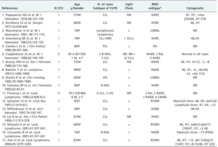
Association of MDS and B-chronic lymphoproliferative disorders (B-CLPD) is an uncommon finding that has been sporadically described (Table 2). We found that 31 cases had been reported since 1974. The median age at presentation was 72 years (range: 49-95) and the male-to-female ratio was
1.2:1. Most cases corresponded to patients with MDS and CLL (19/31) and no significant association with specific subtypes of MDS was observed. Florensa et al. showed a frequency of 1% of B-CLPD in a series of 1198 MDS patients (CLL 0.5%, lymphoplasmacytic lymphoma 0.4% and multi-ple myeloma 0.1%) (7). At present, the general consensus is that these associations may occur randomly (8-10). In line with this idea, we found only one case report supporting the existence of an ontogenic association between both dis-orders: trisomy 8 was detected in 55% of CD13+neutrophils
and in 13% of CD19+/CD5+B lymphocytes in a case with
MDS and systemic vasculitis, suggesting a common stem cell precursor had generated the two neoplastic cell populations (11).
Low numbers of circulating monoclonal B-cells in other-wise healthy individuals has been investigated in the last ten years. A MBL diagnosis is confirmed by the presence of
,56109/L monoclonal B-cells associated with a normal
physical examination and negative history for lymphopro-liferative disease, as observed in our case (12). The progression rate to CLL has been described as approxi-mately 1-2% of MBL cases per year (12). MBL has been observed in a significant number of healthy individuals and in, particular, in elderly people. Despite its frequency, the association of MBL in patients with other hematological neoplastic diseases (e.g., MDS) remains to be established.
Table 1 -Monoclonal antibody reagents used for the immunophenotypic characterization of myelodysplastic syndrome (modified from Matarraz S. et al., Leukemia 2008;22:1175-83) (5).
FITC PE PerCP-Cy5.5 APC
1. HLA-DR CD117 CD45 CD34
2. HLA-DR CD123 CD45 CD34
3. CD11b CD13 CD45 CD34
4. CD36 CD64 CD45 CD34+CD14
5. CD15 CD16 CD45 CD34
6. CD2 CD56 CD45 CD34
7. CD65 NG2 CD45 CD34
8. CD71 Glycophorin-A CD45 CD34
9. CD61 CD33 CD45 CD34
10. CD22 CD25 CD45 CD34
11. nTdT cMPO CD45 CD34
12. cCD3 CD7 CD45 CD34
13. CD19 CD79a CD45 CD34
Figure 1 -Immunophenotypic analysis of bone marrow cell compartments: the blast cells (orange) are CD34+and CD117+with partial
expression of CD7; the erythroblasts (purple) present glycophorin-A and low expression of CD71; the maturing granulocytes (blue) demonstrate an anomalous maturation pattern of CD13/CD11b expression, with increased levels of CD13 and CD11b during intermediate maturation stages; the monocytes (green) express aberrant CD2.
MDS and monoclonal B-cell lymphocytosis
To the best of our knowledge, this is the first report describing the association of MDS and CD5+with CD23+MBL. Interestingly, despite the presence of a mono-clonal B-cell population, no CD34+B-cell precursors were identified in the patient’s BM, as usually occurs in MDS cases. Caballero et al. reported a patient with AML associated with CLL in which the progression of CLL disease was observed after treatment for AML and remission was achieved (13). In contrast, the lymphoid clone remained stable in our case, without change after therapy during the follow-up.
MDS develops in a multistep way. An increasing number of accumulated genetic abnormalities lead to ineffective hemato-poiesis. In addition, it has been noted that immune dysfunction in MDS could also contribute to the development of cytopenia in some groups of patients. Accumulating evidence has demonstrated the association of MDS and autoimmune manifestations, T-cell mediated myelosuppression and cyto-kine-induced cytopenia (14,15). Immunosuppressive therapy in selected MDS patients results in high rates of hematological recovery with improved survival, especially in young patients and in the presence of HLA-DR15 (16). In addition, auto-immune complications are well recognized in CLL, occurring in
10% to 25% of patients at some time during the course of the disease. Autoimmunity in CLL predominantly targets blood constituents, most commonly red cells. The association of MBL and immune dysfunction is uncertain, although Mittal et al. (17) reported a high prevalence (20%) of CLL phenotype lympho-cytes in 31 patients with autoimmune disorders (AIHA, idiopathic thrombocytopenic purpura and Evans’ Syndrome), suggesting the importance of these clones in the pathogenesis of autoimmune blood disorders.
For the reasons stated above, the coexistence of a monoclonal B-cell disorder with MDS deserves special interest. This is particularly true because multiparameter flow cytometry immunophenotyping is the method of choice to detect MBL. Although numerous reports had described the immunophenotypic abnormalities in MDS, it has only recently begun to be applied during the routine work-up for the diagnosis and prognosis of potential MDS cases (18). Moreover, our case illustrates the importance of well-designed flow cytometry panels, capable of analyzing all hematopoietic cell populations because more than one neoplastic disorder may be present in the same case. We suggest the addition of one screening tube in the MDS investigational panel, containing CD19, CD5, anti-k andl
Figure 2 - Immunophenotypic analysis of bone marrow B-cells: monoclonal B-lymphocytes (green) are CD19+, CD5+, CD45++ and
light chains, which could be expanded when necessary. Widespread use of flow cytometry in the routine evaluation of MDS patients will potentially contribute to defining the actual frequency of the association between MBL and MDS and it may provide new insights into the association of these disorders.
ACKNOWLEDGMENTS
This report was supported by grants from FAPESP (proc no. 05/57792-0) and FADA (Unifesp). AFS was supported by CNPq (proc n.142968/2006-4) and CAPES (proc.n. PDEE BEX 1025/05-8).
AUTHOR CONTRIBUTIONS
Sandes AF and Chauffaille ML attended the patient and provided clinical data. Silva MR performed the bone marrow anatomopathological study, and Chauffaille ML was responsible for the cytogenetic experiments and analysis. Sandes AF and Siufi GC were responsible for data acquisition and flow cytometric immunophenotyping analysis. Sandes AF, Orfao A and Yamamoto M planned the study, interpreted the data and drafted the manuscript. All co-authors approved the final version of the manuscript.
REFERENCES
1. Ogata K, Kishikawa Y, Satoh C, Tamura H, Dan K, Hayashi A. Diagnostic application of flow cytometric characteristics of CD34+cells
in low-grade myelodysplastic syndromes. Blood. 2006;108(3):1037-44, http://dx.doi.org/10.1182/blood-2005-12-4916.
2. Ribeiro E, Matarraz SS, de Santiago M, Lima CS, Metze K, Giralt M, et al. Maturation-associated immunophenotypic abnormalities in bone marrow B-lymphocytes in myelodysplastic syndromes. Leuk Res. 2006;30(1):9-16, http://dx.doi.org/10.1016/j.leukres.2005.05.019.
3. Marti G, Abbasi F, Raveche E, Rawstron AC, Ghia P, Aurran T, et al. Overview of monoclonal B-cell lymphocytosis. Br J Haematol 2007; 139(5):701-8, http://dx.doi.org/10.1111/j.1365-2141.2007.06865.x. 4. Nieto WG, Almeida J, Romero A, Teodosio C, Lo´pez A, Henriques AF,
et al. Increased frequency (12%) of circulating chronic lymphocytic leukemia-like B-cell clones in healthy subjects using a highly sensitive multicolor flow cytometric approach. Blood. 2009;114(1):33-7, http://dx. doi.org/10.1182/blood-2009-01-197368.
5. Matarraz S, Lopez A, Barrena S, Fernandez C, Jensen E, Flores J, et al. The immunophenotype of different immature, myeloid and B-cell lineage-committed CD34+ hematopoietic cells allows discrimination between normal/reactive and myelodysplastic syndrome precursors. Leukemia. 2008;22(6):1175-83, http://dx.doi.org/10.1038/leu.2008.49. 6. Sandes AF, Yamamoto M, Matarraz S, Chauffaille MdL, Quijano S,
Lopez A, et al. Altered immunophenotypic features of peripheral blood platelets in myelodysplastic syndromes. Haematologica. 2012;97(6):895-902, http://dx.doi.org/10.3324/haematol.2011.057158.
7. Florensa L, Vallespi T, Woessner S, Domingo A, Crespo N, Rozman M, et al. Incidence and characteristics of lymphoid malignancies in untreated myelodysplastic syndromes. Leuk Lymphoma. 1996;23(5-6):609-2, http:// dx.doi.org/10.3109/10428199609054871.
8. Aviv H, Tang D, Das K, Harrison JS, Hameed M, Varma M. Simultaneous appearance of trisomy 8 and trisomy 12 in different cell populations in a patient with untreated B-cell chronic lymphocytic leukemia and myelodysplasia. Leuk Lymphoma. 2004;45(6):1279-83, http://dx.doi.org/10.1080/10428190310001638869.
Table 2 - Previous reports of MDS and B-chronic lymphoproliferative disorders.
References N (31)
Age y/Gender
N. of cases Subtype of CLPD
Light Chain
MDS
subtype* Cytogenetic
1. Papayannis AG et al. Br J Haematol. 1974;28:125-129.
1 71/M CLL NR RARS 47, XY,+mar
[25]/46, XY (10) 2. Escribano LE et al. Sangre.
1977;22:639-645
1 66/M CLL NR RARS 46, XY
3. Manoharan A et al. Br J Haematol. 1981; 48:111-116.
1 74/F Lymphocytic
Lymphoma
l CMML NR
4. Greenberg BR et al. Br J Haematol. 1983;53:125-133.
1 79/F CLL+MM l(CLL) RARS 46,XX
5. Camba L et al. J Clin Pathol. 1985;38:297-300.
1 80/F PLL l RA NR
6. Copplestone JA et al. Br J Haematol. 1986;63:149-159
5 81.4 (67-95)/ 1 M, 4 F
2 B-NHL; 3 CLL
NR; NRk (3 CLL)
RAEB; 2 RA; 2 RARS
Normal in all cases
7. Bracey AW et al. Am J Hematol. 1989;30:174-180.
1 72/M CLL NR RAEB 46, XY, t(1;7), -7,+8
8. Bastion Y et al. Leukemia. 1991;5:1006-1009.
1 49/M CLL k RAEB 46, XY, -4,+der(4),
-12,+der (12) 9. Shulze R et al. Clin Investig.
1992;70:1082-1084
1 60/M LPL k CMML NR
10. Uematsu M et al. Int J Hematol. 1995;62:45-51.
1 89/F B-NHL k RARS NR
11. Florensa L et al. Leuk Lymphoma. 1996;23:609-612.
11 79.3 (59-86)/ 8 M, 3 F
6 CLL, 5 LPL NR 3 RA, 2 RARS, 2 RAEB, 4 CMML
NR
12. Sylvester LS et al. Leuk Res. 1997;21:619-621.
1 85/F CLL k RCMD Myeloid clone: 46, XX, del(13)/
Lymphoid clone: 47, XX,+12 13. Mitterbauer G et al. Ann
Hematol. 1997;74:193-197.
1 69/F CLL k RAEB 46,XX
14. Lai R et al. Am J Clin Pathol. 1999;111:373-378.
1 67/M CLL NR RARS NR
15. Mossafa H et al. Leuk Lymphoma. 2001;41:337-341.
1 66/M CLL k RCMD 46, XY, add(1),del(11)
(10)/47, XY,+3 (4) 16. Cauwelier B et al. Leuk
Lymphoma. 2001;41:337-341
1 76/F B-NHL l RAEB Myeloid clone:+13 (FISH)
17. Aviv H et al. Leuk Lymphoma. 2004;45:1279-1283.
1 63/M CLL k RCMD 46, XY,+12, del (14)(q21)
(1)/47, XY,+8 (1)/46, XY [23]
*All cases were reclassified according to the WHO criteria; N - number of cases; NR–not reported; CLPD–Chronic lymphoproliferative disorder;
NHL–Non-Hodgkin Lymphoma; LPL–Lymphoplasmacytic Lymphoma; PLL–Prolymphocytic Leukemia; MDS–Myelodysplastic Syndrome; RARS–refractory anemia with ring sideroblasts; CMML–chronic myelomonocytic leukemia; RA–refractory anemia; RCMD- refractory cytopenia with multilineage dysplasia; RAEB– refractory anemia with excess blasts.
APC, allophycocyanin; FITC, fluorescein isothiocyanate; PE, phycoerythrin; PerCP, peridinin chlorophyll protein.
MDS and monoclonal B-cell lymphocytosis
9. Cauwelier B, Nollet F, De Laere E, Van Leeuwen M, Billiet J, Criel A, et al. Simultaneous occurrence of myelodysplastic syndrome and monoclonal B lymphocytes with a different clonal origin. Leuk Lymphoma. 2002;43(1):191-3, http://dx.doi.org/10.1080/10428190210205.
10. Sylvester LS, Nowell PC, Bonner H, Moreau L, Moore JS. Concurrent diagnosis of chronic lymphocytic leukemia and myelodysplastic syn-drome. Leuk Res. 1997;21(7):619-21, http://dx.doi.org/10.1016/S0145-2126(97)00017-9.
11. Billstrom R, Johansson B, Strombeck B, el Rifai W, Larramendy M, Olofsson T, et al. Clonal CD5-positive B lymphocytes in myelodysplastic syndrome with systemic vasculitis and trisomy 8. Ann Hematol 1997;74(1):37-40.
12. Marti G, Abbasi F, Raveche E, Rawstron AC, Ghia P, Aurran T, et al. Overview of monoclonal B-cell lymphocytosis. Br J Haematol 2007;139(5):701-8, http://dx.doi.org/10.1111/j.1365-2141.2007.06865.x. 13. Caballero MD, Gonzalez M, Canizo MC, Orfao A, Nieto MJ, San Miguel
JF. Concomitant chronic lymphocytic leukemia (CLL) and acute myeloid leukemia. Complete remission of CLL achieved with high-dose cytosine arabinoside. Leukemia. 1992;6(8):856-8.
14. Pinheiro RF, Silva MR, Chauffaille ML. The 5q- syndrome and autoimmune phenomena: report of three cases. Leuk Res. 2006;30(4):507-10, http://dx.doi.org/10.1016/j.leukres.2005.08.025.
15. Chamuleau ME, Westers TM, van Dreunen L, Groenland J, Zevenbergen A, Eeltink CM, et al. Immune mediated autologous cytotoxicity against hematopoietic precursor cells in patients with myelodysplastic syn-drome. Haematologica. 2009;94(4):496-506, http://dx.doi.org/10.3324/ haematol.13612.
16. Sloand EM, Wu CO, Greenberg P, Young N, Barrett J. Factors affecting response and survival in patients with myelodysplasia treated with immunosuppressive therapy. J Clin Oncol. 2008;26(15):2505-11, http:// dx.doi.org/10.1200/JCO.2007.11.9214.
17. Mittal S, Blaylock MG, Culligan DJ, Barker RN, Vickers MA. A high rate of CLL phenotype lymphocytes in autoimmune hemolytic anemia and immune thrombocytopenic purpura. Haematologica. 2008;93(1):151-2, http://dx.doi.org/10.3324/haematol.11822.