787
QUANTITATIVE ANALYSIS OF THE DYSTROPHIN GENE BY REAL-TIME PCR
NELA MAKSIMOVIC1, ANA ANDJELKOVIC1, VEDRANA MILIC RASIC 2,
VIDOSAVA RAKOCEVIC STOJANOVIC3, BILJANA KASTRATOVIC KOTLICA4, S. BRANKOVIC5, TATJANA DAMNJANOVIC1, BILJANA JEKIC1, VERA BUNJEVACKI1, LJILJANA LUKOVIC1,
DIJANA PEROVIC1, SUZANA CVJETICANIN1 and IVANA NOVAKOVIC1
1 Institute of Human Genetics, Faculty of Medicine, University of Belgrade, 11000 Belgrade, Serbia 2 Child and Adolescent Neurology and Psychiatry Clinic, Faculty of Medicine, University of Belgrade,
11000 Belgrade, Serbia
3 Clinic of Neurology CCS, Faculty of Medicine, University of Belgrade, 11000 Belgrade, Serbia 4 Clinic of Obstetrics and Gynecology, Faculty of Medicine, University of Belgrade, 11000 Belgrade, Serbia 5 Faculty of Science and Mathematics, University of Prishtina (Kosovska Mitrovica), 28000 Kosovska Mitrovica, Serbia
Abstract - Duchenne and Becker muscular dystrophy (DMD/BMD) are severe X-linked neuromuscular disorders caused by mutations in the dystrophin gene. Our aim was to optimize a quantitative real-time PCR method based on SYBR®
Green I chemistry for routine diagnostics of DMD/BMD deletion carriers. Twenty female relatives of DMD/BMD patients with previously detected partial gene deletions were studied. he relative quantity of the target exons was calculated by a comparative threshold cycle method (ΔΔCt). he carrier status of all subjects was successfully determined. he gene dosage ratio for non-carriers was 1.07±0.20, and for carriers 0.56±0.11. his assay proved to be simple, rapid, reliable and cost-efective.
Key words:Duchenne/Becker muscular dystrophy, Real-time PCR, SYBR® Green, ΔΔCt method, carrier detection
INTRODUCTION
Duchenne and Becker muscular dystrophies (DMD and BMD) are X-linked neuromuscular disorders with incidences of 1:3500 and 1:30000 male births, respectively (Emery, 1991). DMD and BMD are caused by mutations in the dystrophin gene (DMD gene) located at the Xp21.2 region. Approximately 65-70% of DMD and 85% of BMD patients show in-tragenic deletions of one or several exons of the gene. Most of the mutations are located at the proximal (exon 3-7) and central (exon 45-52) regions of the gene (Prior, 2005; Hu, 1990; http://www.umd.be).
hese regions are known as “deletional hotspots”. A small proportion of the mutations (6%) involve du-plications and in the remaining DMD/BMD cases, the disease emerges as a result of small mutations, including point mutations or microdeletions/inser-tions (Hu et al., 1988; Nishino, 2002). Mutations ei-ther are inherited from asymptomatic female carriers (70%), occur de novo or arise from germline mosai-cism (30%) (Worton, 1988).
of an eicient treatment for these progressive disor-ders makes the determination of carrier status very important for genetic counseling and prevention of the disease. Multiplex PCR technique has been widely used for the detection of common deletions in afected males (Beggs et al., 1990; Chamberlain et al., 1988). However, carrier identiication in asymp-tomatic female relatives of deletional DMD/BMD probands is still diicult due to the presence of both normal and mutant copies of the gene. During the past decades, several biochemical and molecular methods have been developed in order to overcome this problem. Such methods include the measure-ment of the serum creatinine phosphokinase (CPK) level, immunohistochemical methods (Panigarhi, 2001), DNA-based linkage analysis (Clemmens et al., 1991), Southern blotting (Den Dunnen et al., 1989), luorescent in situ hybridization (FISH) (Li-gon et al., 2000) and reverse transcriptase-polymer-ase chain reaction (RT-PCR) (Roberts et al., 1991). All these approaches are time-consuming and may sufer from limited sensitivity. Recently, multi-plex ligation-dependent probe ampliication assay (MLPA) (Gatta et al., 2005), multiplex ampliiable probe hybridization assay (MAPH) (White et al., 2002) and quantitative real-time PCR have been suggested as alternative methods to the current di-agnostic approaches.
In the present study, we have optimized the quan-titative real-time PCR assay based on SYBR® Green chemistry for determining the dosage of DMD gene exons 6, 47, 52 for direct DMD/BMD carrier detec-tion. Quantitative real-time PCR is a high precision approach for gene dosage studies that displays digital data corresponding to the changes at gene level (Wil-helm, 2003; Nosaeid, 2009; Ruiz-Ponte et al., 2006).
MATERIALS AND METHODS
Patients
Blood samples were obtained from 2 unrelated ob-ligate female carriers (characterized by two afected DMD sons in one case and by afected BMD father in the other) and 18 possible carriers from 9
fami-lies with sporadic cases (characterized by only one afected son or brother). Afected males had partial dystrophin gene deletions involving either exon 6, 47 or 52, previously detected by multiplex PCR. In par-allel, normal female controls were used as wild-type reference. Informed written consents were obtained from all individuals participating in this study.
DNA extraction
Genomic DNA was extracted from 5ml peripheral blood by standard salting-out (Miller et al., 1988). he concentration and purity of the isolated DNA were determined by measuring the absorbance at 260 and 280 nm. DNAs were diluted in distilled wa-ter to acquire a concentration of 6 ng/µl.
Real-time quantitative PCR
Data analysis
Data evaluation was performed using the 7500 data analysis sotware (version 3.5) and Microsot Excel. Quantitative analysis was performed by comparative Ct. he Ct parameter is deined as the cycle number at which the ampliication plot passes a ixed thresh-old. Mean Ct was the mean Ct value of duplicate ampliication. ΔCt was calculated as the diference between the Ct values of the DMD test exon (exonA) and reference exon (exonB). ΔΔCt values for each tested individual were calculated using ΔCts of the healthy female as a calibrator following the equation: ΔΔCtexonA-exonB=(ΔCtexonA-exonB) targetsample-(ΔCtexonA-exonB) calibrator. he relative fold increase (R) in exon quantity in the tested person was calculated using the equation: RexonA/exonB=2-(ΔΔCtexonA-exonB) (Livak, 2001). Using this method, a ΔΔCt ratio is expected to be about 1 in normal controls, about 0.5 in the females that are carriers of the deletion and 1.5 in females that are carriers of the duplication.
Optimization and validation of real-time PCR assay
In order to validate the ΔΔCt method for the relative quantiication of exons 6, 47 and 52, genomic DNA (gDNA) was isolated from the blood of a healthy fe-male and 2-fold serial dilutions ranging from 50 to 3.125 ng/µl were generated (50, 25,12.5, 6.25, 3.125 ng/µl). For each dilution, three diferent PCR reac-tions were set up. Each reaction contained a pair of primers speciic for one of the three exons of interest, and was run in duplicate. he ΔCt values for each exon pair were calculated (Ctexon6-Ctexon47, Ctexon6-Ctex-on52, Ctexon47-Ctexon52) and plotted against the logarithm of the amount of gDNA in each dilution. Aterwards, trend lines were drawn using Excel sotware (Micro-sot).
RESULTS
Real-time PCR, based on SYBR® Green I chem-istry was optimized for the detection of deletions of exons 6, 47 and 52 within the dystrophin gene. In order to assess the validity of this comparative threshold cycle method for relative quantiication,
standard curves for target and reference exons were prepared over serially diluted genomic DNA sam-ples. he mean Ct values corresponding to each concentration were plotted against the log input of DNA. Trend line slopes were within an accept-able range (-0.1< slope< 0.1), the PCR eiciencies of the target and the reference exons were approxi-mately equal, which was a prerequisite for the ac-curate copy number assessment (Fig. 1). To observe the presence of any possible primer dimer or non-speciically ampliied product formation, which Fig. 1 Validation of the ΔΔCt method for the rela-tive quantiication of exons 6, 47 and 52. he equa-tions of the trend lines are: Ex6-52: y=-0,038X+0,459; Ex 47-52: y= -0,066+0,774; Ex47-6: y=-0,060+0,937.
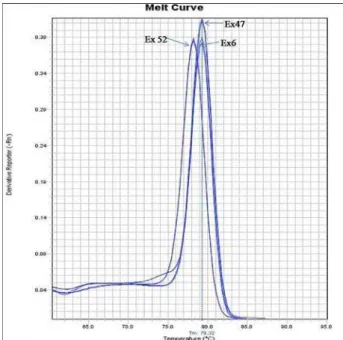
usually creates additional peaks separate from the speciic amplicon, dissociation curve analysis was performed for each ampliication reaction. Results (single PCR product without any ampliication of non-speciic products is seen as one single sharp peak on the melting curve or one single band on agarose gels) showed that no detectable non-specif-ic products were present in the reactions (Fig. 2). he duplicate runs of the exons of interest showed an almost complete overlap of parallel ampliication plots (for all samples, the SD of Ct value was very low range) (Fig. 3). In total, samples from 20 indi-viduals with known or unknown carrier status were tested using real-time PCR. he deletions of target exons were conirmed in 2 obligatory carriers and 8 out of 18 (44.44%) of suspected carriers. he gene dosage ratio (R) for non-carriers was 1.07 ± 0.20 and for carriers 0.56 ± 0.11. he ratio ranges be-tween carriers and non-carriers of the deletions did not overlap, allowing the accurate discrimination of deletion carriers and normal individuals.
DISCUSSION
Due to the lack of eicient rehabilitation and treat-ment of progressive muscular dystrophy, counseling and prenatal diagnosis are the only available options nowadays. However, molecular diagnosis and
car-rier analysis of DMD/BMD is still diicult due to the size of the dystrophin gene, mutation diversity and the presence of a normal allele in female carri-ers. he aim of this study was to develop a rapid and reliable real-time assay for direct DMD/BMD carrier detection that in the future may be used for routine diagnostics. We selected exons 6, 47 and 52 of the dystrophin gene since they are located in two mu-tational hotspots of this gene. he assay is based on SYBR® Green I chemistry. he possibility that SYBR® Green I dye generated false positive signals due to its binding to nonspeciic double stranded DNA was ruled out by performing melting curve analysis or gel electrophoresis of the PCR products. Using real-time PCR assay the carrier status of all subjects was suc-cessfully determined. he gene dosage ratio ranges did not overlap, allowing the accurate discrimination of carriers and non-carriers. However, we had access only to carriers of the deletions, so it has yet to be assessed whether the assay is able to determine the duplication carriers’ status.
In our study, we did not choose an endogenous control outside the dystrophin gene. Instead, one of the unafected dystrophin gene exons was used to quantify the other exon. his ofered the possibility of internal quality control in diagnostics ater the identiication of a deletion and a diferent combina-tion of test and reference exon for conirmacombina-tion of the results (Joncourt et al., 2004).
Multiplex end-point PCR approaches such as MAPH, MLPA and EPFA, can be efectively used for the rapid screening of deletions and duplications in the entire DMD coding region in both afected males and female carriers (Traverso et al., 2006). Once the mutation is known, real-time assay allows rapid identiication of carrier status in a single PCR within few hours. Furthermore, the results obtained by end-point PCR could be conirmed by using an independent set of primers. hus, a combination of these diferent assays may provide secure and cor-rect diagnosis of DMD/BMD.
SYBR® Green-based real-time PCR is highly sen-sitive, speciic and reproducible, requires a minimal Fig. 3 Ampliication curve analysis for DMD exons 6, 47 and
quantity of DNA and is carried out in a single step (Traverso et al., 2006). he simplicity of the protocol allows its easy implementation in diagnostic labora-tories. For routine application of this technique in the diagnosis of DMD/BMD it is necessary to devel-op additional assays for the analysis of other exons frequently involved in DMD rearrangements. Fur-thermore, it may be easily adapted for the screening of other genetic diseases that caused by deletions and duplications.
Acknowledgments - his work was supported by the Serbian
Ministry of Education and Science (grant 175091).
REFERENCES
Beggs, A.H., Koenig, M., Boyce, F.M., and L.M. Kunkel (1990).
Detection of 98% of DMD/BMD gene deletions by poly-merase chain reaction. Hum. Genet. 86, 45–48.
Chamberlain, J.S., Gibbs, R.A., Ranier, J.E., Nguyen, P.N., and C.T.
Caskey (1988). Deletion screening of the Duchenne
mus-cular dystrophy locus via multiplex DNA ampliication.
Nucleic Acids Res. 16, 141–156.
Clemens, P.R., Fenwick, R.G., Chamberlain J.S., Gibbs R.A., de
Andrade M., Chakrabarty R., and C.T. Caskey (1991).
Car-rier detection and prenatal diagnosis in Duchenne and Becker dystrophy families, using dinucleotide repeat poly-morphisms. Am. J. Hum. Genet. 49, 951–960.
Den Dunnen, JT., Grootscholten, PM., Bakker, E., Blonden, L.A., Ginjaar, H.B., Wapenaar, M.C., van Paassen, H.M., van
Broeckhoven, C., Pearson, P.L., and van G.J.Ommen (1989).
Topography of the DMD gene: FIGE and cDNA analysis of 194 cases reveals 115 deletions and 13 duplications. Am.
J. Hum. Genet. 45, 835-847
Gatta, V., Scarciolla, O., Gaspari, A.R., Palka, C., De Angelis, M.V., Di Muzio, A., Guanciali-Franchi, P., Calabrese, G., Uncini, A., and L. Stuppia (2005). Identiication of dele-tions and duplicadele-tions of the DMD gene in afected males and carrier females by multiple ligation probe ampliica-tion (MLPA). Hum. Genet.117, 92–98.
http://www.umd.be/DMD/W_DMD/index.html
Hu, X.Y., Burghes A.H., Ray, P.N., hompson, M.W., Murphy,
E.G., and R.G. Worton (1988). Partial gene duplication
in Duchenne and Becker muscular dystrophies. J. Med.
Genet.25, 369–376.
Hu, X.Y., Ray, P.N., Murphy, E.G., hompson, M.W., and R.G.
Worton, (1990). Duplication mutation at the Duchenne
muscular dystrophy locus: its frequency, distribution,
origin, and phenotypegenotype correlation. Am. J. Hum.
Genet. 46, 682–695.
Joncourt, F., Neuhaus, B., Jostarndt-Foegen, K., Kleinle, S., Steiner,
B., and S. Gallati (2004). Rapid identiication of female
carriers of DMD/BMD by quantitative real-time PCR.
Hum. Mutat. 23, 385-391.
Leiden muscular dystrophy pages (http://dmd.nl)
Ligon, A.H., Kashork C.D., Richards C.S., and L.G. Shafer (2000).
Identiication of female carriers for Duchenne and Becker muscular dystrophies using a FISH-based approach. Eur.
J. Hum. Genet. 8, 293–298.
Liu, J., Yan, M., Wang, Z., Wang, L., Zhou, Y., and B. Xiao (2006).
Molecular diagnosis of alpha-thalassemia by combining real-time PCR with SYBR® Green1 and dissociation curve analysis. Transl. Res. 148, 6–12.
Livak, K. J., and T.D. Schmittgen (2001). Analysis of Relative Gene Expression Data Using Real-Time Quantitative PCR and the 22ΔΔCt Method.Methods. 25,402–408.
Miller, S. A., Dykes, D. D., andH. F. Polesky (1988). A simple
salt-ing out procedure for extractsalt-ing DNA from human nucle-ated cells. Nucleic Acids Res. 16, 1215.
Nishino, I., and E. Ozawa (2002). Muscular dystrophies. Curr.
Opin. Neurol. 15, 539–544.
Nosaeid, M.H., Mahdian, R., Jamali, S., Maryami, F., Babashah,
S., Maryami, F., Karimipoor, M., and S. Zeinali (2009).
Validation and comparison of two quantitative real-time PCR assays for direct detection of DMD/BMD carriers.
Clin. Biochem. 42, 1291-1299.
Panigrahi, I., and B. Mittal (2001). Carrier detection and pre-natal diagnosis in Duchenne/Becker muscular dystrophy.
Indian Pediatr. 38, 631–639.
Prior, TW., and SJ. Bridgeman (2005). Experience and strategy
for the molecular testing of Duchenne muscular dystro-phy. J. Mol. Diagn. 7(3), 317–326.
Roberts, R.G., Barby, T.F., Manners, E., Bobrow, M., and D.R.
Bentley (1991). Direct detection of dystrophin gene
rear-rangements by analysis of dystrophin mRNA in peripheral blood lymphocytes. Am. J. Hum. Genet. 49, 298–310.
Ruiz-Ponte, C., Carracedo, A., and F. Barros (2006). Duplication
and deletion analysis by luorescent real-time PCR-based genotyping. Clin. Chim. Acta. 363, 138–146.
Traverso, M., Malnati, M., Minetti, C., Regis, S., Tedeschi, S.,
Pe-demonte, M., Bruno, C., Biassoni, R., and F.Zara (2006).
Multiplex real-time PCR for detection of deletions and duplications in dystrophin gene. Biochem. Biophys. Res.
Commun.339, 145–150.
M.H., and J.T. den Dunnen (2002). Comprehensive detec-tion of genomic duplicadetec-tions and deledetec-tions in the DMD gene, by use of multiplex ampliiable probe hybridization.
Am. J. Hum. Genet. 71, 365–374.
Wilhelm, J., and A. Pingoud (2003). Real-time polymerase chain
reaction. Chembiochem. 4, 1120-1128.
Worton, R.G., and M.W. hompson (1988). Genetics of