RIGID SPINE SYNDROME
CASE REPORT
VIVIANE H. FLUMIGNAN ZÉTOLA*, ROSANA HERMÍNIA SCOLA**, SALMO RASKIN***, DANIEL MONTE SERRAT PREVENDELLO****,
YLMAR CORREA NETO*, LINEU CESAR WERNECK*****
ABSTRACT - We describe a patient who had difficulty in walking since toddling stage and presented proximal upper and lower member weakness which have evolved to a progressive limitation of neck and trunk flexure, compatible with rigid spine syndrome. The serum muscle enzymes were somewhat elevated and the electromyography showed a myopatic change. The muscle biopsy demonstrated an active and chronic myopathy. The DNA analysis through PCR did not display any abnormality for dystrophin gene. The dystrophin by immnofluorescence was present in all fibers, but some interruptions were found in the plasma membrane giving it the appearance of a rosary. The test for merosin was normal.
KEY WORDS: rigid spine syndrome, merosin, dystrophin.
Síndrome da espinha rígida: relato de caso
RESUMO - Relatamos o caso de um paciente com dificuldade de marcha desde o início da deambulação, com fraqueza proximal de membros, evoluindo com limitação progressiva da flexão do pescoço e tronco, compatível com a síndrome da espinha rígida. As enzimas musculares séricas estavam moderadamente elevadas e a eletromiografia revelou padrão miopático. A biópsia muscular indicou miopatia crônica e ativa. A análise do DNA por PCR não demonstrou alterações no gene da distrofina. A imunofluorescência para distrofina foi positiva em todas as fibras, apresentando interrupções na membrana plasmática, semelhante a um rosário e o teste para merosina mostrou positividade.
PALAVRAS-CHAVE: síndrome da espinha rígida, merosina, distrofina.
First described in 1965 by Dubowitz, the rigid spine syndrome consists of proximal muscular
member weakness as its main symptom; at that time he denominated it “pseudodystrophy” 1. During
the evolution of the disease, five years after its onset, was noted a progressive limitation of trunk and neck flexure and was redefined as “rigid spine syndrome”. Even though other cases have been described, the diagnostic criteria have not yet been established2-12.
With the purpose of contributing to the elucidation of this entity, we relate a case presenting rigid spine syndrome in which we have the opportunity of studing the dystrophin and merosin by immunofluorescence and the dystrophin gene through polymerase chain reaction (PCR).
Division of Neurology and Neuromuscular Disorders, Internal Medicine Department, Hospital de Clínicas da Universidade Federal do Paraná and Genetika, Curitiba, Brazil: *Neurologist, **Assistant Professor, ***Geneticist, ****Medical Student, *****Professor. Aceite: 4-setembro-1998.
CASE REPORT
WPRO, a six years-old male, has walked with great effort and presented frequent falls since toddling stage (1 year and 7 months) tending to tiptoe gait. As the disease developed, he had difficulties in standing up and getting out of a chair. He only climbs stairs with the help of a rail. Gestation and familial history presented no particularities.
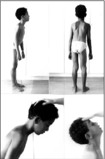
Physical examination: Blood pressure, pulse, cardiac frequency, respiratory frequency and temperature were normal. There was a lordosis (swayback) with thoracic and cervical column flexure limitations, but with preserved cervical extension. The paraspinal muscles had a retraction, mainly at neck and trunk level and he was not being able to turn his trunk. Also he had a bilateral winged scapula and retraction of the Achilles tendon (Fig 1).
Neurological examination: Normal cognitive functions. Cranial nerves were normal. Muscular strength was degree 4+ (MRCM) in the proximal muscles and degree 5 in distal muscles of the upper and lower limbs. There was a slight atrophy of lower limbs. The superficial cutaneous and deep tendon reflexes were absent, except for the Achilles reflex which was normal. Tactile, pain, vibration and position sense were normal, as well as the coordination. He had a myopathic gait and partial Gower’s maneuver.
Table 1. Electromyography.
Muscles (right) biceps quadriceps tibialis
brachii femoris anterior
Insertion activity N N N
Fibrillation potentials A A A
Fasciculation potentials A A A
Positive sharp waves A A A
Myotonic discharges A A A
Duration of motor unit potentials D D D
Amplitude of motor unit potentials D N D
Short polyphasic motor units ++ + ++
Long polyphasic motor units A A A
Recruitment pattern I I I
N, normal; A, absent; D, decreased; I, increased; rare, +; occasional, ++.
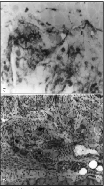
Fig 2. Muscle biopsy. A: Necrotic fibers (magnification x695; hematoxylin and eosin). B. Marked atrophy of the type I and II fibers (magnification x87; ATPase 9.4).
A
Fig 2. Muscle biopsy. C. Increased activity in the interstice and positive fibers (magnification x87; alkaline phosphatase). D. Connective tissue proliferation and excessive internal nuclei (magnification x174; hematoxylin and eosin).
D
C
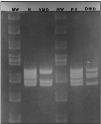
Fig 3. PCR analysis of the dystrophin gene: comparison with Duchenne muscular dystrophy (MW, molecular weight; N, normal; DMD, Du-chenne muscular distrophy; RS, rigid spine).
and amino terminal, as well as to the “rod” segment, demonstrated multiple small and large defects in several sarcolemmas of muscle fibers, with appearance of a rosary. The merosin was studied also with immunofluorescence technique and was normal. The dystrophin gene analysis was done by using DNA extracted from a muscular fiber sample amplified by a PCR technique that analysed 32 exons. No mutation, deletion or duplication were present (Fig 3).
DISCUSSION
The rigid spine syndrome predominates in males and the child can have a history of delayed
development or present the initial symptoms around 7 years of age1-4,7,11. Some reported cases have
recessive autosomal inheritance1-4,7,19. There is proximal muscular weakness of the trunk and neck
with posterior flexure limitation accompanied by progressive scoliosis (crookedness) 10. Reflexes in
general are diminished. Elbow and knee joint contraction were reported in many cases. The disease evolution is slow, without muscular strength reduction. Due to an alteration of the vertebral spine and restrictive respiratory failure, many cases ended in cor pulmonale and other cardiac alterations11.
The serum muscular enzymes are slightly elevated5-7,11. The peripheral nerve conduction velocities
frequently are normal2,4-6. The electromyography shows potentials of short duration and normal
amplitudes or polyphasic potentials 1,2,7. The muscle biopsy reveals nonspecific alterations of the
myopathic type, with fibers in degeneration and regeneration, perimysial and endomysial connective
tissue proliferation, type I fiber disproportion or hypotrophy 4, atrophy and hypertrophy of type II
fiber 5,7. The differences in muscle histology are either most likely due to the fact that the biopsy has
Table 2. Histological alterations in muscle biopsy.
Alteration Abnormality intensity
Connective tissue proliferation +++
Variation of muscular fibers diameter +++
Atrophic rounded fibers dispersed ++
Hypertrophic fibers dispersed ++
Internal nuclei ++
Necrosis ++
Phagocytosis ++
Splitting fibers ++
Basophilic fibers ++
Hipercontractile fibers +
Type I fiber predominate +
Type I fiber hypertrophy +
Type II fiber hypertrophy +
Type I fiber atrophy +
Type II fiber atrophy +
NADH-TR motheaten fibers +++
NADH-TR targets +
NADH-cores (central cores) +
Non-specific esterase increased activity interstice (macrophages) +
Acid phosphatase - focal fibers activity ++
Acid phosphatase - positive fibers ++
Acid phosphatase - increased in mononuclear cells (macrophages) +
Alkaline phosphatase - positive fibers +++
Alkaline phosphatase - increased activity interstice ++
frequent +++ ; occasional ++ ; rare +
initiating their pathological process, which developed asynchronously in different muscles because
of their different activity 12. Ultramicroscopic studies showed the presence of autophagic vacuoles
with membranous tangles with degenerative products in the muscular fiber without specificity 4,5,7,11.
The differential diagnosis includes the muscular dystrophies10 (Duchenne, Becker and
Emery-Dreifuss); congenital myopathies 4,9(central core disease, centro-nuclear myopathy, mitochondrial
and multicore myopathy), nemaline myopathy 13 and ankylosing spondylitis 7.
The reason for the involvement, mainly of paraspinal muscles, is not well known9. It is
suggested that the increase of connective tissue in extensor muscles would result in their shortening which would inhibit the spine flexor muscles. Another possibility could lie on the weakness of the flexor muscles rather than the extensor muscles. The flexors would therefore be unable to resist the stronger group.
Our patient presents symptoms that are in accordance with literature, as well as the histological
and histochemical alterations of muscle biopsies recorded by other authors 4,5,7. The alterations are
similar to the one observed in congenital muscular dystrophy 14. The abnormal immunocytochemistry
for dystrophin in this case is also similar to cases previously described in congenital muscular
dystrophy7. The DNA analysis for the dystrophin gene in this patient did not demonstrate any
alterations, suggesting that the genetic abnormality is not related to the dystrophin gene15-17. However
in some cases of congenital muscular dystrophy is found a merosin (laminin-2 composed of α
2-ß1-γ1 chains) deficiency 18 and one report call the attention for a reduction in the expression of the
laminin ß1 in the early onset autosomal dominant myopathy with rigidity of the spine, but this could
The ‘rigid spine syndrome’ still holds much heterogeneity and efforts are necessary to solve the nosological problem 4,11,20. Future descriptions might add data, which will allow a better recognition
of this syndrome.
REFERENCES
1. Dubowitz V, Brooke MH. Muscle biopsy: a modern approach. London: W.B. Saunders, 1973: 368-371. 2. Seay AR, Ziter FA, Petaja JH. Rigid spine syndrome: a type I fiber myopathy. Arch Neurol 1977;34:119-122. 3. Goto I, Nagasaka S, Nagara H, Kuroiwa Y. Rigid spine syndrome. J Neurol Neurosurg Psychiatry 1979;42:276-279. 4. Bertini E, Marini R, Sabetta G. The spectrum of the so-called rigid spine syndrome: nosological considerations and report
of three female cases. J Neurol 1986;233:248-253.
5. Goebel HH, Lenard HG, Görke W, Kunze K. Fibber type disproportion in the rigid spine syndrome. Neuropädiatrie 1977;8:467-477.
6. Dunn HG. The rigid spine syndrome (Abstract). Can J Neurol Sci 1976, 3:155.
7. Echenne B, Astruc J, Brunel D. Congenital muscular dystrophy and rigid spine syndrome. Neuropediatrics 1983;14:97-101. 8. Merlini L, Granata C, Ballestrozzi A, Marini ML. Rigid spine syndrome and rigid spine sign in myopathies. J Child Neurol
1989;4:274-282.
9. Vanneste JAL, Augustijn PB, Stam FC. The rigid spine syndrome in two sisters. J Neurol Neurosurg Psychiatry 1988;51:131-135.
10. Goto I, Ishimoto S, Yamada T, Hara H, Kuroiwa Y. The rigid spine syndrome and Emery-Dreifuss muscular dystrophy. Clin Neurol Neurosurg 1986;88:293-298.
11. Lotz BP, Stübgen JP. The rigid spine syndrome: a vacuolar variant. Muscle Nerve 1993; 16:530-536.
12. Di Iorio G, Lus G, Cutillo C, Cecio A, Cotrufo R. Histopathological heterogeneity and cytopathological similarity of findings in different muscles of two brothers affected by rigid spine syndrome. J Neurol Sci 1989;94:107-114. 13. Topaloglu H, Gögüs S, Yalaz K, Kücükali T, Serdaroglu A. Two siblings with nemaline myopathy presenting with rigid
spine syndrome. Neuromusc Disord 1994;4:263-267.
14. Werneck LC, Bonilla E. Immunohystochemical alterations of dystrophin in congenital muscular dystrophy. Arq Neuropsiquiatr 1985;53:60-68.
15. Chamberlain JS, Gibbs RA, Ranier JE, Nguyen PN, Caskey CT. PCR for diagnosis of Duchenne muscular dystrophy. In Innis M, Gelfand D, Sninski J, White T (eds.). PCR Protocols and applications - a laboratory manual. New York: Academic Press 1989:272-281.
16. Beggs AH, Koenig M, Boyce FM, Kunkel LM. PCR primers for the dystrophin gene that complement existing ones to detect over 98% of DMD/BMD deletions. Hum Genet 1990;86:45-48.
17. Covone AE, Caroli F, Romeo G. Screening Duchenne and Becker muscular dystrophy patients for deletions in 30 exons of the dystrophyn gene by three-multiplex PCR (Letter). Am J Hum Genet. 1992;51:675-677.
18. Werneck LC, Scola RH, Iwamoto FM. Congenital muscular dystrophy and merosin deficiency. Arq Neuropsiquiat 1997;55:780-787.
19. Taylor J, Muntoni F, Robb S, Dubowitz V, Sewry C. Early onset autosomal dominant myopathy with rigidity of the spine: a possible role for laminin ß1? Neuromusc Disord 1997;7:211-216.