Relationship of
Leishmania
-specific IgG levels and IgG avidity with
parasite density and clinical signs in canine leishmaniasis
Rafael Gonc
¸
alves Teixeira Neto
a,b, Rodolfo Cordeiro Giunchetti
a, Cla´udia Martins Carneiro
a,
Ricardo Wagner de Almeida Vitor
c, Wendel Coura-Vital
a, Patrı´cia Fla´via Quaresma
b,
Henrique Gama Ker
a, Lutiana Amaral de Melo
b, Ce´lia Maria Ferreira Gontijo
b,1,
Alexandre Barbosa Reis
a,d,1,*
aLaborato´rio de Imunopatologia, Nu´cleo de Pesquisas em Cieˆncias Biolo´gicas, Universidade Federal de Ouro Preto, Ouro Preto, Minas Gerais, Brazil bLaborato´rio de Leishmanioses, Instituto Rene´ Rachou, Fundac¸a˜o Oswaldo Cruz, Belo Horizonte, Minas Gerais, Brazil
cDepartamento de Parasitologia, Instituto de Cieˆncias Biolo´gicas, Universidade Federal de Minas Gerais, Belo Horizonte, Minas Gerais, Brazil dLaborato´rio de Imunologia Celular e Molecular, Instituto Rene´ Rachou, Fundac¸a˜o Oswaldo Cruz, Belo Horizonte, Minas Gerais, Brazil
1. Introduction
Visceral leishmaniasis (VL) is the most severe clinical
form of Leishmania chagasi (syn. L. infantum) infection,
giving rise to some 500,000 new cases and 59,000 deaths each year according to data published by the World Health
Organisation (WHO, 2005). The disease is endemic in 87
countries, although 90% of notified cases occur in India,
Sudan, Bangladesh, Nepal and Brazil (WHO, 2005), and its
epidemiological spectrum is broad, extending from tropi-cal and subtropitropi-cal areas through to temperate regions of
the globe (Deane and Deane, 1962; Alvar et al., 2004;
Desjeux, 2004).
Dogs represent important elements in the transmission
of VL and constitute the main domestic reservoirs of L.
chagasi (Deane and Deane, 1954; Molina et al., 1994;
Giunchetti et al., 2006). Indeed, the major focal areas of
human VL are strongly associated with locations that
exhibit a high prevalence of seropositive dogs (Oliveira
A R T I C L E I N F O
Article history:
Received 19 October 2009
Received in revised form 14 January 2010 Accepted 14 January 2010
Keywords:
Canine visceral leishmaniasis Parasite burden
Immunoglobulin avidity IgG profile
A B S T R A C T
The clinical status and tissue parasite burden of the skin and spleen of 40 dogs naturally infected withLeishmania chagasi(syn.Leishmania infantum), together with 5 uninfected control dogs, were assessed. On the basis of the clinical evaluation, infected dogs were classified as asymptomatic (AD) or symptomatic (SD). Infected animals were also grouped according to their parasite load as exhibiting low (LP), medium (MP) and high (HP) parasitism. The results indicated a high parasite load in the skin samples of SD animals in relation to the AD group. The serum immunoglobin isotype profiles of the studied animals revealed increased levels of IgG1in the AD and LP dogs, whereas high levels of IgG2were correlated with SD and HP dogs. The avidity index (AI) of IgGtotalin the SD group was high in comparison of that of the AD group. Moreover, animals with a larger parasite burden either in the spleen or skin showed higher AI values than animals with lower parasitism. Based on these findings, it is suggested that CVL commences with an asymptomatic clinical form with low parasitism, high production of IgG1and low affinity of IgGtotalmolecules, and evolves into a symptomatic clinical form with higher parasitism intensity, higher IgG2 levels, and high affinity of IgGtotal.
ß2010 Elsevier B.V. All rights reserved.
* Corresponding author at: Laborato´rio de Imunopatologia, Nu´cleo de Pesquisas em Cieˆncias Biolo´gicas, Universidade Federal de Ouro Preto, Ouro Preto, Minas Gerais, Brazil. Tel.: +55 31 3559 1694;
fax: +55 31 3559 1680.
E-mail address:alexreis@nupeb.ufop.br(A.B. Reis).
1 These authors have contributed equally to this work.
Contents lists available atScienceDirect
Veterinary Parasitology
j o u r n a l h o m e p a g e : w w w . e l s e v i e r . c o m / l o c a t e / v e t p a r
et al., 2001). In this context, even asymptomatic animals present intense cutaneous parasitism that facilitates the infection of insect vectors. From an epidemiological point of view, therefore, canine visceral leishmaniasis (CVL) is more important than the human form of the disease since efficient control of the former reduces considerably the number of reservoirs and, consequently, the rate of
infection in humans (Ashford, 1996).
Much research effort has been devoted to the elucida-tion of the factors responsible for the resistance or
susceptibility of dogs to L. chagasi infection. Thus, the
natural history of CVL has been well described, particularly with respect to parasite load in different tissues and to the immunopathological changes relating to the progression of
clinical forms of the disease (Reis et al., 2006a,b,c, 2009;
Day, 2007).
In an early report,Keenan et al. (1984)predicted that
the presence of anti-Leishmaniaspecific antibodies would
not be sufficient to confer protection against the disease, although such defence would not be possible in the absence of antibodies. Latter studies have concentrated on correlations between the various classes and sub-classes of immunoglobulin (Ig) and the response pro-duced by the host during the infection process
(Bourdoiseau et al., 1997; Solano-Gallego et al., 2001;
Cordeiro-da-Silva et al., 2003; Quinnell et al., 2003;
Almeida et al., 2005; Reis et al., 2006a). For example,
Deplazes et al. (1995)observed that symptomatic dogs
presented high levels of IgG1, whereas IgG2 was more
abundant in asymptomatic animals. More recent
inves-tigations have demonstrated that IgGtotalconcentrations
were greater in dogs that presented clinical symptoms of
CVL (Solano-Gallego et al., 2001; Almeida et al., 2005;
Iniesta et al., 2005; Reis et al., 2006a). In addition, it has
been reported that the presence of high amounts of anti-Leishmania specific IgG in dogs were correlated with
lower specific immune cells (Fernandez-Perez et al.,
2003), and that levels of IgE were directly correlated with
susceptibility toL. chagasiinfection (Almeida et al., 2005;
Iniesta et al., 2005; Reis et al., 2006a). Such findings have
led to the suggestion that the Ig profile could be used as a biomarker for monitoring resistance or susceptibility to the clinical manifestations of CVL. Additionally, some researchers have described the occurrence in Brazil and Spain of symptomatic dogs presenting elevated
levels of IgM (Rodriguez-Corte´s et al., 2007), although
evidence indicating a lack of correlation between clinical
status and IgM levels has also been reported (Reis et al.,
2006a).
In fact,Quinnell et al. (2003)had already anticipated
that IgG subclasses alone might not constitute satisfactory markers of susceptibility and resistance to CVL. In consideration of this, a new approach was proposed by
Reis et al. (2006a) involving study of the possible
association between parasite load and the clinical/immune response, thus establishing a new strategy for the elucidation of immunopathological aspects of CVL. Within this context, the determination of the IgG avidity index (AI) may contribute to the understanding of CVL. Moreover,
according toRedhu et al. (2006), the degree of avidity of
IgG towards theLeishmaniaantigen is able to predict the
time of infection in VL-affected patients, thus allowing the
discrimination between recent (<6 months) and chronic
(>6 months) patients.
The objective of the present work was to correlate the clinical forms of CVL (asymptomatic and symptomatic)
and the parasite load with IgG subclasses (IgG1and IgG2).
Moreover, the IgG avidity index has been correlated for the first time with the clinical forms of CVL, and possible associations with parasite load in the skin and spleen evaluated. The resulting data open up new prospects for the clinical prognosis of CVL and evaluation of efficacy of vaccines and drugs in the future.
2. Materials and methods
2.1. Animals
The study population consisted of 45 mongrel dogs (Canis familiaris) being 21 male and 24 female of various
ages. Forty of the dogs were naturally infected withL. chagasi
and originated from the Centre for Zoonosis Control, Belo Horizonte, MG, Brazil. The infected animals were subdivided
on the basis of the classification ofMancianti et al. (1988)
into two groups, one of which (n= 20) comprised
asympto-matic dogs (AD) (9 male and 11 female) whilst the other
(n= 20) constituted symptomatic dogs (SD) (10 male and 10
female). The remaining five animals (control group) had been bred and raised in the kennels of the Universidade Federal de Ouro Preto, MG, Brazil, and were considered non-infected even though the area is endemic for CVL. All procedures in this study were according to the guidelines set by the Brazilian Animal Experimental College (COBEA). This study was approved by the ethical Committee for the use of Experimental Animals of the Federal University of Ouro Preto, Minas Gerais state, Brazil (CETEA).
2.2. Confirmation of L. chagasi seropositivity and infection
The presence of anti-L. chagasi antibodies in the
animal population was confirmed by indirect immuno-fluorescence assay (IIFA) and enzyme linked immunosor-bent assay (ELISA), which were performed by the laboratory
of the Centre for Zoonosis Control. Infection byLeishmania
was confirmed by PCR at least one of the tissues evaluated (skin, lymph node, spleen, bone marrow and
liver) using the primers 50(C/G)(C/G)(G/C)CC(C/A)CTAT(T/
A)TTACACCAACCCC30and 50GGGGAGGGGCGTTCTGCGAA30
(Degrave et al., 1994). The determination of theLeishmania
species responsible for infection was performed using a
PCR-RFLP method (Volpini et al., 2004) with L. chagasistrain
MHOM/BR/74/PP75 as control.
2.3. Collection and analysis of samples
Ten milliliters of peripheral blood samples were collected by intravenous puncture in the radial vein of the dogs using disposable 20 mL syringes (21G 25 X 8) and placed into vacuum vials containing clot activator (Vacu-ette, Campinas, SP, Brazil). The resulting serum was stored in 1.8 mL sterile cryogenic vials (Sarstedt, Newton, NC,
USA) at 208C until required for assay.
After euthanasia, skin sample were collected by 5 mm punch biopsy from the right ear. Spleen biopsies, weighing approximately 30 mg, were collected using a sterile scalpel. Tissue fragments were placed onto microscope slides and stained with Giemsa for parasitological diag-nosis. Parasite loads, expressed in Leishman-donovan units (LDU), were determined by counting the number of amastigote forms amongst 1000 nucleated cells according
to the method ofStauber (1955)as modified byReis et al.
(2006a). Following the evaluation of tissue samples, dogs
were grouped according to parasite load as follows: low (LP), medium (MP) and high (HP) parasitism based on spleen and skin specific LDU values categorised
statisti-cally into tertiles according to Reis et al. (2006a) and
Guerra et al. (2008).
2.4. Determination of Ig pattern using ELISA
Anti-Leishmania Ig patterns were determined on the
basis of in-house ELISA tests carried out using soluble promastigote antigen (SLA) obtained according to the
method ofReis et al. (2006b)fromL. chagasi(MHOM/BR/
1972/BH46) grown in axenic culture on LIT medium.
Ninety-six-well microplates (MaxiSorpTM, Nalge Nunc Int.,
Rochester, NY, USA) were coated overnight at 48C with SLA
at a concentration of 10 mg/well. The coated wells were washed and serum samples added at 1:80 dilution. Following further washes, peroxidase-conjugated goat
anti-dog IgG1 (1:1000 dilution), together with sheep
anti-dog IgG (1:8000 dilution) and IgG2 (1:16,000
dilu-tion), all anti-heavy chain specific and purchased from Bethyl Laboratories, Montgomery, TX, USA. Wells were subsequently washed, substrate and chromogen (O-phenylenediamine; Sigma–Aldrich Co., St. Louis, MO, USA) added and absorbances read on an automatic ELISA microplate reader (Multiskan1 MCC 340, Labsystems, Helsinki, Finland) at 492 nm. The conjugate concentrations were determined by a block titration method with positive and negative standard sera.
2.5. IgG avidity test
A modified IgG avidity assays were performed using the
ELISA protocol as described in Section 2.4. except that
wells forming columns 1–6 (left-hand side of the micro-plate) received an extra washing with PBS/Tween follow-ing application of serum samples, whilst wells formfollow-ing columns 7–12 (right-hand side of the microplate) were
washed with 100
mL PBS/Tween and 6 M urea. The plate
was incubated at 378C for 10 min and absorbances read at
490 nm. Similar techniques have been applied in the study
of a number of other parasitic diseases (Suarez-Aranda
et al., 2000; Montoya et al., 2004; Redhu et al., 2006;
Clementino et al., 2007). AI values were determined from
the ratios between the absorbances of urea-treated samples and those of non-treated samples, and the results expressed in percentage form.
Owing to the lack of information regarding IgG avidity inLeishmania-infected dogs, it was necessary to establish an IgG affinity profile during the development of infection in order to obtain appropriate parameters for comparison.
Thus, serum samples from dogs that had been
experi-mentally infected withL. chagasistrain MHOM/BR/1972/
BH46 for between 1 and 19 months were submitted to IgG avidity assay as described above.
2.6. Statistical analyses
Statistic analyses were performed with the aid of Prisma
4.0 and Minitab 13 statistical software. The Studentt-test
or the non-parametric Mann–Whitney test were used to compare parasite loads between the different clinical groups and IgG AI values between groups exhibiting different clinical signs. Analysis of variance (ANOVA) followed by the multiple comparison Tukey test, or the non-parametric Kruskal–Wallis test followed by the multi-ple comparison Dunn test, were used to compare ELISA absorbance values with the clinical profiles of infected dogs. Similar methods were used to compare the ELISA and AI values with the parasite loads of infected dogs. Correlations were established using the Spearman rank test. All tests were carried out at the 95% confidence level.
3. Results
3.1. Clinical and molecular characteristics of the studied dogs
Uninfected control dogs and infected animals of the AD
group presented no clinical signs ofLeishmaniainfection. In
contrast, the SD group exhibited various signs of CVL
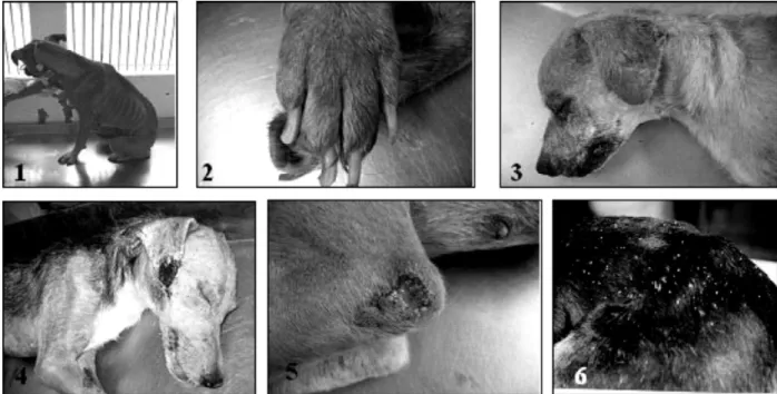
(Fig. 1) such as localized alopecia and onycogryphosis
(present in 70% of dogs), accentuated wasting (50%), furfuraceous dermatitis (35%), skin ulcers (30%), slight wasting, moderate wasting, dull coat (25%), keratocon-junctivitis (20%), generalised alopecia and opaque corneas (15%). All 40 animals forming the infected groups AD and SD were seropositive for CVL as demonstrated by IIFA and ELISA tests. PCR and PCR/RFLP patterns obtained from
samples derived fromLeishmania-infected animals were
similar to those produced byL. chagasistrain MHOM/BR/
74/PP75.
3.2. Parasite loads of skin and spleen tissues
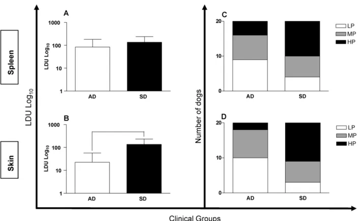
There were no differences between groups AD and SD
regarding the parasite load of the spleen (Fig. 2A), although
the LDU values for the skin of SD animals were greater than
those of dogs in the AD group (Fig. 2B). Animals in the AD
and SD groups were further stratified according to parasite
loads in the studied tissues as represented inFig. 2C and D.
The majority of AD animals were classified as LP for skin
(n= 10) and spleen (n= 9), whereas most of the dogs within
the SD group were classified as HP for skin (n= 11) and
spleen (n= 10). However the differences were significant
(
r
= 0.006) only in skin samples.3.3. IgG profiles
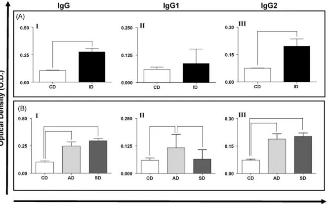
The absorbance values of IgGtotaland IgG2in the serum
of the whole population ofLeishmania-infected dogs were
significantly (
r
<0.05) greater than those of the controlspecifically, the levels of IgGtotal within the AD and SD
groups were significantly larger (
r
<0.05 and 0.001,respectively) than that of the control group (Fig. 3B, panel
I), and the level of IgG1 within the AD group was
significantly larger (
r
<0.05) than those of the SD andcontrol groups (Fig. 3B, panel II). Additionally, the levels of
IgG2 were significantly higher in AD (
r
<0.01) and SD(
r
<0.001) group animals compared with the controlgroup (Fig. 3B, panel III).
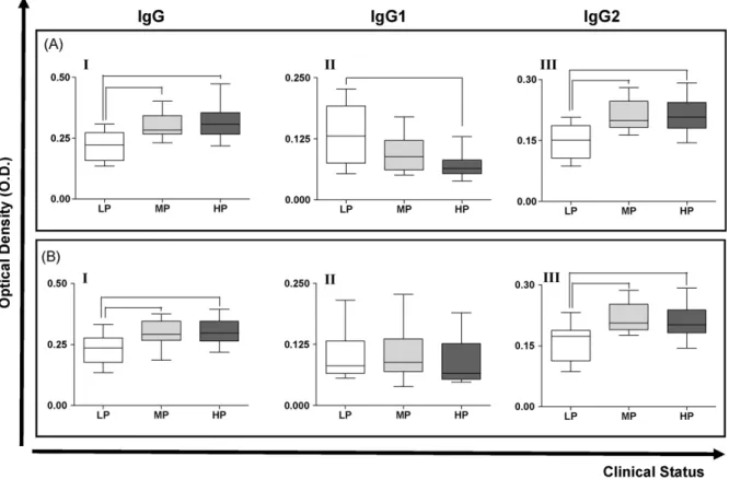
The reactivities of IgG, IgG1and IgG2in serum samples
were also appraised with respect to the parasite loads of skin and spleen tissue samples. Considering spleen
samples (Fig. 4A), the reactivity of serum IgGtotal was
significantly lower (
r
<0.05) in LP dogs compared with MPand HP animals (Fig. 4A, panel I), whilst IgG1reactivity was
significantly smaller (
r
= 0.0232) in HP animals comparedwith LP animals (Fig. 4A, panel II). The results for IgG2were
similar to those observed for IgGtotal, i.e. the reactivity in LP
dogs was smaller than in MP and HP animals (Fig. 4A, panel
III) and the differences were statistically significant
(
r
<0.05 and 0.01, respectively). In the case of skinsamples (Fig. 4B), LP dogs presented significantly lower
levels of IgGtotal and IgG2 compared with MP and HP
animals (
r
<0.05 and 0.01, respectively), but there wereno differences between LP, MP and HP animals regarding the values of IgG1.
3.4. Correlations between IgG reactivities and parasite load (LDU value)
Considering the total population ofLeishmania-infected
dogs, there were weak, but significant, correlations
between IgGtotal(
r
= 0.0164/r= 0.3684), IgG1(r
= 0.0261/r= 0.3563) and IgG2 (
r
= 0.0025/r= 0.4594), and theparasite load of the spleen (Fig. 5A–C). However, when
the different clinical forms of CVL were taken into account, strong positive correlations between the reactivities of
IgGtotal (
r
= 0.0002/r= 0.7404) and IgG2 (r
<0.0001/r= 0.7791) with spleen LDU values were found within
the AD group (Fig. 5D and E).
No correlations were detected between the reactivities of IgG forms and the parasite load of skin samples within
the population of symptomaticLeishmania-infected dogs.
3.5. IgG avidities
The IgG avidity profile determined for dogs
experi-mentally infected withL. chagasirevealed that AI values
tended to increase with the duration of infection. With
respect to the studied population ofLeishmania-infected
dogs, those of the AD group presented significantly smaller
(
r
= 0.008) AI values in comparison with SD group animals(Fig. 6A). When animals were categorised with respect to
parasite load of the spleen, AI values were found to be
significantly lower (
r
<0.01) in LP animals than in MP andHP animals (Fig. 6B). In the case of parasite load of the skin,
it was observed that the AI values in the HP group were
significantly higher (
r
<0.01) than those in the LP group(Fig. 6C).
In order to determine which of the IgG subclasses were associated with AI, the correlations between AI
values and the absorbance values of IgG1 and IgG2
were determined. The results revealed that AI was
strongly correlated (
r
<0.0001/r= 0.7315) with IgG2levels (Fig. 6E) but not with IgG1 levels (
r
= 0.8356/r= 0.03387;Fig. 6D).
Fig. 1.Main clinical signs observed in symptomatic dogs affected by canine visceral leishmaniasis showing: 1. wasting and apathy; 2. onycogryphosis; 3. alopecia/hypotrichosis in the periorbital region and ear tip; 4. lesion in the ear tip; 5. ulcer in the left hock area; 6. furfuraceous dermatitis in the dorsal area.
Fig. 2.Association between the parasite loads of the spleen (A) and skin (B) and the clinical forms of canine visceral leishmaniasis, and their distribution according to the parasite load in the spleen (C;p= 0.102) and skin (D;p= 0.006). AD = asymptomatic dogs, SD = symptomatic dogs, LP = low parasitism, MP = medium parasitism and HP = high parasitism. Bars representing values that are significantly different one from another are shown connected.
R.G.T.
Neto
et
al.
/Veterinary
Parasitology
169
(2010)
4. Discussion
CVL is a multisystemic disease with chronic evolution that mainly affects visceral and cutaneous tissue. The development of the various clinical forms of CVL depends upon complex interactions between the parasite and the immune system of the host. This means that some infected animals are susceptible and develop an active form of the infection, whereas others are resistant and remain asymptomatic. In addition, the nutritional status of the dog may directly influence the development of the disease. It is of interest to note that, following a study carried out in
a region that was endemic for CVL, Cabral et al. (1998)
reported that a large proportion (60–80%) of dogs presenting specific antibody and immune cell responses
showed no signs ofLeishmaniainfection.
The categorization of animals according to clinical manifestations and parasite load has been very helpful in understanding the immunopathological aspects of the disease. Such an approach has been used in the present investigation, in which the animal population was divided
into AD and SD groups as suggested byMancianti et al.
(1988). The high incidence (85%) of cutaneous alterations,
such as alopecia and dermatitis, observed in SD dogs agreed with those given in earlier reports (i.e. 81–89%;
Koutinas et al., 1999; Ciaramella et al., 1997), whilst the
frequencies of ocular alterations, such as keratoconjuncti-vitis (20%) and opaque corneas (15%), also corresponded
with published values (i.e. 24%; Pen˜a et al., 2000). In
contrast, however, the incidence of onycogryphosis (70%) amongst SD dogs was much higher in the present study
than has been observed earlier (i.e. 20–30%;Koutinas et al.,
1999; Baneth et al., 2008).
Various sophisticated techniques based on LDU values
(Reis et al., 2006a), quantitative nucleic acid
sequence-based amplification (QT-NASBA; Van Der Meide et al.,
2005), immunohistochemistry (Tafuri et al., 2004;
Giunchetti et al., 2006) and real time PCR (qPCR;Rola˜o
et al., 2004; Vitale et al., 2004) have been applied to the
quantitative evaluation of tissue parasitism in animals affected by CVL. Since skin and spleen appear to be the
tissues most densely parasitised byLeishmania
indepen-dent of the clinical forms of the disease (Reis et al., 2006a),
the present study attempted to correlate the humoral immune response of infected animals with clinical signs and the parasite density of these tissues. One of the most interesting findings was the lower parasite load of AD dogs
compared with SD animals (Fig. 2B), suggesting that
cutaneous parasitism evolves concomitantly with the
clinical manifestations of CVL. Indeed,Molina (1997)has
reported that, whilst AD animals presented positive xenodiagnosis, the proportions of infected phlebotomines were highest in those that had fed from dogs with severe signs of CVL, probably due to the intense parasitism
in such animals. Furthermore, according to
Rodriguez-Corte´s et al. (2007), the clinical profiles of experimentally
infected dogs were directly correlated with the degree
of parasitism. In contrast, Solano-Gallego et al. (2004)
Fig. 3.IgGtotal (I), IgG1 (II) and IgG2 (III) reactivities in (A) the serum of dogs naturally infected withL. chagasiand (B) distributed according to clinical form. CD = control dogs, ID = infected dogs, AD = asymptomatic dogs and SD = symptomatic dogs. Bars representing values that are significantly different one from another are shown connected.
Fig. 4.IgGtotal, IgG1 and IgG2 reactivities in the serum of dogs naturally infected withL. chagasicategorised according to the parasite load of (A) the spleen, and (B) the skin. LP = low parasitism, MP = medium parasitism and HP = high parasitism. Bars representing values that are significantly different one from another are shown connected.
observed that the numbers of parasites in the skin of symptomatic animals were no larger than those in asymptomatic animals, hence emphasising the impor-tance of AD animals in the maintenance and transmission of CVL.
In respect of the potential prognostic value of IgG
isotypes, some authors (Deplazes et al., 1995; Iniesta
et al., 2005) have demonstrated that high levels of IgG1
were correlated with symptomatic infection, whereas
high levels of IgG2were related with asymptomatic CVL.
These authors concluded that such a pattern could represent a dichotomous response of CVL. Conflicting results have, however, been reported by other
research-ers (Nieto et al., 1999; Boceta et al., 2000; Almeida et al.,
2005; Reis et al., 2006a; Cardoso et al., 2007) in which
high levels of IgG2 were associated with active clinical
infection. Such inconsistent results highlight the diffi-culty in establishing a typical immunological response in CVL and emphasize the importance of continued investigation.
The IgG1 levels of the AD animals presently studied
were found to be higher in comparison with those of the SD
group, in agreement with the findings ofBourdoiseau et al.
(1997)andReis et al. (2006a), thus suggesting a possible
association with mechanisms of immunoprotection
against CVL. Conversely, serum IgGtotaland IgG2appeared
to be associated with CVL morbidity since higher levels were found in animals presenting severe clinical forms. On
the other hand,Solano-Gallego et al. (2001)claimed, on the
basis of a cross-sectional study involving 280 animals in different stages of infection, that there was no correlation between clinical status and IgG subclass. Furthermore,
Quinnell et al. (2003)suggested that IgG subclasses were
not satisfactory markers of resistance or susceptibility towards CVL.
In the present study, IgG profiles were evaluated in
relation to LDU values revealing that increased IgGtotaland
IgG2reactivities were associated with the enhancement of
parasite load in the skin and spleen. Although the
correlations between IgGtotal and IgG2and parasite load
Fig. 6.IgG avidity index (AI) in the serum of dogs naturally infected withL. chagasistratified according to clinical form (A), and the degrees of parasitism in the spleen (B) and skin (C). Correlations between the IgG AI and the absorbances of IgG1 and IgG2 are shown in panels D and E, respectively. AD = asymptomatic dogs, SD = symptomatic dogs, LP = low Parasitism, MP = medium parasitism and HP = high parasitism. Bars representing values that are significantly different one from another are shown connected.
were positive, there was a negative correlation with IgG1, strengthening the hypothesis that this IgG is associated
with immunoprotection mechanisms during L. chagasi
infection. Indeed, animals with high concentrations of IgG1 were asymptomatic and presented low parasitism,
whereas animals with high levels of IgG2were associated
with more severe clinical signs and high parasitism, hence with greater susceptibility to infection. These results
support previous claims (Reis et al., 2006a) that some
serum anti-LeishmaniaIgGs could be of value as markers of
clinical status and parasite load of tissues of naturally infected dogs, thus representing a good indicator of disease
morbidity. On the other hand,Nieto et al. (1999)stated
that the presence of anti-Leishmaniaantibodies cannot be
considered as conclusive markers of CVL progression. IgG avidity tests were established in the present study with a view to expanding the debate over the role of IgG subtypes in CVL. Similar techniques have been applied in
the study of a number of other parasitic diseases (Montoya
et al., 2004; Clementino et al., 2007). In the case of infection
byToxoplasma gondii,Suarez-Aranda et al. (2000)
demon-strated that AI values >50% indicated chronic
toxoplas-mosis whilst AI values<50% suggested an acute form of the
disease. One of the few studies that have employed AI in the differentiation of chronic from acute VL was conducted
byRedhu et al. (2006), and these authors demonstrated
that the IgG avidity test could be employed to estimate the
time of development ofLeishmaniainfection in the host.
Results from the present study demonstrate that SD animals exhibit higher AI values compared with the AD group, and that animals showing elevated LDU values in the skin and spleen also present high AI values compared with those exhibiting low parasitism. Moreover, dogs with
high IgG2reactivity also presented high AI values. These
findings open up the possibility of applying AI values in the characterisation of CVL-affected dogs, although the limit-ing values of the index that differentiate recent and old forms of infection have not yet been fully established.
The results presented in this study support the idea that L. chagasi-infected dogs exhibit immunological character-istics that suggest a gradual evolution of CVL. Initially the disease is characterised by an absence of clinical signs (asymptomatic form), but a variety of symptoms (oligo-symptomatic form) emerge during CVL progression, and finally numerous clinical signs appear simultaneously that are characteristic of the classical symptomatic form of the disease, which probably leads to death.
In the present study, the IgG avidity test has been used for the first time in the diagnosis of CVL and shows promise as a biomarker for determining the progression of Leishmaniainfection. It has been demonstrated that the combined analysis of the various aspects connected with CVL enables the various clinical forms of the disease to be characterised. Thus, AD animals presented lower tissue parasitism (mainly in the skin), low affinity for IgG and a humoral immunoresponse with high levels of IgG1, all of which characterise the initial phase of infection. On the other hand, SD animals exhibited greater tissue parasitism, particularly in the skin, high affinity for IgG, and a humoral immunoresponse with high levels of IgG2, characterising the chronic phase of infection. It is thus concluded that
determination of AI values could provide an alternative tool with which to evaluate the morbidity of CVL, thus increasing the accuracy of prognosis for the animal.
Acknowledgments
The study was supported by the Fundac¸a˜o de Amparo a`
Pesquisa do Estado de Minas Gerais, Brazil (PRONEX 2007). CMC, RWAV, CMFG and ABR thank CNPq for fellowships. The authors wish to express their appreciation of the hard
work carried out by the staff of the Fundac¸a˜o Nacional da
Sau´de during the execution of this project. The authors are also grateful for the use of facilities at CEBIO, Universidade Federal de Minas Gerais, Centro de Pesquisas Rene´ Rachou (Fiocruz, Minas), Prefeitura Municipal de Belo Horizonte and Rede Mineira de Bioterismo (FAPEMIG), and for support with the provision of experimental animals.
References
Almeida, M.A.O., Jesus, E.E.V., Sousa-Atta, M.L.B., Alves, L.C., Berne, M.E.A., Atta, A.M., 2005. Antileishmanial antibody profile in dogs naturally infected withLeishmania chagasi. Vet. Immunol. Immunopathol. 106, 151–158.
Alvar, J., Canavate, C., Molina, R., Moreno, J., Nieto, J., 2004. Canine leishmaniasis. Adv. Parasitol. 57, 1–88.
Ashford, R.W., 1996. Leishmaniasis reservoirs and their significance in control. Clin. Dermatol. 14, 523–532.
Baneth, G., Koutinas, A.F., Solano-Gallego, L., Bourdeau, P., Ferrer, L., 2008. Canine leishmaniasis—new concepts and insights on an expanding zoonosis: part one. Trends Parasitol. 24, 324–330.
Boceta, C., Alonso, C., Jimenez-Ruiz, A., 2000. Leucine rich repeats are the main epitopes inLeishmania infantumPSA during canine and human visceral leishmaniasis. Parasite Immunol. 22, 55–62.
Bourdoiseau, G., Bonnefont, C., Hoareau, E., Boheringer, C., Stolle, T., Chabanne, L., 1997. Specific IgG1and IgG2antibody and lymphocyte
subset levels in naturallyLeishmania infantum-infected treated and untreated dogs. Vet. Immunol. Immunopathol. 145, 171–176. Cabral, M., O’Grady, J.E., Gomes, S., Souza, J.C., Thompson, H., Alexander, J.,
1998. The immunology of canine leishmaniasis: strong evidence for a developing disease spectrum from asymptomatic dogs. Vet. Parasitol. 76, 173–180.
Cardoso, L., Schallig, H.D.F.H., Cordeiro-da-Silva, A., Cabral, M., Alunda, J.M., Rodrigues, M., 2007. Anti-Leishmania humoral and cellular immune responses in naturally infected symptomatic and asympto-matic dogs. Vet. Immunol. Immunopathol. 117, 35–41.
Ciaramella, P., Oliva, G., Luna, R.D., Gradoni, L., Ambrosio, R., Cortese, L., Scalone, A., Persechino, A., 1997. A retrospective clinical study of canine leishmaniasis in 150 dogs naturally infected byLeishmania infantum. Vet. Rec. 141, 539–543.
Clementino, M.M., Souza, M.F., Andrade-Neto, V.F., 2007. Seroprevalence andToxoplasma gondii—IgG avidity in sheep from Lajes, Brazil. Vet. Parasitol. 146, 199–302.
Cordeiro-da-Silva, A., Cardoso, L., Arau´jo, N., Castro, H., Toma´s, A., Rodri-gues, M., Cabral, M., Vergnes, B., Sereno, D., Ouaissi, A., 2003. Identi-fication of antibodies toLeishmaniasilent information regulatory 2 (SIR2) protein homologue during canine natural infections: patholo-gical implications. Immunol. Lett. 68, 155–162.
Day, M.J., 2007. Immunoglobulin G subclass distribution in canine leish-maniosis: a review and analysis of pitfalls in interpretation. Vet. Parasitol. 147, 2–8.
Deane, M.P., Deane, L.M., 1954. Infecc¸a˜o experimental doPhebotomus longipalpisem raposas (Lycalopex ventulus) naturalmente infectadas pelaLeishmania donovani. O Hospital 46, 651–653.
Deane, L.M., Deane, M.P., 1962. Leishmaniases in Brazil: Geographical distribution and transmission. Rev. Inst. Med. Trop. Sa˜o Paulo 4, 198– 212.
Degrave, W., Fernandes, O., Campbell, D., Bozza, M., Lopes, U., 1994. Use of molecular probes and PCR for detection and typing ofLeishmania—a mini review. Mem. Inst. Oswaldo Cruz 89, 463–469.
Deplazes, P., Smith, N.C., Arnold, P., Lutz, H., Ecker, J., 1995. Specific IgG1
and IgG2antibody response of dogs toLeishmania infantumand other
Desjeux, P., 2004. Leishmaniasis: current situation and new perspectives. Comp. Immunol. Microbiol. Infect. Dis. 27, 305–318.
Fernandez-Perez, F.J., Gomez-Munoz, T., Mendez, S., Alunda, J.M., 2003. Leishmania-specific lymphoproliferative response and IgG1/IgG2
immunodetection patterns by western blot in asymptomatic, symp-tomatic and treated dogs. Acta Trop. 86, 83–91.
Giunchetti, R.C., Mayrink, W., Genaro, O., Carneiro, C.M., Correˆa-Oliveira, R., Martins-Filho, O.A., Marques, M.J., Tafuri, W.L., Reis, A.B., 2006. Relationship between canine visceral leishmaniasis and the Leishma-nia (LeishmaLeishma-nia) chagasiburden in dermal inflammatory foci. J. Comp. Pathol. 135, 100–107.
Guerra, L.L., Teixeira-Carvalho, A., Giunchetti, R.C., Martins-Filho, O.A., Reis, A.B., Correˆa-Oliveira, R., 2008. Evaluation of the influence of tissue parasite density on hematological and phenotypic cellular parameters of circulating leukocytes and splenocytes dur-ing ongodur-ing canine visceral leishmaniasis. Parasitol. Res. 104, 611– 622.
Iniesta, L., Ga´llego, M., Portu´s, M., 2005. Immunoglobulin G and E responses in various stages of canine leishmaniasis. Vet. Immunol. Immunopathol. 103, 77–81.
Keenan, C.M., Hendricks, L.D., Lightner, L., Johnson, A.J., 1984. Visceral leishmaniasis in the German Shepherd Dog - II. Vet. Pathol. 21, 80– 86.
Koutinas, A.F., Polizopoulou, Z.S., Saridomichelakis, M.N., Argyriadis, D., Fytianou, A., Plevraki, K.G., 1999. Clinical considerations on canine visceral leishmaniasis in Greece: a retrospective study of 158 cases (1989–1996). J. Am. Anim. Hosp. Assoc. 35, 376–383.
Mancianti, F., Gramiccia, M., Gradoni, L., Pieri, S., 1988. Studies on canine leishmaniasis control. I. Evolution of infection of different clinical forms of canine leishmaniasis following antimonial treatment. Trans. R. Soc. Trop. Med. Hyg. 82, 566–567.
Molina, R., 1997. Dogs infectivity and control. In: Workshop on New Trends in Leishmaniasis Epidemiology and control in the Mediterra-nean Area, Instituto Zooprofilattico Sperimentale della Sicilia/Medi-terranean Zoonoses Control Programme/World Health Organization, Palermo.
Molina, R., Amela, C., Nieto, J., San-Andres, M., Gonzales, F., Castillo, J.A., Lucientes, J., Alvar, J., 1994. Infectivity of dogs naturally infected with Leishmania infantumto colonizedPhlebotomus perniciosus. Trans. R. Soc. Trop. Med. Hyg. 88, 491–493.
Montoya, J.G., Huffman, H.B., Remington, J.S., 2004. Evaluation of the immunoglobulin G avidity test for diagnosis of toxoplasmic lympha-denopathy. J. Clin. Microbiol. 42, 4627–4631.
Nieto, C.G., Garcia-Alonso, M., Requena, J.M., Miro´n, C., Soto, M., Alonso, C., Navarrete, I., 1999. Analysis of the immune response against total and recombinant antigens ofLeishmania infantum: correlation with dis-ease progression in canine experimental leishmaniasis. Vet. Immunol. Immunopathol. 67, 117–130.
Oliveira, C.L., Assuncao, R.M., Reis, I.A., Proietti, F.A., 2001. Spatial dis-tribution of human and canine visceral leishmaniasis in Belo Hor-izonte, Minas Gerais State, Brasil, 1994–1997. Cad. Sau´de Pu´blica 17, 1231–1239.
Pen˜a, M.T., Roura, X., Davidson, M.G., 2000. Ocular and periocular man-ifestations of leishmaniasis in dogs: 105 cases (1993–1998). Vet. Ophthalmol. 3, 35–41.
Quinnell, R.J., Courtenay, O., Garcez, L.M., Kaye, P.M., Shaw, M.A., Dye, C., Day, M.J., 2003. IgG subclass responses in a longitudinal study of canine visceral leishmaniasis. Vet. Immunol. Immunopathol. 91, 161–168. Redhu, N.S., Dey, A., Balooni, V., Singh, S., 2006. Use of immunoglobulin G
avidity to determine the course of disease in visceral and post-Kala-Azar dermal leishmaniasis patients. Clin. Vaccine Immunol. 13, 969– 971.
Reis, A.B., Teixeira-Carvalho, A., Vale, A.M., Marques, M.J., Giunchetti, R.C., Mayrink, W., Guerra, L.L., Andrade, R.A., Correˆa-Oliveira, R., Martins-Filho, O.A., 2006a. Isotype patterns of immunoglobulins: Hallmarks
for clinical status and tissue parasite density in Brazilian dogs natu-rally infected byLeishmania (Leishmania) chagasi. Vet. Immunol. Immunopathol. 112, 1012–1116.
Reis, A.B., Martins-Filho, O.A., Teixeira-Carvalho, A., Carvalho, M.G., Mayr-ink, W., Franc¸a-Silva, J.C., Giunchetti, R.C., Genaro, O., Correˆa-Oliveira, R., 2006b. Parasite density and impaired biochemical/hematological status are associated with severe clinical aspects of canine visceral leishmaniasis. Res. Vet. Sci. 81, 68–75.
Reis, A.B., Teixeira-Carvalho, A., Giunchetti, R.C., Guerra, L.L., Carvalho, M.G., Mayrink, W., Genaro, O., Correˆa-Oliveira, R., Martins-Filho, O.A., 2006c. Phenotypic features of circulating leucocytes as immunologi-cal markers for cliniimmunologi-cal status and bone marrow parasite density in dogs naturally infected byLeishmania chagasi. Clin. Exp. Immunol. 146, 303–311.
Reis, A.B., Martins-Filho, O.A., Teixeira-Carvalho, A., Giunchetti, R.C., Carneiro, C.M., Mayrink, W., Tafuri, W.L., Correˆa-Oliveira, R., 2009. Systemic and compartmentalized immune response in canine visc-eral leishmaniasis. Vet. Immunol. Immunopathol. 128, 87–95. Rodriguez-Corte´s, A., Ojeda, A., Lo´pez-Fuertes, L., Timo´n, M., Altet, L.,
Solano-Gallego, L., Sanchez-Robert, E., Francino, O., Alberola, J., 2007. A long term experimental study of canine visceral leishmania-sis. Int. J. Parasitol. 37, 683–693.
Rola˜o, N., Cortes, S., Rodrigues, O.R., Campino, L., 2004. Quantification of Leishmania infantumparasites in tissue biopsies by real-time poly-merase chain reaction and polypoly-merase chain reaction-enzyme-linked immunosorbent assay. J. Parasitol. 90, 1150–1154.
Solano-Gallego, L., Riera, C., Roura, X., Iniesta, L., Gallego, M., Valladares, J.E., Fisa, R., Castillejo, S., Alberola, J., Ferrer, L., Arboix, M., Portu´s, M., 2001. Leishmania infantum specific IgG, IgG1 and IgG2 antibody
responses in healthy and ill dogs from endemic areas: evolution in the course of infection and after treatment. Vet. Parasitol. 96, 265– 276.
Solano-Gallego, L., Fernandez-Bellon, H., Morell, P., Fondevila, D., Alber-ola, J., Ramis, A., Ferrer, L., 2004. Histological and immunohistochem-ical study of clinimmunohistochem-ically normal skin ofLeishmania infantum—infected dogs. J. Comp. Pathol. 130, 7–12.
Stauber, L.A., 1955. Leishmaniasis in the hamster. In: Cole, W.H. (Ed.), Some Physiological Aspects and Consequence of Parasitism. Rugers University Press, New Brunswick, NJ, pp. 77–90.
Suarez-Aranda, F., Galisteo, A.J., Hiramoto, R.M., Cardoso, R.P.A., Meireles, L.R., Miguel, O., Andrade, H.F., 2000. The prevalence and avidity of Toxoplasma gondiiIgG antibodies in pigs from Brazil and Peru. Vet. Parasitol. 91, 23–32.
Tafuri, W.L., Santos, R.L., Arantes, R.M.E., Gonc¸alves, R., Melo, M.N., Michalick, M.S.M., Tafuri, W.L., 2004. An alternative immunohisto-chemical method for detecting Leishmania amastigotes in paraffin-embedded canine tissues. J. Immunol. Methods 292, 17–23.
Van Der Meide, W.F., Schoone, G.J., Faber, W.R., Zeegelaar, J.E., De Vries, H.J., Ozbel, Y., Lai, A., Fat, R.F., Coelho, L.I., Kassi, M., Schallig, H.D., 2005. Quantitative nucleic acid sequence-based assay as a new molecular tool for detection and quantification ofLeishmania parasites in skin biopsy samples. J. Clin. Microbiol. 43, 5560– 5566.
Vitale, F., Reale, S., Vitale, M., Petrotta, E., Torina, A., Caracappa, S., 2004. TaqMan-based detection of Leishmania infantum DNA using canine samples. Ann. N.Y. Acad. Sci. 1026, 139–143.
Volpini, A.C., Passos, V.M., Oliveira, G.C., Romana, A.J., 2004. PCR-RFLP to identifyLeishmania(Viannia)braziliensisandL.(Leishmania) amazo-nensis causing American cutaneous leishmaniasis. Acta Tropica 90, 31–37.
World Health Organization (WHO), 2005. Leishmaniasis Seventeenth Programme Report. Progress 2003–2004, Geneva. URL http:// www.who.int/tdr/publications/publications/pdf/pr17/leishmaniasis. pdf.