Acute Zonal Occult Outer Retinopathy in
Japanese Patients: Clinical Features, Visual
Function, and Factors Affecting Visual
Function
Saho Saito1, Wataru Saito1,2*, Michiyuki Saito1, Yuki Hashimoto1, Shohei Mori1, Kousuke Noda1,2, Kenichi Namba1, Susumu Ishida1,2
1Department of Ophthalmology, Hokkaido University Graduate School of Medicine, Sapporo, Hokkaido, Japan,2Department of Ocular Circulation and Metabolism, Hokkaido University Graduate School of Medicine, Sapporo, Hokkaido, Japan
Abstract
Purpose
To evaluate the clinical features and investigate their relationship with visual function in Jap-anese patients with acute zonal occult outer retinopathy (AZOOR).
Methods
Fifty-two eyes of 38 Japanese AZOOR patients (31 female and 7 male patients; mean age at first visit, 35.0 years; median follow-up duration, 31 months) were retrospectively collect-ed: 31 untreated eyes with good visual acuity and 21 systemic corticosteroid-treated eyes with progressive visual acuity loss. Variables affecting the logMAR values of best-corrected visual acuity (BCVA) and the mean deviation (MD) on Humphrey perimetry at initial and final visits were examined using multiple stepwise linear regression analysis.
Results
In untreated eyes, the mean MD at the final visit was significantly higher than that at the ini-tial visit (P= 0.00002). In corticosteroid-treated eyes, the logMAR BCVA and MD at the final visit were significantly better than the initial values (P= 0.007 andP= 0.02, respectively). The final logMAR BCVA was 0.0 or less in 85% of patients. Variables affecting initial visual function were moderate anterior vitreous cells, myopia severity, and a-wave amplitudes on electroretinography; factors affecting final visual function were the initial MD values, female sex, moderate anterior vitreous cells, and retinal atrophy.
Conclusions
Our data indicated that visual functions in enrolled patients significantly improved spontane-ously or after systemic corticosteroids therapy, suggesting that Japanese patients with OPEN ACCESS
Citation:Saito S, Saito W, Saito M, Hashimoto Y, Mori S, Noda K, et al. (2015) Acute Zonal Occult Outer Retinopathy in Japanese Patients: Clinical Features, Visual Function, and Factors Affecting Visual Function. PLoS ONE 10(4): e0125133. doi:10.1371/journal.pone.0125133
Academic Editor:Keisuke Mori, Saitama Medical University, JAPAN
Received:October 20, 2014
Accepted:March 11, 2015
Published:April 28, 2015
Copyright:© 2015 Saito et al. This is an open access article distributed under the terms of the Creative Commons Attribution License, which permits unrestricted use, distribution, and reproduction in any medium, provided the original author and source are credited.
Data Availability Statement:All relevant data are within the paper.
Funding:The authors have no support or funding to report.
AZOOR have good visual outcomes during the follow-up period of this study. Furthermore, initial visual field defects, gender, anterior vitreous cells, and retinal atrophy affected final vi-sual functions in these patients.
Introduction
Acute zonal occult outer retinopathy (AZOOR), first described by Gass in 1993, is an
idiopath-ic syndrome of acute outer retinal impairment [1]. AZOOR is characterized by funduscopically
normal or minimally abnormal retinal appearances at the early stage [1,2], which may
occa-sionally be followed by retinal pigment epithelium (RPE) degeneration in the affected area [2,3].
Visual field defects in AZOOR patients are caused by outer retinal impairment which is
demonstrated by impaired responses in multifocal electroretinography (ERG) [4] and a
dis-rupted ellipsoid zone (originally called the inner segment/outer segment junction) detected by
optical coherence tomography (OCT) [5,6], corresponding to AZOOR retinal lesions.
Associ-ated RPE damage can be more clearly visible by fundus autofluorescence (FAF) [3,7].
Regarding the pathogenesis, we recently demonstrated using laser speckle flowgraphy that the choroidal blood flow velocity at AZOOR lesions significantly increased as visual function
and outer retinal morphology improved [8]. Changes in the blood flow showed“inflammatory”
pattern in the choroid, similar to our previous findings in patients with choroiditis,
Vogt-Koya-nagi-Harada disease [9], and serpiginous choroiditis [10]. From these observations, we
hypoth-esized that inflammation in the choroid caused secondary photoreceptor impairment in AZOOR and may explain the absent or minimally abnormal retinal appearance during early stages of AZOOR, although an alternative explanation is considered as follows; the outer retina is primarily affected by inflammation and destruction of photoreceptor outer segments leads to
secondary reduction of the choroidal blood flow [11].
In 2002, Gass et al. reported the following clinical features of 51 primarily Caucasian AZOOR patients over more than three years of follow-up: young myopic women predomi-nated; 76% had bilateral involvement at the final visit; the recurrence rate was 31%; subsequent retinal atrophy was observed in 48% of patients; visual field impairment ceased in 78% of pa-tients 6 months after the initial visit, but partially improved in only 24% of papa-tients; and the logarithm of minimal angle of resolution (logMAR) value of the best-corrected visual acuity
(BCVA) at the final visit was 1.0 or more in 27% of patients [2]. These results showed that
AZOOR patients did not have a good visual prognosis. Few studies examining the clinical char-acteristics or prognostic factors in a large case series have since been reported because of the condition’s rarity.
Only several single case reports and a few case series with small patient populations have
de-scribed visual outcomes in Asian patients with AZOOR [12]. Among 13 Chinese AZOOR
pa-tients, the final logMAR BCVA was less than 0.35 in 57% of patients and more than 1.0 in only
one patient [13]. However, in a study examining six Japanese patients, the final logMAR
BCVA was 1.0 or more in 50% of the patients [14]. Apparently, AZOOR progression is variable
[12], and reports of patients with good visual outcomes improving spontaneously [8,13,15,16]
or showing poor visual prognosis [14,17] have been described. Thus, the visual prognosis in
Materials and Methods
Subjects
In total, 52 eyes of 38 patients diagnosed with AZOOR who visited the vitreo-retina clinic at Hokkaido University Hospital from 2002 to 2012 and were followed up for more than six months were enrolled and their records were retrospectively reviewed. The study was approved by the ethics committee of Hokkaido University Hospital (approval ID: 014–0042) and fol-lowed the tenets of the Declaration of Helsinki. Written informed consent was obtained from all subjects after the nature and possible consequences of the study had been explained.
AZOOR was diagnosed using the following criteria [2]: acute visual field or vision loss usually
with concurrent photopsia; one or more visual field defect regions that could not be explained by funduscopic examination or fluorescein angiography (FA); decreased multifocal ERG re-sponses corresponding to the retinal sites with visual field defects; and, beginning in 2007, outer retinal morphologic abnormalities including an absent or discontinuous ellipsoid zone
on OCT [5,6,18]. Patients who involved funduscopic and angiographic findings consistent
with white dots observed in AZOOR complex (e.g., multiple evanescent white dot syndrome) were excluded. Patients aged over 50 years or with medical history of uncontrolled systemic hy-pertension at disease onset were also excluded so that cancer-associated retinopathy or hyper-tensive chorioretinopathy could not be ruled out.
Treatment
Twenty-four patients (31 eyes) with no central visual acuity loss at the initial visit who did not progress clinically were not treated and were followed thereafter. Fourteen patients (21 eyes) with progressive central visual acuity loss were treated as follows: intravenous methyl-prednis-olone (PSL) at 1,000 mg/day for 3 days, oral PSL at 30 mg/day for 7 days, and intravenous methyl-PSL at 1,000 mg/day for 3 days. The PSL dose was then tapered as follows: 1 month at 30 mg/day, 1 month at 20 mg/day, 1 month at 15 mg/day, 1 month at 10 mg/day, and 1 month at 5 mg/day. In patients experiencing worsened visual field defects on perimetry or subjective symptoms at re-examination, the schedule was prolonged, or the dose was temporarily in-creased. Two patients experienced multiple recurrences when the oral PSL dose was decreased and were prescribed oral azathioprine at 50 mg/day in addition to corticosteroids.
Ophthalmologic Examinations
During the initial visit, all patients underwent thorough ophthalmic examinations including a decimal BCVA assessment with a Japanese standard Landolt visual acuity chart, indirect oph-thalmoscopy, FA, indocyanine green angiography (ICGA), OCT (OCT Ophthalmoscope C7, RS-3000, or RS-3000 Advance; Nidek, Gamagori, Japan), and 20J scotopic single-flash ERG, followed several days later by visual field testing (Goldmann perimetry and/or the Humphrey 30–2 Swedish interactive threshold algorism standard test) and multifocal ERG. Multifocal ERG was recorded with the Visual Evoked Response Imaging System (EDI, San Mateo, CA). The extent of anterior vitreous cells was quantified using the general grading score for aqueous
inflammatory cells [19] and was classified as mild (occasional~1+ cells), moderate (2~3+ cells),
anti-neutrophil cytoplasmic antibodies, angiotensin-converting enzyme, anti-thyroid peroxidase antibody, and anti-phospholipid antibody were performed as well. During follow-up, BCVA assessment, Humphrey perimetry, and OCT were performed to quantitatively evaluate the ac-tivity of AZOOR. Patients included in this study were classified into non-treated eyes and corti-costeroid-treated eyes, and BCVA and mean deviation (MD) values on Humphrey perimetry were statistically analyzed between the worst initial (before treatment) and the final values in both groups.
Statistics
The BCVA was converted to the logMAR scale for statistical analysis. The Wilcoxon signed-rank test was used to compare changes in the logMAR values of BCVA and MD values on Humphrey perimetry in eyes diagnosed with AZOOR. Simple linear regression analysis and a multiple stepwise linear regression analysis determined the independent variables affecting
log-MAR BCVA and MD values at the initial and final visits.Pvalues less than 0.05 were
consid-ered statistically significant in all analyses. A significant improvement or deterioration in MD values during follow-up was defined as MD value changes of 30% or more.
Results
Patient Demographics
The Japanese patient population comprised 7 male (18.4%) and 31 female (81.6%) patients. Bi-lateral involvement was diagnosed in 7 patients (18.4%) at the initial visit and in 14 patients (36.8%) at the last visit. The mean presumed age of AZOOR onset was 33.2 ± 8.7 years (range, 15–47 years), and the mean age at the initial visit was 35.0 ± 10.6 years (range, 15–61 years). Presumed duration from the onset to the initial visit ranged from 0 to 122 months (mean, 14.1 ± 30.1; median, 1 month). The follow-up duration ranged from 6 to 132 months (mean, 37.7 ± 31.3; median, 31 months).
Medical and Family History
Systemic diseases were noted in the medical history in 13 cases (34.2%). Pregnancy, Hashimoto disease, and hyperthyroidism were diagnosed in two cases each, and cervical cancer (after con-firming no metastasis), thyroid cancer (after surgical excision, no metastasis), well-controled schizophrenia, rheumatoid arthritis, suspected Sjögren’s syndrome, atopic dermatitis, and chronic pancreatitis (idiopathic, no medication) were diagnosed in one case each. Two eyes in a single patient have been administered topical latanoprost for ocular hypertension. Seven cases (18.4%) had one or more family members with Type 1 diabetes mellitus, retinitis pigmen-tosa, systemic lupus erythematosus, Behçet's disease, Basedow’s disease, or polygrandular autoimmune syndrome.
Subjective Symptoms
Refraction
Of the 52 eyes, 48 (92.3%) were diagnosed with myopia: less than -3D in 13 eyes (25.0%), -3D
to -5.75D in 11 eyes (21.2%), and greater than or equal to -6.0D in 24 eyes (46.1%) (Fig 1).
Hy-peropia or emmetropia was diagnosed in four eyes (7.7%).
Ophthalmic Examinations
Relative afferent pupillary defect was positive in 12 (23.5%) of 51 eyes examined at the initial visit. Slit-lamp examination did not reveal any cells in the anterior chamber of any eye and mild (14 eyes) or moderate (8 eyes) cells in the anterior vitreous in 22 (44.9%) of the 49 eyes ex-amined. Seven of 8 eyes with moderate anterior vitreous cells were administered systemic
corti-costeroid therapy. The retina was funduscopically normal in 43 eyes (82.7%) (Fig 2A), but
myopia-associated punctuate or patchy chorioretinal atrophic lesions at the posterior pole in four eyes, a few punctate chorioretinal scars outside the posterior retinal pole in two eyes, and a lacquer crack lesion in two eyes were found. A white line at the margin of the affected area was
Fig 1. Frequency of spherical equivalent in 52 eyes with acute zonal occult outer retinopathy (AZOOR).Myopia of -6.0D or over was the most frequent (46.1%).
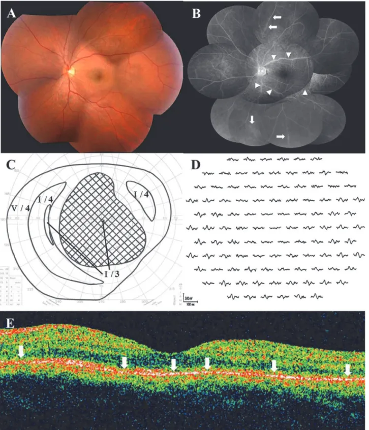
Fig 2. Photographs of the left eye at the initial visit in a 36-year-old male patient with AZOOR.(A) Fundus photograph shows normal appearance, except for retinal arterial narrowing. (B) Late-phase fluorescein angiography shows staining of the retinal vein walls (arrows) and leakages from the retinal vessels (arrowheads). (C) Goldmann perimetry reveals a central scotoma of 80 × 70°. LogMAR value of the best-corrected visual acuity (BCVA) decreased to 2.0. (D) Multifocal electroretinography (ERG) shows reduced responses corresponding to the visual field defect. (E) Horizontal optical coherence tomography through the fovea shows diffuse ellipsoid zone loss (arrows) in the macular area.
observed in one eye with a large scotoma; the line spontaneously disappeared. Myopic tempo-ral conus was found in 15 eyes, morning glory-like optic disc with no retinal schisis in one eye, and the sheathing, narrowing, and tortuosity of retinal arteries were observed in two eyes. Dur-ing follow-up examinations, five eyes (9.6%) developed an area of zonal retinal degeneration at
the RPE level with or without pigmentation, despite an initially normal retinal appearance (Fig
3A); however, none of the patients had an apparent demarcating line at the margin of the
retinal degeneration.
Fundus Angiography
Initially, FA showed no abnormalities in 28 (56.0%) of 50 eyes examined. The optic disc stain-ing and retinal vascular wall stainstain-ing with leakage were observed on the late phase in 15 eyes
(30.0%) and 13 eyes (26.0%), respectively (Fig 2B), and hyperfluorescent punctate lesions were
identified in 4 eyes (8.0%). Of the 44 eyes examined with ICGA, 10 eyes (22.7%) appeared nor-mal, but 29 eyes (65.9%) had a diffuse choroidal hyperfluorescence at the posterior pole to the mid-peripheral region during the middle phase. In addition, punctate-patchy hypofluorescence
at the posterior pole to mid-peripheral region was observed in 17 eyes (38.6%) (Fig 4B), and
the hyperfluorescence along with the choroidal middle or large vessels was seen in 6 eyes
(13.6%) (Fig 4B).
ERG Examination
In the 47 eyes examined, single-flash ERG showed a normal amplitude in 28 eyes (59.6%), duced a-wave amplitude in 7 eyes (14.9%), reduced b-wave amplitude in 5 eyes (10.6%), re-duced a- and b-wave amplitudes in 3 eyes (6.4%), and non-recordable responses in 4 eyes (8.5%). There were noticeably reduced multifocal ERG responses corresponding to visual field
loss in all of the eyes examined (Figs2Dand4D).
OCT Examination
Forty-four of the 48 eyes that underwent OCT had ellipsoid zone irregularities corresponding
to retinal sites with the visual field defect (Figs2Eand3C). Among the eyes examined with
SD-OCT, the interdigitation zone (originally called the cone outer segment tip line) was lost in
four eyes [18], despite a visually normal ellipsoid zone.
Systemic Screenings
Infectious disease tests showed normal results in all patients. Serum autoantibodies were
de-tected in 8 patients (21.1%) as follows: anti-nucleus antibody (≧160×) in 5 patients, high levels
of anti-thyroglobulin and/or thyroid peroxidase autoantibodies in 4 patients, rheumatoid fac-tor in 3 patients, and anti-SS-A antibody in 1 patient (there is some overlap). Imaging of the orbit and brain revealed no abnormal findings in all 16 patients examined.
Visual Acuity Changes
The logMAR BCVA (N= 52) at the initial visit or before treatment was 0.0 or less in 34 eyes
(65.3%),>0.0–<0.35 in 7 eyes (13.5%),≧0.35–<1.0 in 7 eyes (13.5%), and 1.0 or more in 4
eyes (7.7%) (Fig 5). The BCVA at the final visit was 0.0 or less in 44 eyes (84.7%),>0.0–<0.35
in 4 eyes (7.7%),≧0.35–<1.0 in 2 eyes (3.8%), and 1.0 or more in 2 eyes (3.8%). In all eyes
(N= 52), the mean BCVA logMAR values at the initial and final visits were 0.10 ± 0.42 and
-0.0035 ± 0.36, respectively. There was no significant difference between these phases
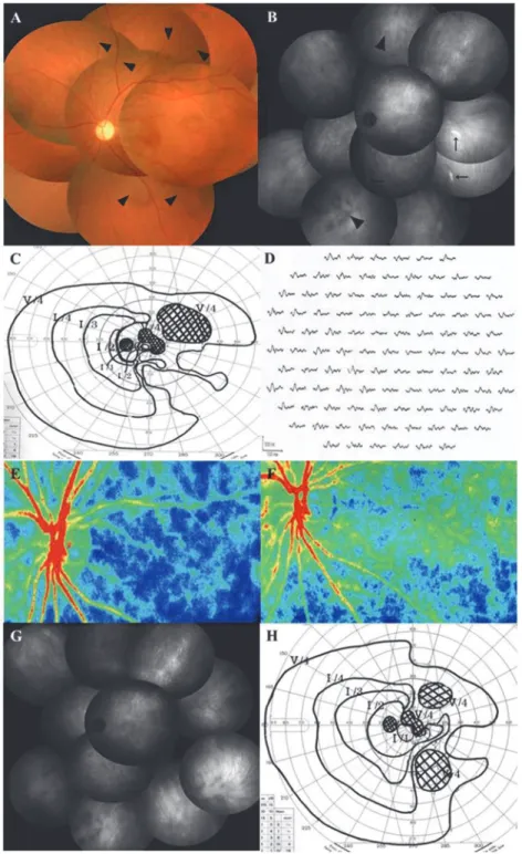
Fig 4. Photographs of the left eye before (A-E) and after (F-H) systemic corticosteroids therapy in a 39-year-old woman with AZOOR.(A) Fundus photograph shows normal retinal appearance except for narrowing and tortuosity of the retinal arteries (arrowheads). The patient’s logMAR BCVA was 0.52. (B) Indocyanine green angiography (ICGA) in the late phase shows hyperfluorescence along with choroidal middle or large vessels (arrows) and hypofluorescent patches (arrowheads). (C) Goldmann perimetry shows paracentral and isolated scotomata together with peripheral contraction. (D) Multifocal ERG reveals decreased responses corresponding to the visual field defects. (E, F) Composite color map of laser speckle flowgraphy. Mean blur rate (MBR), an index of relative blood flow velocity, increased three weeks after corticosteroid pulse therapy (F) compared to the pretreatment level (E), suggesting an improvement in choroidal circulation impairment after treatment. Warm color indicates high MBR and cool color low MBR. (G) On late-phase ICGA three weeks after treatment, hyperfluorecence along with choroidal vessels
(-0.10 ± 0.11) and final logMAR BCVA (-0.11 ± 0.07) (P= 1.0). In contrast, in the 21 eyes ad-ministered systemic corticosteroids, the final BCVA (0.16 ± 0.52) was significantly better than
the pre-treatment value (0.56 ± 0.60) (P= 0.007).
Perimetry
Goldmann perimetry at AZOOR onset revealed an enlarged blind spot often associated with vi-sual field defects of the other types in 26 eyes (50.0%), ring scotoma in 13 eyes (25.0%), arcuate scotoma in 4 eyes (7.6%), central scotoma (CS) in 3 eyes (5.8%), and other factors in 6 eyes
(11.5%) (Figs2C,4CandTable 1).
Of 32 AZOOR eyes that received Humphrey perimetry at 6 month after baseline, the 6-month MD values increased by 30% or more in 23 eyes (71.9%) and remained unchanged in
9 eyes (28.1%), as compared to the baseline MD values (Fig 6A). None of the eyes showed a
worsening of 30% or more in this short-term period. These 23 eyes achieving the early-phase improvement of visual field loss comprised 15 of 18 untreated eyes (83.3%) and 8 of 14 treated
Fig 5. BCVA changes in 52 eyes with AZOOR.The final logMAR BCVA was 0.0 or less in 84.7% and 1.0 or more in only 3.8%.
eyes (57.1%). The MD values at the final visit increased by 30% or more in 24 eyes (63.2%), re-mained unchanged in 13 eyes (34.2%), and decreased by 30% or more in 1 eye (2.6%) of all 38
eyes followed up using Humphrey perimetry (Fig 6B). In all eyes (N= 38), the mean MD at the
final visit (-5.30 ± 8.51 dB) was significantly higher than the baseline (-9.31 ± 8.39 dB)
(P= 0.000004). In the untreated eyes (N= 21), the mean MD at the final visit (-1.65 ± 2.85 dB)
was significantly higher than the baseline (-5.80 ± 4.32 dB) (P= 0.00002). In eyes administered
corticosteroids (N= 17), the mean MD at the final visit (-9.82 ± 10.74 dB) was significantly
higher than the initial value (-13.66 ± 10.73 dB) (P= 0.02).
Visual Function
As shown inTable 2, the initial visual function in AZOOR patients was positively correlated
with the ERG a-wave amplitude at the initial visit, but negatively with the presence of moderate anterior vitreous cell infiltration at the initial visit. Visual function at the final visit was posi-tively correlated with BCVA and MD at the initial visit and the ERG a-wave amplitude; it nega-tively correlated with moderate anterior vitreous cell infiltration, the presence of retinal
atrophy at the final visit, and the administration of systemic corticosteroid therapy (Table 3).
Multiple stepwise linear regression analysis revealed that moderate anterior vitreous cells, myo-pic severity, and a-wave amplitude significantly correlated with visual functions at the initial
visit (Table 2). On the other hand, the initial MD, female sex, moderate anterior vitreous cells,
and retinal atrophy significantly correlated with visual functions at the final visit (Table 3).
Recurrences and Ocular or Systemic Complications
AZOOR recurrence occurred in 9 eyes (17.3%) of 7 patients (18.4%) during follow-up. Six of these 7 patients had received systemic corticosteroid therapy. Four of the 6 treated patients ex-perienced recurrences when oral PSL was tapered to less than 15 mg/ day; 2 cases were subse-quently administered a second corticosteroid pulse therapy. The mean number of recurrences was 1.7 (range, 1–4), and the median duration from the onset to the recurrences was 6.5 months (3–52 months). Three patients were diagnosed with steroid-induced diabetic mellitus, and 1 patient experienced a pulmonary thrombosis and a lower extremity vein thrombosis. There were no ocular complications in the AZOOR eyes during follow-up. In 1 patient, idio-pathic choroidal neovascularization occurred in the contralateral eye during follow-up.
Table 1. Visual Field Findings at Onset of AZOOR in 52 Eyes with AZOOR.
Types of Scotomata Number of Eyes %
BSE+isolated scotoma 10 19.2
BSE+central scotoma 7 13.5
BSE 6 11.5
BSE+contraction 3 5.8
Ring scotoma 13 25.0
Central scotoma 3 5.8
Arcuate scotoma 2 3.8
Arcuate scotoma+PCS 2 3.8
The others 6 11.5
Total 52 100
AZOOR = acute zonal occult outer retinopathy
BSE = blind spot enlargement; PCS = paracentral scotoma
Discussion
In the present study, the clinical features and visual functional changes in a relatively large pop-ulation of Japanese AZOOR patients with a follow-up duration of more than 6 months were retrospectively examined. Eyes with good visual acuity at the initial visit spontaneously showed good visual outcomes with significantly improved MD values. Eyes that developed progressive visual deterioration also showed significantly improved visual function after receiving systemic
Fig 6. Changes in mean deviation (MD) values on Humphrey perimetry from baseline to 6 months (A) and the final visit (B).(A) MD values increased by 30% or more in 71.9% of all eyes (83.3% of untreated eyes and 57.1% of treated eyes). None of the eyes showed a worsening of 30% or more. (B) MD values increased by 30% or more in 63.2% of all eyes (76.2% of untreated eyes and 47.1% of treated eyes), but decreased by 30% or more in only 2.6% of all eyes.
doi:10.1371/journal.pone.0125133.g006
Table 2. Variables Affecting Visual Functions at Initial Visit in Patients with AZOOR.
Initial BCVA (logMAR) Initial MD
Simple linear regression analysis
Multiple linear regression analysis
Simple linear regression analysis
Multiple linear regression analysis
cor P value β P value cor P value β P value
Age 0.20 0.1450 - - -0.028 0.8664 -
-Sex -0.0057 0.9681 - - -0.27 0.1055 -
-Moderate ant.vit.cells 0.42 0.0024** - - -0.61 0.0001** -0.32 0.0216*
Degree of myopia -0.19 0.1886 - - -0.14 0.3981 -0.24 0.0338*
Retinal vasculitis 0.029 0.8430 -0.20 0.1270 -0.042 0.8030 0.17 0.1445
a-wave in ERG -0.60 6.9E-6** -0.65 1.0E-5** 0.73 8.5E-7** 0.67 1.7E-5**
BCVA = best-corrected visual acuity; MD = mean deviation; ant.vit.cells = anterior vitreous cells; ERG = electroretinography *P<0.05;
**P<0.01
corticosteroid therapy. The final logMAR BCVA was<0.35 in 92.4% and≧1.0 in 3.8% of
pa-tients. Retinal atrophy later developed in 9.6% of AZOOR eyes. A decreased MD at the initial visit, female sex, moderate anterior vitreous cells, and retinal atrophy were prognostic factors affecting visual function at the final visit. This is the first report that not only characterized the clinical features and visual function of Japanese AZOOR patients, but also elucidated the vari-ables affecting visual functions in AZOOR.
Patients with good visual function at the initial visit accounted for 60% of the study popula-tion, and their perimetry results significantly improved without treatment, with visual acuity remaining stable during follow-up. These results suggest that visual function improves sponta-neously in a certain percentage of Japanese AZOOR patients. Furthermore, in the remaining 40% of patients with progressive visual impairment, visual functions significantly improved after long-term high-dose systemic corticosteroid therapy. In a subset of these patients, chronic corticosteroid therapy was required because AZOOR recurred when the dose was decreased or the therapy was discontinued. It is difficult to determine the therapeutic efficacy of systemic corticosteroids in the enrolled patients because spontaneous visual improvement could not be ruled out in this retrospective study. However, recent reports found that AZOOR was
success-fully treated with immunosuppressive therapy including systemic corticosteroids [6,8,20] and
adalimumab [21]. Randomized prospective studies of a large patient population are needed to
clarify the efficacy of systemic corticosteroid therapy for AZOOR patients with progressive vision loss.
In this study, the ERG a-wave amplitudes at the initial visit were significantly correlated to the initial visual function. This result supports that AZOOR causes visual impairment by fecting the photoreceptor. Moreover, a moderate level of anterior vitreous cells negatively af-fected the initial visual function. Gass et al. reported that patients with anterior vitreous cells frequently have a high incidence of large visual field defect sites, subsequent retinal
degenera-tion, and a lack of visual improvement [2]. Our results seem to verify these observations by
Gass et al. and suggest that these patients had worse visual functions due to severe activity at the initial visit. Conversely, the presence of this finding positively affected the final visual
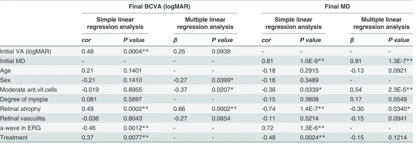
Table 3. Variables Affecting Visual Functions at Final Visit in Patients with AZOOR.
Final BCVA (logMAR) Final MD
Simple linear regression analysis Multiple linear regression analysis Simple linear regression analysis Multiple linear regression analysis
cor P value β P value cor P value β P value
Initial VA (logMAR) 0.48 0.0004** 0.25 0.0939 - - -
-Initial MD - - - - 0.81 1.0E-9** 0.91 1.3E-7**
Age 0.21 0.1401 - - -0.18 0.2915 -0.13 0.0921
Sex -0.21 0.1410 -0.27 0.0399* -0.16 0.3489 -
-Moderate ant.vit.cells -0.019 0.8955 -0.37 0.0207* -0.36 0.0339* 0.54 2.3E-5**
Degree of myopia 0.081 0.5697 - - -0.15 0.3808 0.17 0.0549
Retinal atrophy 0.49 0.0002** 0.66 0.0002** -0.74 1.4E-7** -0.30 0.0340*
Retinal vasculitis -0.036 0.8043 -0.27 0.0654 -0.11 0.5214 -0.15 0.0941
a-wave in ERG -0.46 0.0012** - - 0.72 1.3E-6** -
-Treatment 0.37 0.0077** - - -0.48 0.0024** -0.15 0.1214
BCVA = best-corrected visual acuity; MD = mean deviation; ant.vit.cells = anterior vitreous cells; ERG = electroretinography *P<0.05;
**P<0.01
functions. Interestingly, most of the eyes with moderate anterior vitreous cells received system-ic cortsystem-icosteroid therapy, leading to a signifsystem-icant improvement in visual functions. These seem-ingly contradictory results may possibly represent the efficacy of our systemic corticosteroid therapy regimen for AZOOR patients with severe activity.
Associated retinal atrophy also correlated with the severity of visual field impairment at the final visit. Gass et al. observed that visual acuity seemed to improve when the fundus
main-tained a normal appearance during follow-up [2]. Our result suggests that patients with this
finding involve permanent visual field disorders following irreversible damage to the outer reti-na due to having severe activity.
Clinical features of the present study examining Japanese patients and the previous study of
primarily Caucasian patients [2] were summarized inTable 4. There were no differences in the
age at the onset and the gender ratio between both studies. The frequency of myopia was higher in Japanese patients; however, this may have reflected the higher incidence of myopia in Japan than in the United States. Notably, Japanese patients had a lower rate of retinal atrophy devel-opment, fewer patients with poor visual outcomes at final visit, and more patients with good vi-sual outcomes at final visit, although the substantial gaps in the follow-up period (median: 31 months vs. 96 months) and the duration from the onset to the recurrence (median: 6.5 months
vs. 39 months) between this study and the previous report [2] may have influenced the
differ-ences in these clinical parameters. Nevertheless, the early-phase improvement (over 70%) of vi-sual field loss in our patients, whether treated or untreated, contrasted sharply with the tendency (roughly less than 30%) of short-term (within 6 months) amelioration of the visual
field shown in the previous report [2]. Thus, Japanese AZOOR patients appear to present good
visual functions at least in the relatively early course of the disease. Several mechanisms (i.e., ethnic difference, efficacy of systemic corticosteroids, and possibility of more frequent inclu-sion of milder cases in our study) would be speculated as the reason(s) of the differences ob-served. Further studies are needed to make a proper comparison of the clinical features and
Table 4. Clinical Features of the Present Study and a Previous Study.#
Variables Saito et al. Gass et al.
Patients Number 38 Patients (52 Eyes) 51 Patients (90 Eyes)
Race Japanese Caucasian
Follow-up duration (months)* 31 (6–132) 96 (36–420)
Age at onset (years)* 35 (15–47) 33 (13–63)
Female patients 31/38 (82%) 37/51 (73%)
Unilateral involvement at initial visit 31/38 (82%) 31/51 (61%) Unilateral involvement atfinal visit 24/38 (63%) 12/51 (24%)
Myopia 48/52 (92%) 59/90 (66%)
Photopsia 31/52 (60%) 69/90 (77%)
Recurrences 7/38 (18%) 16/51 (31%)
Normal or AZOOR-unrelated fundus at initial visit 49/52 (94%) 82/90 (91%)
Retinal atrophy atfinal visit 5/52 (10%) 43/90 (48%)
Final logMAR BCVA<0.35 48/52 (92%) 61/90 (68%)
Final logMAR BCVA≧1.0 2/52 (4%) 24/90 (27%)
#Gass JDM et al., Am J Ophthalmol 2002;
*Median (range);
BCVA = best-corrected visual acuity
visual prognosis between Japanese and Caucasian AZOOR patients by matching the follow-up duration and treatment regimens.
Conclusions
In the present study, approximately 60% of enrolled Japanese patients with AZOOR had good visual acuity at the initial visit and these most showed spontaneous regression. In patients experiencing progressive visual impairment, visual function significantly improved after sys-temic corticosteroid therapy. The final logMAR BCVA was 0.0 or less in 85% of patients. Initial MD value, gender, moderate anterior vitreous cells, and retinal atrophy were prognostic indica-tors of the final visual function. Thus, Japanese patients with AZOOR appear to have good vi-sual outcomes during the follow-up period of this study.
Author Contributions
Conceived and designed the experiments: SS WS MS SI. Performed the experiments: SS WS YH SM K. Noda K. Namba. Analyzed the data: SS WS MS YH SM K. Noda K. Namba SI. Wrote the paper: SS WS MS YH K. Noda SI. Statistical analysis: MS. Critical revision of the manuscript: WS SM K. Noda K. Namba SI.
References
1. Gass JD. Acute zonal occult outer retinopathy. Donders Lecture: The Netherlands Ophthalmological Society, Maastricht, Holland, June 19, 1992. J Clin Neuroophthalmol. 1993; 13: 79–97. PMID: 8340485
2. Gass JD, Agarwal A, Scott IU. Acute zonal occult outer retinopathy: a long-term follow-up study. Am J Ophthalmol. 2002; 134: 329–339. PMID:12208243
3. Fujiwara T, Imamura Y, Giovinazzo VJ, Spaide RF. Fundus autofluorescence and optical coherence to-mographic findings in acute zonal occult outer retinopathy. Retina. 2010; 30: 1206–1216. doi:10.1097/ IAE.0b013e3181e097f0PMID:20661173
4. Arai M, Naoi N, Sawada A, Hayashida T. Multifocal electroretinogram indicates visual field loss in acute zonal occult outer retinopathy. Am J Ophthalmol. 1998; 126: 466–469. PMID:9744389
5. Li D, Kishi S. Loss of photoreceptor outer segment in acute zonal occult outer retinopathy. Arch Ophthalmol. 2007; 125: 1194–1200. PMID:17846358
6. Spaide RF, Koizumi H, Freund KB. Photoreceptor outer segment abnormalities as a cause of blind spot enlargement in acute zonal occult outer retinopathy-complex diseases. Am J Ophthalmol. 2008; 146: 111–120. doi:10.1016/j.ajo.2008.02.027PMID:18439564
7. Mrejen S, Khan S, Gallego-Pinazo R, Jampol LM, Yannuzzi LA. Acute zonal occult outer retinopathy: a classification based on multimodal imaging. JAMA Ophthalmol. 2014; 132:1089–1098. doi:10.1001/ jamaophthalmol.2014.1683PMID:24945598
8. Saito M, Saito W, Hashimoto Y, Yoshizawa C, Shinmei Y, Noda K, et al. Correlation between de-creased choroidal blood flow velocity and the pathogenesis of acute zonal occult outer retinopathy. Clin Experiment Ophthalmol. 2014; 42: 139–150. doi:10.1111/ceo.12143PMID:23777505
9. Hirose S, Saito W, Yoshida K, Saito M, Dong Z, Namba K, et al. Elevated choroidal blood flow velocity during systemic corticosteroid therapy in Vogt-Koyanagi-Harada disease. Acta Ophthalmol. 2008; 86: 902–907. doi:10.1111/j.1755-3768.2008.01384.xPMID:19016661
10. Takahashi A, Saito W, Hashimoto Y, Saito M, Ishida S. Impaired circulation in the thickened choroid of a patient with serpiginous choroiditis. Ocul Immunol Inflamm. 2014; 22: 409–413. doi:10.3109/ 09273948.2014.902075PMID:24724811
11. Fagan XJ. A new insight into an old mystery. Clin Experiment Ophthalmol. 2014; 42: 103–104. doi:10. 1111/ceo.12295PMID:24606329
12. Monson DM, Smith JR. Acute zonal occult outer retinopathy. Surv Ophthalmol. 2011; 56: 23–35. doi: 10.1016/j.survophthal.2010.07.004PMID:21056448
14. Takai Y, Ishiko S, Kagokawa H, Fukui K, Takahashi A, Yoshida A. Morphological study of acute zonal occult outer retinopathy (AZOOR) by multiplanar optical coherence tomography. Acta Ophthalmol. 2009; 87: 408–418. doi:10.1111/j.1755-3768.2008.01269.xPMID:18778338
15. Chai Y, Yamazaki H, Fujinami K, Tsunoda K, Yamamoto S. Case of acute zonal occult outer retinopa-thy with abnormal pattern visual evoked potentials. Clin Ophthalmol. 2011; 5: 1235–1241. doi:10. 2147/OPTH.S23194PMID:21966193
16. So K, Shinoda K, Matsumoto CS, Satofuka S, Imamura Y, Mizota A. Focal functional and microstructur-al changes of photoreceptors in eyes with acute zonmicrostructur-al occult outer retinopathy. Case Rep Ophthmicrostructur-almol. 2011; 2: 307–313. doi:10.1159/000332734PMID:22125531
17. Saito A, Saito W, Furudate N, Ohno S. Indocyanine green angiography in a case of punctate inner chor-oidopathy associated with acute zonal occult outer retinopathy. Jpn J Ophthalmol. 2007; 51: 295–300. PMID:17660991
18. Tsunoda K, Fujinami K, Miyake Y. Selective abnormality of cone outer segment tip line in acute zonal occult outer retinopathy as observed by spectral-domain optical coherence tomography. Arch Ophthal-mol. 2011; 129: 1099–1101. doi:10.1001/archophthalmol.2011.217PMID:21825201
19. Hogan MJ, Kimura SJ, Thygeson P. Signs and symptoms of uveitis. I. Anterior uveitis. Am J Ophthal-mol. 1959; 47: 155–170. PMID:13649855
20. Kitakawa T, Hayashi T, Takashina H, Mitooka K, Gekka T, Tsuneoka H. Improvement of central visual function following steroid pulse therapy in acute zonal occult outer retinopathy. Doc Ophthalmol. 2012; 124: 249–254. doi:10.1007/s10633-012-9318-1PMID:22402912