323 Clinical findings in 16 patients with tomographic diagnosis of schizencephaly
Radiol Bras 2006;39(5):323–326 Original Article
CLINICAL FINDINGS IN 16 PATIENTS WITH TOMOGRAPHIC
DIAGNOSIS OF SCHIZENCEPHALY*
Maria do Carmo de Souza Rodrigues1
, Alexandra Maria Vieira Monteiro2
, Juan Clinton Llerena Junior3
, Alexandre Ribeiro Fernandes4
OBJECTIVE: To establish a correlation between clinical features in a group of children with tomographic diag-nosis of schizencephaly and clefts extent and localization. MATERIALS AND METHODS: Retrospective study of dossiers from the archives of Neurology and Medical Genetics Services at Instituto Fernandes Figueira/Fiocruz and Hospital Municipal Jesus, Rio de Janeiro, RJ, Brazil, in the period between 2000 and 2003. The study included 16 patients, nine female and seven male, with tomographic diagnosis of schizencephaly investigated for clinical findings, psychomotor development, motor/cognitive deficits and epilepsy. RESULTS: Predominance of bilateral clefts in 10:16 patients, open-lip schizencephaly type in 23:27 patients, and small lips in 11:27 patients. As regards anomalies associated with schizencephaly, pellucid septum absence was the most fre-quent one (10:16 patients). As regards clinical findings, 15 patients presented with developmental delay and motor deficit, six patients with cognitive deficit and ten with epilepsy. In three patients, we observed discor-dant clinical findings and cleft sizes, although the clefts were small, the clinical features severity was high because of other cerebral anomalies. CONCLUSION: The clinical features of schizencephaly are related to the size of the clefts, regardless laterality, presenting higher severity when associated with other cerebral anomalies.
Keywords: Schizencephaly; Computed tomography; Clinical findings.
Aspectos clínicos em 16 pacientes com diagnóstico tomográfico de esquizencefalia.
OBJETIVO: Correlacionar o quadro clínico de um grupo de crianças com diagnóstico tomográfico de esquizen-cefalia com a extensão e localização das fendas. MATERIAIS E MÉTODOS: Estudo retrospectivo de prontuá-rios do arquivo dos serviços de Neurologia e Genética Médica do Instituto Fernandes Figueira e Hospital Mu-nicipal Jesus, Rio de Janeiro, RJ, Brasil, no período de 2000 a 2003. Foram incluídos 16 pacientes, nove do sexo feminino e sete do sexo masculino, com diagnóstico tomográfico de esquizencefalia e analisados quanto a aspectos da tomografia computadorizada, desenvolvimento neuropsicomotor, déficit motor e cognitivo e epi-lepsia. RESULTADOS: Predominaram as fendas bilaterais em 10:16 pacientes, lábios abertos em 23:27 fendas e pequenas em 11:27 fendas. Das anomalias associadas à esquizencefalia, a ausência de septo pelúcido foi a mais freqüente (10:16 pacientes). Dos aspectos clínicos, 15 pacientes apresentaram atraso do desenvolvi-mento e déficit motor; seis apresentaram déficit cognitivo e dez apresentaram epilepsia. Em três pacientes observamos discordância entre o quadro clínico e o tamanho das fendas: embora as fendas fossem pequenas, o quadro clínico foi intenso, em virtude de presença de outras anomalias cerebrais. CONCLUSÃO: O quadro clínico guarda relação com o tamanho das fendas, independentemente da lateralidade, sendo mais intenso quando há associação com outras anomalias cerebrais.
Unitermos: Esquizencefalia; Tomografia computadorizada; Aspectos clínicos. Abstract
Resumo
* Study developed at Centro de Genética Médica Dr. José Carlos Cabral de Almeida/Instituto Fernandes Figueira/Fundação Oswaldo Cruz/RJ and with Program of Post-Graduation in Medi-cal Sciences of Universidade do Estado do Rio de Janeiro, Rio de Janeiro, RJ, Brazil. Funding Source: Faperj (E-26/171.077/ 2002-APQ1).
1. Master in Medicine – Program of Post-Graduation in Medi-cal Sciences at Universidade do Estado do Rio de Janeiro, MD, Geneticist at Hospital Universitário Cassiano Antonio Moraes – Universidade Federal do Espírito Santo.
2. Doctor in Medicine, Adjunct Professor of Radiology for Courses of Post-Graduation at Faculdade de Ciências Médicas da Universidade do Estado do Rio de Janeiro.
3. Doctor in Biological Sciences, MD, Clinical Geneticist, Chief for Centro de Genética Médica Dr. José Carlos Cabral de Almeida, Instituto Fernandes Figueira/Fundação Oswaldo Cruz.
4. Master in Child and Women’s Health, Professor of Pediat-rics at Universidade Gama Filho, MD, at Department of Neuropediatrics – Hospital Municipal Jesus.
Mailing address: Dra. Maria do Carmo de Souza Rodrigues. Rua Itaporanga, 26, Itaparica. Vila Velha, ES, Brazil 29102-270. E-mail: [email protected]
Received November 1, 2005. Accepted after revision November 16, 2005.
INTRODUCTION
Schizencephaly(1) is an extremely rare
congenital disorder characterized by a full-thickness cleft within the cerebral hemi-spheres, delimited by an abnormal cor-tex(2–4), extending from the ventricular
surface to the arachnoid space(3,4).
Fre-quently, schizencephaly involves the perisylvian regions(2,5) and large portions
of the cerebral hemispheres may be absent and replaced by fluid(2).
Presentations are highly variable, de-pending on the clefts extent and localiza-tion, but patients present from a normal in-telligence to convulsions and severe
neu-rological involvement(2,3,5,6). The
differen-tial diagnosis should take into consider-ation holoprosencephaly(7),
porence-phaly(7–9), hydranencephaly(7,9,10) and
sub-arachnoid cysts(9,10).
Dubey et al.(7) have reported the
324
Rodrigues MCS et al.
Radiol Bras 2006;39(5):323–326 The majority of cases described are of
a sporadic nature, although there are re-ports on familial cases(3,6,11). Some
au-thors(2,3,6) have described homeotic gene
EMX2 (Empty Spiracles, Drosophila, 2, Homolog of) mutations in patients with schizencephaly.
Many features of schizencephaly still remain obscure, such as etiology, develop-mental mechanisms and stages involved in the disorder pathogenesis. Data reported in specialized scientific publications indicate towards a heterogeneously anomalous pathogenesis and etiology, describing ge-netic and environmental causes, abnormal cerebral morphogenesis resulting from dis-ruptive factors, and cell proliferation and/ or neuronal migration defects, neuronal migration and/or cortical organization fects and cortical areas specification de-fects.
Amongst the diagnostic imaging meth-ods, computed tomography (CT) may de-tect the characteristic findings, although magnetic resonance imaging (MRI) is the gold standard method for a more detailed anatomical evaluation(9). However, MRI
disadvantages are its high cost and inacces-sibility for the general population(12).
The objective of the present study was to establish a correlation between clinical features in a group of children with tomo-graphic diagnosis of schizencephaly and clefts extent and localization.
MATERIALS AND METHODS
Retrospective study of dossiers of pa-tients with tomographic diagnosis of schizencephaly from the archives of Neu-rology and Medical Genetics Services at Instituto Fernandes Figueira/Fiocruz (IFF-Fiocruz) and Hospital Municipal Jesus (SUS/RJ), Rio de Janeiro, RJ, Brazil, in the period between 2000 and 2003. Of an ini-tial group of 28 patients, 12 were excluded due impossibility of contact or refusal from the part of the patients´ family to include them in the study. Of the remaining 16 pa-tients, nine were female and seven were male. Terms of Free and Informed Consent were signed by all the persons responsible for the patients and the research was ap-proved by IFF/Fiocruz National Commit-tee of Ethics in Research (process no. 208/
2002) and Comissão Nacional de Ética em Pesquisa (Conep) (process no. 4912/2002). The following clinical parameters were analyzed: neuropsychomotor development, motor deficit, cognitive deficit (in school-aged children) and the presence of epilepsy (type of convulsive crisis, refractoriness to antiepileptic drugs).
Cranial CT studies of each patient were independently analyzed by two examiners and only those presenting diagnostic agree-ment were included in the present study. The Barkovich & Kjos(5) criterion was
adopted for classification of schizence-phalic clefts, considering the cleft type (open-lip or closed-lip) and size (small, medium or large).
RESULTS
The data on the clinical-tomographic correlation are summarized in Table 1. Twenty-seven clefts were observed in the 16 patients. As regards the sizes of the clefts, 14:27 were small (Figure 1), 11:27
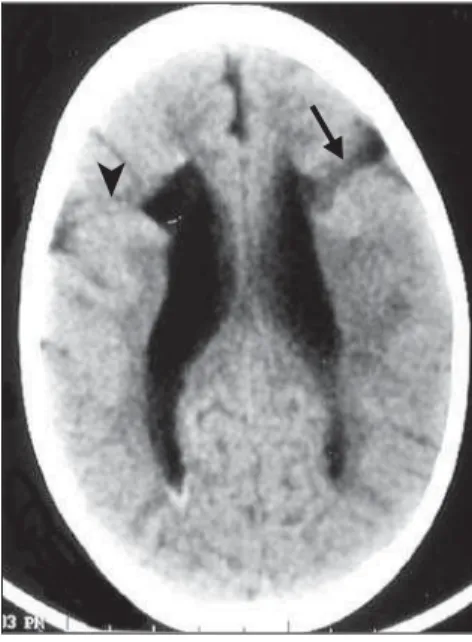
were large (Figure 1) and 2:27 were me-dium (Figure 2). As regards localization, parietal clefts predominated (16:27), fol-lowed by frontoparietotemporal clefts (5:27). As regards laterality, bilateral clefts predominated (Figures 1, 2 and 3) in 10:16 patients, 5:10 patients presenting open-lip clefts and 5:10 with open and closed (or fused)-lip clefts (Figure 3). Of six patients with unilateral clefts (Figure 4), five pre-sented open-lip clefts.
Periventricular calcifications (Figure 4) were observed in 3:16 patients, all of them presenting negative serology for congeni-tal TORCH infection.
Thirteen of 16 patients presented other central nervous system anomalies associ-ated with schizencephaly, the pellucid sep-tum absence (Figure 1) and cortical dyspla-sias (Figure 5) being the most frequent findings respectively in 10:13 patients and in 4:13 patients.
Neuropsychomotor development delay was present in 15 patients and six school-aged patients also presented cognitive
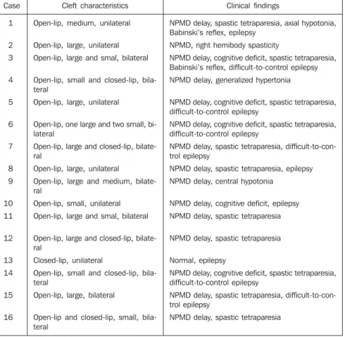
defi-Table 1 Clinical-tomographic correlation. . Case 1 2 3 4 5 6 7 8 9 10 11 12 13 14 15 16 Cleft characteristics
Open-lip, medium, unilateral
Open-lip, large, unilateral Open-lip, large and smal, bilateral
Open-lip, small and closed-lip, bila-teral
Open-lip, large, unilateral
Open-lip, one large and two small, bi-lateral
Open-lip, large and closed-lip, bilate-ral
Open-lip, large, unilateral
Open-lip, large and medium, bilate-ral
Open-lip, small, unilateral Open-lip, large and smal, bilateral
Open-lip, large and closed-lip, bilate-ral
Closed-lip, unilateral
Open-lip, small and closed-lip, bila-teral
Open-lip, large, bilateral
Open-lip and closed-lip, small, bila-teral
Clinical findings
NPMD delay, spastic tetraparesia, axial hypotonia, Babinski’s reflex, epilepsy
NPMD, right hemibody spasticity
NPMD delay, cognitive deficit, spastic tetraparesia, Babinski’s reflex, difficult-to-control epilepsy NPMD delay, generalized hypertonia
NPMD delay, cognitive deficit, spastic tetraparesia, difficult-to-control epilepsy
NPMD delay, cognitive deficit, spastic tetraparesia, difficult-to-control epilepsy
NPMD delay, spastic tetraparesia, difficult-to-con-trol epilepsy
NPMD delay, spastic tetraparesia, epilepsy NPMD delay, central hypotonia
NPMD delay, cognitive deficit, epilepsy NPMD delay, spastic tetraparesia
NPMD delay, spastic tetraparesia
Normal, epilepsy
NPMD delay, cognitive deficit, spastic tetraparesia, difficult-to-control epilepsy
NPMD delay, spastic tetraparesia, difficult-to-con-trol epilepsy
NPMD delay, spastic tetraparesia
325 Clinical findings in 16 patients with tomographic diagnosis of schizencephaly
Radiol Bras 2006;39(5):323–326 cit. Neurological examination detected anomalies in 15:16 patients, the most fre-quent finding being pyramidal release signs (spastic tetraparesia) in 11:15 patients. Amongst the 16 patients, ten presented epilepsy, with the first crisis onset in the first year of life of 4:10 patients. The most frequent type of seizure was the general-ized tonic-clonic one, reported in 5:10 pa-tients. Epilepsy was difficult to control in 6:10 patients.
DISCUSSION
The clinical feature severity was related to the affected cortical area, both in patients with bilateral and unilateral clefts, accord-ing data reported in the specialized scien-tific literature(2,3,5,6). Although clefts in
three of our patients were small, the clini-cal feature was severe as a result of the presence of other cerebral anomalies asso-ciated with schizencephaly. Therefore, our data suggest that the clinical feature is also related to the presence of other central ner-vous system anomalies, corroborating the findings of Granata et al.(13).
The clinical feature was in agreement with data reported by other authors(6,9,14,15),
with the majority of patients presenting motor deficit and neuropsychomotor devel-opment delay(8,9).
Figure 1. Bilateral schizencephaly. Large parietal open-lip cleft at left (arrowheads), small parietal open-lip cleft at right (black arrow), pellucid sep-tum absence (white arrow)(case 11).
Figure 3. Bilateral schizencephaly. Parietal closed-lip cleft at right (arrowhead), small parietal open-lip cleft at left (arrow)(case 16).
Figure 2. Bilateral schizencephaly. Large fronto-temporoparietal open-lip cleft at left (arrowheads), medium parietal open-lip cleft at right (arrow)(case 9).
Figure 4. Unilateral schizencephaly – large parietal open-lip cleft at left (arrowheads), bilateral periven-tricular calcifications (arrows)(case 2).
Figure 5. Bilateral schizencephaly. Small parietal open-lip cleft at left (arrow), parietal closed-lip cleft at right (arrowhead). Observe bilateral heterotopia on the corona radiata and bilateral frontotemporo-parietal cortical dysplasia (case 4).
Also, we have observed that epilepsy was more frequent and severe in patients with a more significant loss of cortical area, an aspect that has not been described in the studies of Barkovich & Kjos(5), Granata et
al.(14) and Denis et al.(15).
In a comparative analysis of the tomo-graphic data, there was an agreement with data reported by other authors as regards
laterality(9), cleft type(5,9) and localization
and size(5,14,15). Amongst associated
anoma-lies, pellucid septum absence was our most frequent finding, similarly to the findings reported by other authors(9,10,13).
Although the presence of cleft(s) is sig-nificant for determination of the neurologi-cal picture, in some cases there was dis-agreement between the cleft extent and
➤
➤
➤
➤
➤
➤
➤
326
Rodrigues MCS et al.
Radiol Bras 2006;39(5):323–326 clinical findings, as result of the presence
of other cerebral anomalies associated with schizencephaly.
REFERENCES
1. OMIM. On-line Mendelian Inheritance in Man. Johns Hopkins University [cited 2002 April 15]. Available from: http://www.ncbi.nlm.nih.gov 2. Brunelli S, Faiela A, Capra V, et al. Germline
mutation in the homeobox gene EMX2 in patients with severe schizencephaly. Nature Genet 1996;12:94–96.
3. Faiella A, Brunelli S, Granata T, et al. A number of schizencephaly patientes including 2 brothers are heterozygous for germline mutations in the homeobox gene EMX2. Eur J Hum Gen 1997;-5:186–190.
4. Guerrini R, Carrozzo R. Epilepsy and genetic
malformations of the cerebral cortex. Am J Med Genet 2001;106:160–173.
5. Barkovich AJ, Kjos BO. Schizencephaly: corre-lation of clinical findings with MR characteris-tics. AJNR Am J Neuroradiol 1992;13:85–94. 6. Granata T, Farina L, Faiella A, et al. Familial
schizencephaly associated with EMX2 mutation. Neurology 1997;48:1403–1406.
7. Dubey AK, Gupta RK, Sharma P, Sharma RK. Schizencephaly type-I. Indian Pediatr 2001;38:-1049–1052.
8. Packard AM, Miller VS, Delgado MR. Schizen-cephaly: correlations of clinical and radiologic features. Neurology 1997;480:1427–1434. 9. Amaral JG, Yanaga RH, Geissler HJ, Carvalho
Neto A, Bruck I, Antoniuk SA. Schizencephaly: report of eleven cases. Arq Neuro-Psiquiat 2001; 59:244–249.
10. al-Alawi AM, al-Tawil KI, al-Hathal MM, Amir I. Sporadic neonatal schizencephaly associated
with brain calcification. Ann Trop Paediatr 2001; 21:34–37.
11. Robinson RO. Familial schizencephaly. Dev Med Child Neurol 1991;33:1010–1012.
12. Montandon C, Ribeiro FAS, Lobo LVB, Montan-don Júnior ME, Teixeira KISS. Disgenesia do corpo caloso e más-formações associadas: acha-dos de tomografia computadorizada e ressonân-cia magnética. Radiol Bras 2003;36:311–316. 13. Granata T, Freri E, Caccia C, Setola V, Taroni F,
Battaglia G. Schizencephaly: clinical spectrum, epilepsy, and pathogenesis. J Child Neurol 2005;20:313–318.
14. Granata T, Battaglia G, D’Incerti L, et al. Schizen-cephaly: neuroradiologic and epileptologic find-ings. Epilepsia 1996;37:1185–1193.