190
Srp Arh Celok Lek. 2015 Mar-Apr;143(3-4):190-194 DOI: 10.2298/SARH1504190A
ПРИКАЗ БОЛЕСНИКА / CASE REPORT UDC: 616.716.4-006
Correspondence to:
Jean Nunes DOS SANTOS Avenida Araújo Pinho, 62 Canela, Salvador-BA Brazil 40110-150
SUMMARY
Introduction Ameloblastic fibroma (AF) is a rare benign odontogenic tumor that usually occurs in the first two decades of life. It affects adolescents and young adults and is found in the mandible and with a higher frequency in the posterior region of this segment. There are rare case reports with a long-term follow-up.
Case Outline We report the case of a 6-year-old boy with extensive ameloblastic fibroma in the mandi-ble. Treatment consisted of enucleation and bone curettage, with the preservation of permanent teeth adjacent to the tumor. Clinical and radiographic follow-up of the patient over a period of 7 years showed no signs of recurrence or malignant transformation.
Conclusion Patients with AF should be under follow-up for prolonged periods of time, even in cases exhibiting a low proliferation index, because of the potential for recurrence and malignant transforma-tion of this tumor.
keywords: ameloblastic fibroma; odontogenic tumors; jaws; follow-up
Ameloblastic Fibroma: A Rare Case Report with
7-Year Follow-Up
Leonardo de Araújo Melo1, Adna Conceição Barros1, Sandra de Cássia Santana Sardinha2,
Arlei Cerqueira2, Jean Nunes dos Santos3
1School of Dentistry, Federal University of Bahia, Salvador, Bahia, Brazil;
2Division of Oral and Maxillofacial Surgery, School of Dentistry, Federal University of Bahia, Salvador,
Bahia, Brazil;
3Laboratory of Oral Surgical Pathology, School of Dentistry, Federal University of Bahia, Salvador,
Bahia, Brazil
INTRODUCTION
Ameloblastic fibroma (AF) is a rare tumor which accounts for 0.9 to 2.4% of all odontogenic tu-mors involving the jaw bones [1-4]. AF mainly affects adolescents and young adults and has a slight male predominance. Most tumors are found in the mandible, while a higher in-cidence is observed in the posterior region. Patients usually seek treatment because of a swelling in the jaw bones and delayed tooth eruption, although some cases are diagnosed incidentally during routine radiographic ex-amination [5]. Radiographically, AF appears as a unilocular lesion with well-defined margins. Multilocular lesions are occasionally observed in more extensive cases. The lesions are fre-quently associated with impacted teeth [6, 7].
Histologically, AF is characterized by epithe-lial islands and cords whose center resembles the stellate reticulum of the enamel organ. The islands and cords are immersed in ectomes-enchyme that mimics the dental papilla. The presence of a capsule is considered an uncom-mon feature of AF [8].
AF shows a high rate of recurrence [5], while malignant transformation is observed in some cases [9, 10]. These findings highlight the im-portance of the study of this tumor and the need for long-term follow-up. This study re-ports a case of extensive AF in the mandible of a child. No signs of recurrence were observed after 7 years of follow-up.
CASE REPORT
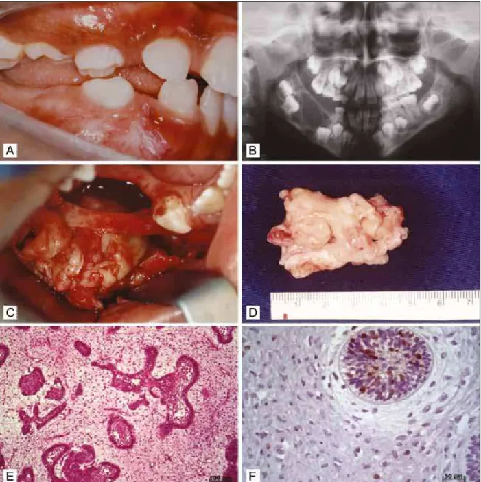
A 6-year-old boy was referred to a private clinic in Salvador, Bahia, Brazil, for evaluation of a swelling on the right side of the face. The parents reported no eating difficulties. The medical history revealed that the patient had thalassemia minor, but no clinical signs or symptoms were observed.
tu-mor specimen, which measured 45×40×10 mm, revealed an irregular surface and fibroelastic consistency (Figure 1D). Histopathological analysis confirmed the diagnosis of AF (Figure 1E). Immunohistochemistry detected few Ki-67-positive nuclei, especially in the epithelial paren-chyma, indicating a low proliferation index of the tumor (Figure 1F).
No complications were observed during the postopera-tive period. The patient was followed up over a period of 7 years and no clinical or radiographic signs of recurrence were observed. Control panoramic radiographs, after 2 months, 1 and 5 years, showed the formation of new bone in the right posterior region of the mandibular body and complete eruption of teeth 46 and 47 (Figure 2).
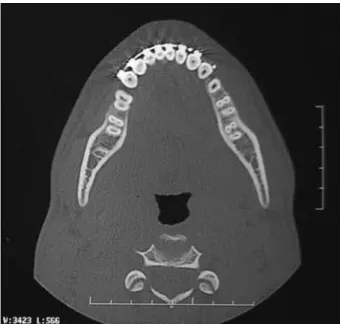
Com-puted tomography scans obtained after 7 years revealed cortical bone integrity, complete formation of medullary bone, and complete eruption of tooth 46 which had been preserved during surgery. In addition, the bone architec-ture in the right posteroinferior region was preserved and no radiolucency was observed (Figure 3).
DISCUSSION
This study reported a case of AF involving the body and angle of the mandible in a 6-year-old child. Philipsen et al. [11] and Tomich [6] suggested that AF corresponds to a stage of maturation of ameloblastic fibro-odontoma and
192
de Araújo Melo L. et al. Ameloblastic Fibroma: A Rare Case Report with 7-Year Follow-Up
odontoma. This hypothesis has been refuted since recur-rent AFs show no evidence of maturation to ameloblastic fibro-odontoma or odontoma. In addition, AFs usually affect patients in the third decade of life when the process of odontogenesis is already completed [12].
Clinically, the patient presented with a mandibular swelling extending from the right deciduous first molar to the mandibular angle on the same side, causing a delay in tooth eruption. According to Kim and Jang [8], this swell-ing is caused by gradual expansion of the cortical plate during growth of the tumor. In addition, these authors have reported that AF shows slower growth than other odontogenic tumors, such as ameloblastoma, and does not tend to infiltrate bone. However, Pereira et al. [13] reported a case of rapidly growing AF in an 11-month-old infant, which was attributed to the small size of the jaw.
The patient had thalassemia minor, the heterozygous state of a hereditary genetic disease characterized by a re-duction in beta globin chain synthesis, causing mild and silent microcytic and hypochromic anemia [14, 15]. There are no reports in the literature correlating this hereditary disease with AF, but this odontogenic tumor has been diag-nosed in other diseases such as Goldenhar syndrome [16]. The histopathological features of the present case met the criteria for AF [12, 17]. The lesion consisted of odon-togenic epithelial islands and cords amidst a cellular and embryonic stroma resembling the dental papilla. Usubu-tun et al. [18] also found extensive cystic formation as a result of a degenerative process, but this was not observed in the present case.
According to Carnelio and Vij [19] and Rao et al. [20], AF exhibits a low proliferation rate. This was confirmed in the present study by sparse immunostaining for Ki-67, especially in the parenchymal compartment. Evaluation of the proliferative potential of AF using immunohistochemi-cal markers contributes to the understanding of tumor ag-gressiveness and to adequate surgical planning [8], as in the present case.
The initial diagnosis of the case included a dentigerous cyst, odontogenic keratocystic tumor and ameloblastoma due to the clinical and radiographic similarities between these lesions. The differential diagnosis of AF should in-clude odontogenic lesions such as the dentigerous cyst, ameloblastoma, myxoma, and other mixed tumors [21, 22]. According to Nelson and Folk [23], despite many similarities, it is essential to differentiate AF from other mixed odontogenic tumors in view of its true neoplastic potential, possibility of recurrence and malignant trans-formation potential.
The present patient was submitted to conservative treat-ment considering that the lesion was a primary tumor and to avoid possible esthetic deformities. Consensus exists regarding the preference of conservative treatment of AFs, including enucleation of the tumor and removal of all teeth involving bone curettage [13, 24, 25]. According to these authors, en bloc resection results in significant morbidity and esthetic deformity and should be reserved for exten-sive tumors invading soft tissue and for recurrent cases.
Figure 2. Control panoramic radiographs, after 2 months (A), 1 year (B) and 5 years (C), respectively, showing the formation of new bone in the right posterior region of the mandibular body and complete eruption of teeth 46 and 47.
The present patient was followed up for 7 years and no clinical or radiographic signs of recurrence or malignant transformation were observed after this period, demon-strating the success of treatment. Long-term follow-up of AF as done in the present study is important due to the possibility of recurrence and malignant transformation.
Follow-up of cases for up to 12 years has been reported [8, 13, 21, 25, 26].
In conclusion, patients with AF should be followed up for prolonged periods of time, even cases exhibiting a low proliferation index, because of the potential for recurrence and malignant transformation of this tumor.
1. Tawfik MA, Zyada MM. Odontogenic tumors in Dakahlia, Egypt: analysis of 82 cases. Oral Surg Oral Med Oral Pathol Oral Radiol Endod. 2010; 109:e67-73.
2. Osterne RL, Brito RG, Alves AP, Cavalcante RB, Sousa FB. Odontogenic tumors: a 5-year retrospective study in a Brazilian population and analysis of 3406 cases reported in the literature. Brasil Oral Res. 2011; 111:474-81.
3. Servato JP, de Souza PE, Horta MC, Ribeiro DC, de Aguiar MC, de Faria PR, et al. Odontogenic tumours in children and adolescents: a collaborative study of 431 cases. Int J Oral Maxillofac Surg. 2012; 41:768-73.
4. Siriwardena BS, Tennakoon TM, Tilakaratne WM. Relative frequency of odontogenic tumors in Sri Lanka: analysis of 1677 cases. Pathol Res Pract. 2012; 208:225-30.
5. Chen Y, Wang JM, Li TJ. Ameloblastic fibroma: a review of published studies with special reference to its nature and biological behavior: Oral Oncol. 2997; 43:960-9.
6. Tomich CE. Benign mixed odontogenic tumors. Semin Diagn Pathol. 1999; 16:308-16.
7. Cohen DM, Bhattacharyya I. Ameloblastic fibroma, ameloblastic fibro-odontoma, and odontoma. Oral Maxillofac Surg Clin North Am. 2004; 16:375-84.
8. Kim SG, Jang HS. Ameloblastic fibroma: report of a case. J Oral Maxillofac Surg. 2002; 60:216-8.
9. Kobayashi K, Murakami R, Fujii T, Hirano A. Malignant transformation of ameloblastic fibroma to ameloblastic fibrosarcoma: case report and review of the literature. J Craniomaxillofac Surg. 2005; 33:352-5.
10. DeLair D, Bejarano PA, Peleg M, El-Mofty SK. Ameloblastic carcinosarcoma of the mandible arising in ameloblastic fibroma: a case report and review of the literature. Oral Surg Oral Med Oral Pathol Oral Radiol Endod. 2007; 103:516-20.
11. Philipsen HP, Reichart PA, Praetorius F. Mixed odontogenic tumours and odontomas. Considerations on interrelationship. Review of the literature and presentation of 134 new cases of odontomas. Oral Oncol. 1997; 33:86-99.
12. Chen Y, Li TJ, Gao Y, Yu SF. Ameloblastic fibroma and related lesions: a clinicopathologic study with reference to their nature and interrelationship. J Oral Pathol Med. 2005; 34:588-95.
13. Pereira KD, Bennett KM, Elkins TP, Qu Z. Ameloblastic fibroma of the maxillary sinus. Int J Pediatr Otorhinolaryngol, 2004; 68:1473-7. 14. Succar J, Musallam KM, Taher AT. Thalassemia and venous
thromboembolism. Mediterr J Hematol Infect Dis. 2011; 3:e2011025.
15. Park N, Lazow S, Berger J. β-Thalassemia: medical and surgical considerations in managing facial deformities: case report and review of the literature. J Oral Maxillofac Surg. 2012; 70:e284-9. 16. Naidoo LC, Stephen LX. Congenital ameloblastic fibroma in
association with oculoauriculovertebral spectrum. Int J Pediatr Otorhinolaryngol. 1988; 43:283-8.
17. Slootweg PJ. Ameloblastic fibroma/fibrodentinoma. In: Barnes L, Evenson JW, Reichart P, Sidransky D, editors. The World Health Organization Classification of Tumours. Pathology and Genetics of Head and Neck Tumours. Lyon: IARC Press; 2005; p.308.
18. Usubütün A, Atayar C, Basal N, Araz K. Cystic ameloblastic fibroma. Br J Oral Maxillofac Surg, 2002; 40:512-4.
19. Carnelio S, Vij H. Expression of tenascin and nucleolar organizer region in ameloblastoma and ameloblastic fibroma. J Oral Pathol Med. 2010; 39:223-9.
20. Rao SP, Srivastava G, Smitha B. Ameloblastic fibroma. J Oral Maxillofac Pathol. 2012; 16:444-5.
21. Pitak-Arnnop P, Chaine A, Dhanuthai K, Bertrand JC, Bertolus C. Extensive ameloblastic fibroma in an adolescent patient: a case report with a follow-up of 4 years. Eur J Dent. 2009; 3:224-8. 22. Scholl RJ, Kellett HM, Neumann DP, Lurie AG. Cysts and cystic
lesions of the mandible: clinical and radiologic-histopathologic review. Radiographics. 1999; 19:1107-24.
23. Nelson BL, Folk GS. Ameloblastic fibroma. Head Neck Pathol. 2009; 3:51-3.
24. Mosby EL, Russell D, Noren S, Barker BF. Ameloblastic fibroma in a 7-week-old infant: a case report and review of the literature. J Oral Maxillofac Surg. 1998; 56:368-72.
25. Vasconcelos BC, Andrade ES, Rocha NS, Morais HH, Carvalho RW. Treatment of large ameloblastic fibroma: a case report. J Oral Sci. 2009; 51:293-6.
26. Martín-Granizo López R, Ortega L, González Corchón MA, Berguer Sández A. Ameloblastic fibroma of the mandible. Report of two cases. Med Oral. 2003; 8:150-3.
194
КРАТАК САДРжАЈ
Увод Аме ло бласт ни фи бром је ре дак, бе ниг ни, одон то ге ни ту мор ко ји се обич но ја вља до двадесете го ди не жи во та. Обо ље ње по га ђа адо ле сцен те и мла де од ра сле осо бе, а за хва та до њу ви ли цу, и то че шће по сте ри ор ну ре ги ју овог сег мен та. При ка зи бо ле сни ка са ду го роч ним кли нич ким пра ће њем су рет ки.
При каз бо ле сни ка При ка зу је мо ше сто го ди шњег де ча ка са екс тен зив ним аме ло бласт ним фи бро мом до ње ви ли це. Бо-ле сник је Бо-ле чен при ме ном хи рур шке ре сек ци је и ки ре та же
ко сти, уз очу ва ње стал них зу ба у бли зи ни ту мо ра. Кли нич-ко и ра ди о ло шнич-ко ис пи ти ва ње бо ле сни ка то нич-ком сед мо го ди-шњег пе ри о да ни је по ка за ло зна ке ре ци ди ва или ма лиг не тран сфор ма ци је.
За кљу чак Бо ле сни ке обо ле ле од аме ло бласт ног фи бро ма по треб но је ду го роч но над гле да ти, чак и у слу ча је ви ма с ни-ским ин дек сом про ли фе ра ци је, с об зи ром на мо гу ћу по ја ву ре ци ди ва и ма лиг ну тран сфор ма ци ју овог ту мо ра.
Кључ не ре чи: аме ло бласт ни фи бром; одон то ге ни ту мо ри; ви ли ца; кли нич ко пра ће ње
Амелобластни фибром: приказ ретког случаја са седмогодишњим периодом
клиничког праћења
Леонардо де Араужо Мело1, Адна Консеисао Барос1, Сандра де Касиа Сантана Сардиња2, Арлеи Серквеира2,
Жеан Нунес дос Сантос3
1Стоматолошки факултет, Државни универзитет Баије, Салвадор, Баија, Бразил;
2Катедра за оралну и максилофацијалну хирургију, Стоматолошки факултет, Државни универзитет Баије, Салвадор, Баија, Бразил; 3Лабораторија за оралну патологију, Стоматолошки факултет, Државни универзитет Баије, Салвадор, Баија, Бразил
Примљен • Received: 28/04/2014 Прихваћен • Accepted: 01/08/2014