cop
yr
ight
© ABE&M todos os direitos reser
v
ados
clinical case report
LETICIA G. SILVEIRA EDUARDO P. DIAS BRUNA C. G. MARINHO RICARDO S. GOMEZ LUIZ DE MARCO MARTA S. SARQUIS
Departamentos de Farmácia (LGS, EPD, BCGM, LDM, MSS), Medicina (MSS), Cirurgia Oral e Patologia (RSG), Universidade Federal de Minas Gerais; Instituto Felice Rosso de Pesquisa e Educação Continuada (Iferpec – Felício Rocho Hospital) (EPD, BCGM, LDM, MSS); Belo Horizonte, MG, Brasil.
Received in 23/8/2008 Accepted in 31/10/2008 ABSTRACT
It is still debatable which is the best management to familial forms of hyper-parathyroidism. Conservative, minimally invasive or aggressive surgical ap-proaches have been proposed from different groups around the world. Our objective was to study the gene mutation, expression of HRPT2 and the clini-cal outcome after 32 years of follow-up in one Brazilian kindred with familial isolated hyperparathyroidism (FIHP). Clinical and biochemical data, direct se-quencing of the HRPT2 gene, analysis of parafi bromin expression using RT-PCR, and immunohistochemistry were done. A nonsense mutation was found in exon 1 (c.96G>A)(p.Trp32X) in all affected members studied. Using RT-PCR, mRNA transcription was altered with complete absence of both transcripts in tumor tissue. Immunohistochemical analysis of tumors showed loss of para-fi bromin immunoreactivity. In this kindred there was a high prevalence of recurrence (75%), or persistence after less than subtotal parathyroidectomy that led us to consider a more aggressive surgical approach should be dis-cussed among the affected family members, once surgical criteria was met. We concluded that it is necessary to individualize the surgical approach for HRPT2-related hyperparathyroidism until we can gather a better phenotype-genotype correlation in larger series, to best defi ne their treatment. (Arq Bras Endocrinol Metab 2008; 52/8:1211-1220)
Keywords: HRPT2; Parafi bromin; Parathyroid; Jaw tumor; FIHP; Surgery
RESUMO
Formas Familiares de Hiperparatireoidismo Relacionadas a Mutações no Gene HRPT2: Estudos Moleculares Poderiam Direcionar Procedimentos Cirúrgicos? A melhor conduta nas formas familiares de hiperparatireoidismo relacionadas a mutações no gene HRPT2 ainda é controvertida. Cirurgias conservadoras, mini-mamente invasivas ou mais agressivas já foram propostas por diferentes gru-pos. Objetivamos estudar a seqüência e a expressão do gene HRPT2, além do desfecho clínico, após seguimento de até 32 anos de uma família brasileira com hiperparatireodismo familiar isolado (FIHP). Utilizamos dados clínicos e bio-químicos, seqüenciamento direto do HRPT2 além de análise da expressão da parafi bromina através da RT-PCR e imunohistoquímica. Foi identifi cada mutação nonsense no éxon 1 (c.96G>A)(p.Trp32X) em todos os membros afetados que foram estudados. A análise do mRNA transcrito, através da RT-PCR, demonstrou ausência do transcrito no tecido tumoral. A imunohistoquímica também eviden-ciou ausência da parafi bromina. Nessa família houve alta (75%) prevalência de recorrência ou persistência da doença após paratireoidectomia parcial o que nos levou a considerar fundamental discutir uma abordagem cirúrgica mais agres-siva com os outros familiares portadores da mutação caso critérios de indicação cirúrgica sejam atingidos. Dessa maneira, até que estudos mais amplos esta-beleçam uma correlação genótipo-fenótipo no hiperparatireoidismo familiar relacionado a mutações no HRPT2, a abordagem cirúrgica deverá ser individual-izada. (Arq Bras Endocrinol Metab 2008; 52/8:1211-1220)
cop
yr
ight
© ABE&M todos os direitos reser
v
ados
INTRODUCTION
F
amilial hyperparathyroidism (HPT) encompasses a clinically and genetically heterogeneous group of disorders. These heritable forms are responsible for ap-proximately 10% of primary HPT. They are usually seen in the context of syndromes, such as multiple endocri-ne endocri-neoplasia types 1 and 2 (MEN 1, MIM #131100 and MEN 2, MIM #171400, respectively), hyperpara-thyroidism-jaw tumor syndrome (HPT-JT, MIM #145001), familial benign hypocalciuric hypercalcemia (FHH, MIM #145980) and familial isolated hyperpa-rathyroidism (FIHP, #145000) (1).FIHP is a rare autosomal dominant condition, cha-racterized by the occurrence of familial hyperpara-thyroidism in the absence of other associated lesions or endocrinopathies, with a clinical and genetically hete-rogeneous presentation (2-6). Most FIHP families do not have identifi able mutations, although some patients have been found to harbor germline mutations in MEN1, CASR, HRPT2 and, more recently, studies have also linked a FIHP locus to chromosome 2p14-p13.3 (HRPT3) (2,3).
In patients with FIHP, genetic screening for MEN1 and CASR is most likely to identify mutations in those with young age at onset and multigland parathyroid involvement, whereas HRPT2 mutations should be considered in those families with parathyroid carcino-ma, cystic parathyroid tumors, or jaw tumors - present in up to 30% of the affected patients (4-7).
The majority of FIHP with MEN 1 mutations pre-sented with mild hypercalcemia and multiglandular di-sease or parathyroid hyperplasia, similar to what is usually seen in MEN1 (8-13). On the other hand, in FIHP families where HRPT2 gene mutations have been detected, the clinical presentation is more severe and in all cases the histopathological diagnosis was pa-rathyroid carcinoma or adenoma, frequently associated with atypical or cystic features similar to those seen in HPT-JT syndrome (13-15). These observations sug-gest that FIHP could be subdivided in at least two ma-jor genetic subsets: a MEN1-related FIHP, characterized by milder forms of hyperparathyroidism, typically pre-senting as multiglandular disease; and HRPT-related variety, characterized by an aggressive disease, para-thyroid adenomas or carcinomas and HRPT2 gene mutations that could be regarded as a variant of HPT-JT syndrome.
In view of the atypical behavior of adenomas, and the high prevalence of parathyroid carcinomas in fami-lies with syndromes associated with HRPT2 mutations in contrast to the extremely rare occurrence of sporadic parathyroid malignancy, the HRPT2 gene has been in-vestigated in parathyroid carcinoma. Inactivating so-matic mutations of HRPT2 were found in the majority of sporadic parathyroid cancers and even germline mu-tations were detected in some patients with apparently sporadic tumors (16,17). On the other hand, intrage-nic HRPT2 somatic mutations were detected only in 0.8 to 1.8% of sporadic adenomas, depending on the inclusion criteria used, supporting the view that HRPT2 inactivation is not an important contributor to para-thyroid tumorigenesis (18).
The HRPT2 gene is located on chromosome 1q31.2 and consists of 17 exons that span 1.3 Mb of genomic DNA (6) encoding a protein named parafi -bromin, with 531 amino acids. Recently, parafi bromin was identifi ed as a component of the human Paf1 (pe-roxisome assembly factor-1) complex which is an acces-sory factor to the RNA polymerase II suggesting, as has been shown for its yeast homologue (Cdc 73), which it may have a role in transcription regulation (19). Ove-rexpression of parafi bromin in mammalian cell lines inhibits cell proliferation and blocks expression of Cyclin D1, which has been implicated in parathyroid neoplasia (20). Tan et al. (21) demonstrated loss of pa-rafi bromin nuclear immunoreactivity in 96% of para-thyroid carcinomas and in eight of nine adenomas associated to HPT-JT syndrome. Furthermore, normal expression of parafi bromin has been demonstrated in the majority of the sporadic parathyroid adenomas so far studied (20-22).
The management of familial hyperparathyroidism differs among the specifi c syndromes and is generally complex due to the underlying causative genetic abnor-mality that can predispose patients to persistent and recurrent hyperparathyroidism. Therefore it is of ut-most importance to identify these syndromes before the surgical procedure is planned. Besides a detailed fa-mily history and search of the well-known co-morbidi-ties present in those syndromes, molecular analyses might also infl uence the therapeutic decision.
cop
yr
ight
© ABE&M todos os direitos reser
v
ados
SUBJECTS AND METHODS
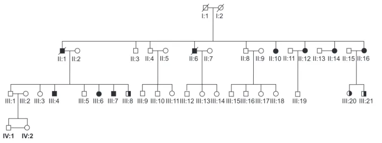
The present study was approved by the Felicio Rocho Hospital Ethics Committee and all participating patients and family members signed a written informed consent. Most of the patients, except for II:10, have been follo-wed by the Endocrinology Center, at least once a year. Hyperparathyroidism diagnosis was established by the detection of hypercalcemia associated with elevated se-rum PTH levels (23). Multiple endocrine neoplasia type 1 was excluded by detecting normal levels of serum glu-cose, insulin, calcitonin, gastrin, IGF1, and prolactin as well as sequencing all coding regions of the MEN1 gene. The family’s pedigree is shown in Figure 1. Peripheral blood samples for DNA extraction were obtained from individuals II:1, II:10, II:12, II:14, II:16, III:4, III:6, III:7, III:8, III:20 and III:21.
Fresh samples of parathyroid adenomas were ob-tained during surgical removal of the tumor, for RNA and DNA extraction from III:6 and III:7 in this kin-dred. In each case, a portion of the tumor was resec-ted, immediately snap frozen and stored at –80 oC.
For immunohistochemistry, sections were obtained from paraffi n-embedded tissue blocks. DNA was ex-tracted as previously described (24).
CASE REPORT
This is a four generation family, in which nine members were diagnosed with isolated hyperparathyroidism and
underwent parathyroidectomy (Figure 1). No other endocrinopathies were reported in any affected indivi-dual after 32 years of follow-up since the diagnosis of the index case. Maxillary and/or jaw tumors were ruled out in all patients by clinical examination and plain X-ray of the jaws. Lack of consanguineous marriages and pedigree analysis (Figure 1) suggested autosomal do-minant mode of inheritance. Biochemical and molecu-lar screening for MEN1 were negative. Clinical and biochemical details of affected members of this family are shown in Table 1.
The propositus (II:12), 32 years of age, was refer-red in 1976 because of a maxillary lesion diagnosed as brown tumor. She complained of generalized bone pain, polyuria, polydipsia and weight loss (13 kg in one year). After biochemical diagnosis of primary hyperparathyroidism, she underwent surgical removal of a single 1-cm left inferior parathyroid adenoma. She became normocalcemic, needing calcium supplements for about 5 months. The “brown tumor” disappeared and she became asymptomatic until 1981, when she returned with recurrent hypercalcemia and raised para-thyroid hormone levels. Once again she underwent cervical surgery, with removal of two more parathyroid adenomas (0.5 and 1.2 cm). Her serum calcium nor-malized, calcium supplements were needed for a short period of time and her bone mass density improved dramatically. She remained asymptomatic with no la-boratory abnormalities for 24 years, when a knee le-sion was detected during an orthopedic investigation
I:1 I:2
II:1 II:2 II:3 II:4 II:5 II:6 II:7 II:8 II:9 II:10 II:11 II:12 II:13 II:14 II:15 II:16
III:1 III:2 III:3 III:4
IV:1
III:5 III:6 III:7 III:8 III:9 III:10 III:11III:12 III:13III:14 III:15III:16 III:17III:18 III:19 III:20 III:21
IV:2
cop
yr
ight
© ABE&M todos os direitos reser
v
ados
(Figure 2). The patient declined another bone biopsy. Biochemical diagnosis of recurrent hyperparathyroi-dism was again established. Recently, another adeno-ma has been localized by ultrasound, after continuous imaging investigations three years after the recurrence diagnosis was made. She is now 65 years old and was recently (for the third time), referred to another surgi-cal procedure.
Surgical outcomes
Except for one patient (II:16) who had three and a half parathyroid glands removed, each of the eight other affected siblings had only one or two adenomas excised at their fi rst surgical procedure (Table 1). Seven of the-se patients have being followed by our group. This follow-up ranges from 32 months to 32 years, and six
out of seven had recurrence/persistence of their disease during this period (Table 1). Patient III:6 was surgi-cally treated in December 2005, when two adenomas were excised and to date, 32 months later, have not had any disease recurrence.
Mutational screening of the HRPT2 gene
DNA isolated from the tumors and peripheral blood was used to amplify the 17 exons of the HRPT2 gene,
using specifi c intronic primer pairs as previously descri-bed (25). PCR reactions were performed in a fi nal vo-lume of 25 µl containing 100-300 ng of template DNA, 10 pmol of each primer, 1.5 to 4 mM MgCl2, 1.5 mM of dNTPs, 2.5 µl of 10x PCR buffer, and 1.25 U Taq DNA polymerase (Invitrogen Life Technologies, Carl-sbad, CA) in a Mastercycler gradient thermocycler
Table 1. Clinical and biochemical details of affected members.
Case Sex Age (y) at Diagnose
Serum Calcium
(8.5-10.5mg/dL) PTH (10-65pg/ml)
Parathyroid Histologyc
Associated Lesions
Recurrence Persistenced
II:1 M 65 11.9 mg/dL 664 pg/mL Adenoma (1) Bilateral renal
cysts Yes (5y)
II:6 M 52 11.9 mg/dL 525 pg/mL Adenoma (1) Bilateral renal
cysts Yes (persistence)
II:10 F 34 11.7 mg/dL 1500 pg/mL Atypical adenomas (2)
Uuterine
leiomyomata Yes (11y)
II:12 F 32 12.4 mg/dL 1600 pg/mL Cystic adenomas (2) Uterine
leiomyomata Yes (5 and 30y)
II:14 F 38 13.0 mg/dL 1.1mg/dL
NV(<1mg/dL) Cystic adenomas (2)
Renal tumor and uterine leiomyomata
?
II:16 F 37 11.9 mg/dL 1,100 pg/mL Adenomas (2)
PTX (3 ½)
Renal tumor and uterine leiomyomata
No
III:4 M 27 1.93 mmol/Lb 354a pg/mL Adenoma (1) Yes (1y)
III:7 M 26 11.9 mg/dL 132a pg/mL Adenomas (2) Yes
(persistence)
III:6 F 25 11.9 mg/dL 73a pg/mL Adenomas (2) Uterine
leiomyomata No
III:20e F 19 1.19 mmol/L b 22a pg/ml
III:8e M 26 1,26 mmol/Lb
III:21e M 17 1,24 mmol/Lb 28a pg/ml
IV:IIe,f F 19 1,30 mmol/Lb 49a pg/ml
II:12, propositus; M: male; F: female; PTX (3 ½): parathyroidectomy of 3 and ½ glands; a: PTH nomal value: 7-53pg/ml; b Ionized calcium nomal value: 1.17-1.32mmol/L; c
Histology of the fi rst surgery and number of glands excised in parenthesis, dTime to recurrence/persistence (in years), after the fi rst surgery; e Mutation carrier without
cop
yr
ight
© ABE&M todos os direitos reser
v
ados
(Eppendorf AG, Hamburg). Amplifi cations were car-ried out as follows: an initial denaturation for 8 min at 95 oC followed by 35 cycles of 94 oC for 40 s, 55-59 oC
for 40 s, 72 oC for 1 min, and a fi nal extension at 72 oC
for 5 min. All products were analyzed by electrophore-sis on 6.5% polyacrylamide gel followed by silver stai-ning, purifi ed using the GFX PCR DNA and Gel Band Purifi cation Kit (Amersham Biosciences, Piscataway, NJ) and sequenced on the ABI PRISM 310 Genetic Analyzer (Applied Biosystems, Foster City, CA). In addition, the amplifi ed PCR products from exon 1 were digested with HaeIII restriction enzyme as
re-commended by the manufacturer (New England Bio-Labs, Ipswich, MA). This enzyme has a restriction site in exon 1 that after digestion results in fragments of 255, 156 and 57 bp. The products obtained by
diges-tion were electrophoresed on 6.5% polyacrylamide gel followed by silver stain.
Reverse transcription-PCR analysis
Total RNA was extracted from parathyroid tumor cells with Trizol reagent (Invitrogen Life Technologies, Carlsbad, CA) according to the manufacturer’s recom-mendations and treated with DNAse (Invitrogen Life Technologies, Carlsbad, CA). First-strand cDNA was prepared from 1 μg of total RNA treated with DNAse using the Superscript fi rst strand synthesis system (Invi-trogen Life Technologies, Carlsbad, CA). After reverse transcription, the cDNA was used as a template for PCR amplifi cation of the full length transcript of the human
HRPT2 cDNA, using three primer pairs: exon 1 to 7,
forward primer 5’-GCGACAAGAGAAGAAGGAG-3’ (5’ UTR region) and reverse primer 5’-TCCACTGA-CATAGCTTCAGACAA-3’, exon 5 to 12, forward pri-mer 5’-GTCAAACGAGCTGCAGATGA-3’ and reverse primer 5’-CTTCTGATTTGGGGGAGGTC-3’, exon 10 to 17, forward primer 5’-TTGACACTATGGGA-ACCTACCA-3’ and reverse primer 5’-CCTTGAAG-CACA AAGCATCA-3’ (3’ UTR region). The ampli-fi cations were performed in a volume of 25 μl containing 10 pmol of each primer, 2.5 mM MgCl2, 1.5 mM of dNTPs, 10x PCR buffer, and 1.25 unit of Taq DNA Polymerase, Recombinant (Invitrogen Life Technolo-gies, Carlsbad, CA). Amplifi cations were carried out in a Mastercycler gradient thermocycler (Eppendorf AG, Hamburg) as follows: an initial denaturation for 5 min at 95oC followed by 35 cycles of 94oC for 40 s, 59oC for
40 s, 72oC for 1 min, and a fi nal extension for 5 min at
72oC. Glyceraldehyde-3-phosphate dehydrogenase
(GAPDH) cDNA was amplifi ed as control for cDNA quality. The amplifi ed products were submitted to elec-trophoresis on a 6.5% polyacrylamide gel, thereafter following the same protocol described above for HRPT2
mutational analyses.
Parafi bromin immunohistochemistry
Tissue sections from the lesions were immunostained with parafi bromin antiserum. Briefl y, 4 μm paraffi n-embedded sections were dewaxed in xylene and hydra-ted with graded ethanol. Endogenous peroxidase activity was blocked with 3% H2O2 in water for 10 min. Heat-induced epitope retrieval was performed with 10 mM citrate buffer pH 6.0 for 30 min in a steamer at 96
oC; avidin D solution for 15 minutes (Vector Labs,
Burlingame, CA); biotin solution for 15 minutes
cop
yr
ight
© ABE&M todos os direitos reser
v
ados
tor Labs); and primary monoclonal antibody to parafi -bromin (targeting the peptide RRPDRKDLLGYLNC) (26) at 10 μg/mL for 18 h at 4 oC. This was followed by
incubation with the labeled streptavidin-biotin (LSAB) Kit (DakoCytomation California Inc, Carpinteria, CA). Peroxidase activity was developed with DAB (Sigma, St Louis, MI) with timed monitoring using a positive con-trol sample. The sections were then counterstained with hematoxylin, dehydrated and mounted. Two experienced independent pathologists examined multiple fi elds and scored tissue sections for the extent of nuclear staining regardless of staining intensity. A primary parathyroid hyperplasia and one sporadic parathyroid adenoma were used as positive controls. Data not showen.
RESULTS
The entire coding sequence of the HRPT2 gene was
am-plifi ed and sequenced from tumor DNA obtained from patients III:4, III:6 and III:7 from this kindred. Geno-mic DNA was also screened in patients II:10, II:12, II:14, III:4, III:6, III:7, III:8, III:20 and III:21 to de-termine the somatic or germline origin of the mutation. In this kindred a G to A transversion in exon 1 (c.96G>A) was identifi ed in germline and tumor tissue from the proposita (II:12), leading to a stop codon (W32X), demonstrating loss of heterozygozity in the tumor tissue (data not shown).
The same mutation was found in heterozygous sta-te in the germline DNA from patients II:10, II:14, III:4, III:6 and III:7. Germline DNA was also available from individuals III:8 (age 26), III:20 (age 19) and III:21 (age 17) who were found to carry the mutation without any clinical or biochemical symptoms of the disease. They have been closely followed with calcium and PTH screening twice a year.
RNA extracted from the parathyroid adenomas un-derwent RT-PCR amplifi cation to assess the presence of aberrant transcripts, originating from the mutated gene. In the adenoma from patient III:4, who carried the W32X mutation, no HRPT2 transcript was detec-ted (data not shown). At another parathyroid adeno-ma, from a patient with HPT-JT syndrome used for comparison, we only found the normal transcript. Spo-radic adenomas used as controls showed normal HRPT2 transcript after RT-PCR.
Parathyroid adenomas showed diffuse loss of para-fi bromin immunostaining, compared with normal
pa-rathyroid or sporadic papa-rathyroid adenoma (data not shown).
DISCUSSION
While surgical management of sporadic hyperpara-thyroidism is currently well established, management of familial forms requires a careful approach. Sporadic hyperparathyroidism is typically caused by a single ade-noma and excision of the affected gland is the treat-ment of choice, as recurrence is rarely seen (27,28).
On the other hand, surgical procedure in familial ca-ses is still controversial in the literature, especially concer-ning HRPT2-related FIHP (29-32). To allow a proper management of the parathyroid tumors, it is essential that we better comprehend the natural history of familial syn-dromes associated with hyperparathyroidism.
Although in the majority of FIHP cases no genetic alterations can be identifi ed, it is important to distin-guish MEN1 from HRPT2 associated FIHP, whenever possible.
FIHP associated with germline mutations of the MEN1 gene is thought to render the parathyroids sus-ceptible to develop a tumor after a somatic mutation (the second hit). It has been proposed that the initial surgical procedure in patients with MEN1 syndrome or FIHP who have MEN1 mutations is either subtotal pa-rathyroidectomy, leaving a remnant of approximately 20-30 mg of one of the glands or total parathyroidec-tomy with heterotopic autotransplantation of resected parathyroid tissue. Previous studies of the parathyroid histology in MEN1 report, simultaneous and asymme-tric tumors involving 3–4 parathyroid glands (33), and less than subtotal parathyroidectomy is associated with an unacceptably high frequency of persistent and recur-rent hyperparathyroidism in patients with mutations of the MEN1 gene (32).
interven-cop
yr
ight
© ABE&M todos os direitos reser
v
ados
tion with ipsilateral hemithyroidectomy and total parathyroidectomy may be warranted.(29,34,35).
Most of the already reported good results using mi-nimally invasive parathyroidectomy (MIP) in FIHP, re-quired intraoperative parathyroid hormone (IOPTH) assay combined with accurate and expensive preoperati-ve and intraoperatipreoperati-ve identifi cation of the parathyroid glands, including those that may exist in ectopic loca-tions (30). However, citaloca-tions available in PubMed when HRPT2 AND hyperparathyroidism AND surgery are searched, show that the time of follow-up, the scarcity of molecular data and the small number of patients repor-ted in several series are not suffi cient to lead to a defi nive conclusion (Table 2) (25,29,31,36-42). Furthermore, when multiglandular disease is present, minimally invasi-ve approach, based solely on preoperatiinvasi-ve imaging stu-dies, may result in treatment failure (43).
In a study of 36 kindreds with familial hyperpara-thyroidism, it has been noticed that the number of ope-rations per case was higher in the HPT-JT subgroup (2) due to the high prevalence of parathyroid cancer in these families (36%) (2). Nevertheless the authors con-sidered unnecessary the indication for total parathyroi-dectomy as the initial operation, if compliance with long-term follow-up was likely to happen, given that there were patients who remained normocalcemic up to 29 years after 1 or 2 glands were removed (2). It is important to emphasize that HRPT2 had not been clo-ned at that time.
In 2007, Iacobone et al. (31) reported 3 kindreds (12 patients) with germline HRPT2 mutations (not depicted). They described a single gland involvement in all cases and a recurrence rate of 25% (3 cases: 2 ade-nomas and 1 carcinoma) after a disease free interval of 5, 9 and 27 years, respectively. They proposed that li-mited parathyroidectomy could be an adequate proce-dure in HRPT2-related hyperparathyroidism, especially when preoperative imaging techniques concordantly localize a single affected gland (60% in their study), allowing focused and minimally invasive parathyroidec-tomy, with the potential advantage of causing lower risk of hypoparathyroidism and minimal tissue trauma, facilitating reoperations in case of recurrent hyperpara-thyroidism.
The family herein described has up to 32 years of follow up since its index case had her fi rst surgical proce-dure performed. Out of nine patients, only one has been followed by another Hospital, but we have been infor-med that she is asymptomatic and normocalcemic.
Patient II:1 died from metastatic prostate carcino-ma and recurrence of hyperparathyroidism after 5 years of follow-up. Patient II:6 died following a myocardial infarction associated with renal failure and persistent hyperparathyroidism. The proposita had now her third recurrence diagnosed after 32 years of follow-up. This long follow-up allowed us to perceive a very high (75%) prevalence of recurrence or persistence of the disease after less than subtotal parathyroidectomy. This percen-tage could be even higher as one of the patients un-derwent surgery only 32 months ago (III:6). Taking this family as an example, we would like to emphasize that the follow-up time is of the utmost importance to determine the recurrence rates. Along with our data, PubMed search showed a high frequency (80%) of per-sistence or recurrence considering the minimal follow-up of 120 months (Table 2). Finally, treatment in patients with FIHP has to be individualized and the options of a conservative approach or a more radical initial surgery, with three and a half parathyroidectomy, should be discussed with the patients.
Another point to be considered is the importance of identifying and reporting all the HRPT2 mutations and/or polymorphisms, so that an attempt to establish a genotype-phenotype correlation be made, as previou-sly done with RET and medullar thyroid carcinoma. Unfortunately, descriptions of the mutations are usu-ally unavailable for further analyses (Table 2).
cop
yr
ight
© ABE&M todos os direitos reser
v
ados
Table 2. Surgical, clinical and molecular data from literature review of HRPT12– related hyperparathyroidism.
Number
of Patients Surgery
HRPT2 germline mutation
HRPT2 somatic mutation
Recurrence
Persistence Percentage
Time of Follow-up
(months) Pathology Findings Reference
1 PTX
a
(1 gland)
c.686delGAGT
(exon 7) 1 100 60
Adenoma /
Carcinomac 29
1 PTX (1 gland)
c.679delAG
(exon 7) 1 100 96 Adenoma 29
1 PTX (1 gland)
c. 685-688delAGAG
(exon 7) 1 100 180 Atypical adenoma 34, 36
2 ? c. 685-688delAGAG (exon 7) ? ? ? Atypical adenoma and carcinomac 34, 36
1 PTX c. 415C>T
(exon 5) 1 100 60 Carcinoma 34, 36
1 PTX c. 518-521delTCTC (exon 7) 0 0 24 Carcinoma 34, 36
1 PTX (1 gland)
c.343G>C
(exon 4) 1 100 180
Atypical adenoma / Carcinomac 34, 36
1 PTX
(1 gland)
R91P (exon 3)
CGA>CCA A2S and Y54X 1 100 204 Adenoma 37
11 limited ?d 3 27 113 (average) Adenoma 31
1
bloc PTX and thyroid lobectomy
?d 1 100 29 Carcinoma 31
7 radioguided PTX ?d 0 0 32 (average) Adenoma /
Hyperplasia 38
1 PTX (2 glands)
c.140-144del5 (exon 2)
c.61-64del4
(exon 1) 1 100 24 Atypical adenoma 39
1 PTX (1 gland)
c.140-144del5
(exon 2) 1 100 216
Adenoma /
Carcinomac 39
1 PTX (1 gland)
c.140-144del5
(exon 2) 0 0 6 Adenoma 39
4 ? 518-521del (exon 7)
70-73del, 95-102del, 518-521del
1 25 102 Atypical adenomas2 adenomas and 2 25
1 PTX (1 gland)
62-66del
(exon 1) 1 100 108 Adenoma 25
1 ? 39delC
(exon 1) 0 0 ? Adenoma 25
2 PTX (1 gland)
IVS2-1G>A (intron 2)
85delG and
13-30del 0 0 24 and 72 Carcinoma 40
3 ? IVS2-1G>A
(intron 2) 0 0 50 (average)
1 Adenoma and 2 Atypical adenomas 25
4 less than total IVS2-1G>C
(intron 2) 2 50 120
1 Hyperplasia and 3 Adenomas 41
5 less than total 191T>C (L64P)
(exon 2) 1 20 ? Adenoma 41
10
PTX (resection guided by
IOPTH)
?e 2 20 > 6 Adenoma 42
1
PTX (resection guided by
IOPTH)
?de 0 0 9 Carcinoma 42
3
PTX (resection guided by
bIOPTH)
?e 0 0 < 6 Adenoma 42
a PTX: parathyroidectomy; b IOPTH: intra-operative PTH assay; c Carcinoma recurrence after previous adenoma excision; d Confi rmed HRTP2 mutations without their
cop
yr
ight
© ABE&M todos os direitos reser
v
ados
It is important to note that HRPT2 mutations and the fi nding of loss of parafi bromin immunostaining are associated with more aggressive forms of hyperpara-thyroidism and a high risk of parathyroid carcinoma. It has been shown that HRPT2 mutation is an early event that may lead to parathyroid malignancy and, moreo-ver, it has been suggested that intragenic mutation of HRPT2 should be considered as a marker of malignant potential in both familial and sporadic parathyroid tu-mors (16). Therefore it is essential to be aware of this diagnostic possibility, so that adequate genetic or clini-cal screening could be undertaken in both patients and relatives, enabling earlier diagnosis and treatment (17). Parafi bromin immunostaining, a rather simple diagnos-tic test, should be widely used to analyze parathyroid tumors especially in the context of aggressive behavior or young age (26). The fi nding of loss of parafi bromin immunoreactivity would further implicate a high like-lihood of familial cases and would also help to select more subjects for genetic analysis.
We conclude that it is necessary to gather additio-nal data related to the molecular aspects of HRPT2-related FIHP, and better follow the outcomes in order to fi nd risk factors for recurrence or malignancy. Until this is done it is premature to defi ne the best surgical approach to these patients and this matter must be kept open for further discussions.
Acknowledgements:We thank the patients and their families for their cooperation. This work was supported by partial grants from FAPEMIG and CNPq (Brasil). We are in debt with Dr. Bin T. Teh for kindly sending us the antibody used in the immunos-taining. We are also indebted to Dr. Stephen Marx, Dr. Eitan Friedman and Dr. Charis Eng for critical review of earlier drafts of this manuscript. No potential confl ict of interest relevant to this article was reported.
REFERENCES
1. Miedlich S, Krohn K, Paschke R. Update on genetic and clinical aspects of primary hyperparathyroidism. Clin Endocrinol. 2003;59(5):539-54.
2. Simonds WF, James-Newton LA, Agarwal SK, Yang B, Skarulis MC, Hendy GN et.al. Familial isolated hyperparathyroidism: clinical and genetic characteristics of 36 kindreds. Medicine. 2002;81(1):1-26.
3. Warner JV, Nyholt DR, Busfi eld F, Epstein M, Burgess J, Stranks S et al. Familial isolated hyperparathyroidism is linked to a 1.7 Mb region on chromosome 2p13.3 14. J Med Genet. 2006;43(3):e12.
4. Evans DB, Rich TA, Cote GJ. Surgical management of familial hyperparathyroidism. Ann Surg Oncol. 2007;14(2):1525-27.
5. Szabo J, Heath B, Hill VM, Jackson CE, Zarbo RJ, Mallette LE, et al. Hereditary hyperparathyroidism-jaw tumor syndrome: the endocrine tumor gene HRPT2 maps to chromosome 1q21-q31. Am J Hum Genet. 1995;56(4):944-50.
6. Carpten JD, Robbins CM, Villablanca A, Forsberg L, Presciutti-ni S, Bailey-Wilson J et al. HRPT2, encoding parafi bromin, is mutated in hyperparathyroidism-jaw tumor syndrome. Nat Genet. 2002;32(4):676-80.
7. Jackson CE, Norum RA, Boyd SB, Talpos GB, Wilson SD, Tag-gart RT, et al. Hereditary hyperparathyroidism and multiple ossifying jaw fi bromas: a clinically and genetically distinct syndrome. Surgery. 1990;108(6):1006-12.
8. Huang SM, Duh QY, Shaver J, Siperstein AE, Kraimps JL, Clark OH. Familial hyperparathyroidism without multiple endocrine neoplasia. World J Surg. 1997;21(1):22-8.
9. Teh BT, Esapa CT, Houlston R, Grandell U, Farnebo F, Nor-denskjold M, et al. A family with isolatedhyperparathyroidism segregating a missense MEN1 mutation and showing loss of the wild-type alleles in the parathyroid tumors. Am J Hum Ge-net. 1998;63(5):1544-9.
10. Honda M, Tsukada T, Tanaka H, Maruyama K, Yamaguchi K, Obara T, et al. A novel mutation of the MEN 1 gene in a Japa-nese kindred with familial isolated hyperparathyroidism. Eur J Endocrinol. 2000;142(2):138-43.
11. Kassem M, Kruse TA, Wong FK, Larsson C, Teh BT. Familial isolated hyperparathyroidism as a variant of multiple endocri-ne endocri-neoplasia type 1 in a large Danish pedigree. J Clin Endocri-nol Metab. 2000;85(1):165-7.
12. Cetani F, Pardi E, Giovannetti A, Vignali E, Borsari S, Golia F, et al. Genetic analysis of the MEN1 gene and HPRT2 locus in two Italian kindreds with familial isolated hyperparathyroidism. Clin Endocrinol. 2002;56(4):457-64.
13. Mizusawa N, Uchino S, Iwata T, Tsuyuguchi M, Suzuki Y, Mi-zukoshi T, et al. Genetic analyses in patients with familial iso-lated hyperparathyroidism and hyperparathyroidism-jaw tumour syndrome. Clin Endocrinol. 2006;65(1):9-16.
14. Teh BT, Farnebo F, Twigg S, Hoog A, Kytola S, Korpi-Hyovalti E, et al. Familial isolated hyperparathyroidism maps to the hyperparathyroidism-jaw tumor locus in 1q21-q32 in a subset of families. J Clin Endocrinol Metab. 1998;83(6):2114-20. 15. Haven CJ, Wong FK, van Dam EW, van der Juijt R, van
Aspe-ren C, Jansen J, et al. A genotypic and histopathological study of a large Dutch kindred with hyperparathyroidism-jaw tumor syndrome. J Clin Endocrinol Metab. 2000;85(4):1449-54. 16. Howell VM, Haven CJ, Kahnoski K, Khoo SK, Petillo D, Chen
J,et. al. HRPT2 mutations are associated with malignancy in sporadic parathyroid tumors. J Med Genet. 2003;40(1):657-63. 17. Shattuck TM, Valimaki S, Obara T, Gaz RD, Clark OH, Shoback
D, et al. Somatic and germ-line mutations of the HRPT2 gene in sporadic parathyroid carcinoma. N Engl J Med. 2003;349 (18):1722-9.
18. Krebs LJ, Shattuck TM, Arnold A. HRPT2 mutational analysis of typical sporadic parathyroid adenomas. J Clin Endocrinol Metab. 2005;90(9):5015-7.
19. Rozenblatt-Rosen O, Hughes CM, Nannepaga SJ, Shanmu-gam KS, Copeland TD, Guszczynski T, et al. The parafi bromin tumor suppressor protein is part of a human paf1 complex. Mol Cell Biol. 2005;25(2):612-20.
cop
yr
ight
© ABE&M todos os direitos reser
v
ados
21. Tan MH, Morrison C, Wang P, Yang X, Haven CJ, Zhang C, et al. Loss of parafi bromin immunoreactivity is a distinguishing feature of parathyroid carcinoma. Clin Cancer Res. 2004;10 (19):6629-37.
22. Juhlin C, Larsson C, Yakoleva T, Leibiger I, Leibiger B, Alimov A, et al. Loss of parafi bromin expression in a subset of para-thyroid adenomas. Endocr Relat Cancer. 2006;13(2):509-23. 23. Marx SJ. Hyperparathyroid and hypoparathyroid disorders. N
Engl J Med. 2000;343(25):1863-75.
24. Boom R, Sol C, Beld M, Weel J, Goudsmit J, Wertheim-van Dillen P. Improved silica-guanidinium thiocyanate DNA isola-tion procedure based on selective binding of bovine alpha-casein to silica particles J Clin Microbiol. 1999;37(3):615-9. 25. Cetani F, Pardi E, Borsari S, Viacava P, Dipollina G, Cianferotti
L, et al. Genetic analyses of the HRPT2 gene in primary hyper-parathyroidism: germline and somatic mutations in familial and sporadic parathyroid tumors. J Clin Endocrinol Metab. 2004;89(11):5583-91.
26. Tan MH, Morrison C, Wang P, Yang X, Haven CJ, Zhang C, et al. Loss of parafi bromin immunoreactivity is a distinguishing feature of parathyroid carcinoma. Clin Cancer Res. 2004;10 (19):6629-37.
27. Riss P, Kaczirek K, Heinz G, Bieglmayer C, Niederle B. A “defi ned baseline” in PTH monitoring increases surgical success in pa-tients with multiple gland disease. Surgery. 2007;142(3):398-404. 28. Shabtai M, Ben-Haim M, Muntz Y, Vered I, Rosin D, Kuriansky
J, et al. 140 consecutive cases of minimally invasive, radio-guided parathyroidectomy: lessons learned and long-term re-sults. Surg Endosc. 2003;17(5):688-91.
29. Sarquis MS, Silveira LG, Pimenta FJ Dias EP, Teh BT, Friedman E et al. Familial hyperparathyroidism: surgical outcome after 30 years of follow-up in three families with germline HRPT2 mutations. Surgery. 2008;143(5):630-40.
30. Evans DB, Rich TA, Cote GJ. Surgical management of familial hyperparathyroidism. Ann Surg Oncol. 2007;14(5):1525-7.
31. Iacobone M, Barzon L, Porzionato A, Masi G, Macchi V, Marino F, et al. Parafi bromin expression, single-gland involvement, and limited parathyroidectomy in familial isolated hyperpara-thyroidism. Surgery. 2007;142(6):984-91.
32. Carling T, Udelsman R. Parathyroid surgery in familial hyper-parathyroid disorders. J Intern Med. 2005;257(1):27-37. 33. Pannett AA, Kennedy AM, Turner JJ Forbes SA, Cavaco BM,
Bassett JH, et al. Multiple endocrine neoplasia type 1 (MEN1) germline mutations in familial isolated primary hyperpara-thyroidism. Clin Endocrinol. 2003;58(5):639-46.
34. Guarnieri V, Scillitani A, Muscarella LA, Battista C, Bonfi tto N, Bisceglia M, et al. Diagnosis of parathyroid tumors in familial isolated hyperparathyroidism with HRPT2 mutation: implica-tions for cancer surveillance. J Clin Endocrinol Metab. 2006;91(8):2861-3.
35. VanderWalde LH, Haigh PI. Surgical approach to the patient with familial hyperparathyroidism. Curr Treat Options Oncol. 2006;7(4):326-32.
36. Scillitani A, Guarnieri V, Bisceglia M, Bonfi too N, Cetani F, Mar-cocci C, et al. Familial hyperparathyroidism: surgical outcome after 30 years of follow-up in three families with germline HRPT2 mutations. Letter to the Editor – Surgery (ahead of pu-blication)
37. Cetani F, Pardi E, Ambrogini E, Viacava P, Borsari S, Lemmi M, et al. Different somatic alterations of the HRPT2 gene in a pa-tient with recurrent sporadic primary hyperparathyroidism carrying an HRPT2 germline mutation. Endocr Relat Cancer. 2007;14(2):493-9.
38. Lal A, Bianco J, Chen H. Radioguided parathyroidectomy in patients with familial hyperparathyroidism. Ann Surg Oncol. 2007;14(2):739-43.
39. Kelly TG, Shattuck TM, Reyes-Mugica M, Stewart AF, Simonds WF, Udelsman R, et. al. Surveillance for early detection of ag-gressive parathyroid disease: carcinoma and atypical adenoma in familial isolated hyperparathyroidism associated with a ger-mline HRPT2 mutation. J Bone Min Res. 2006;21(10):1666-71. 40. Moon SD, Park JH, Kim EM, Kim JH, Han JH, Yoo SJ, et al. A
novel IVS2-1GOA mutation causes aberrant splicing of the HRPT2 gene in a family with hyperparathyroidism–jaw tumor syndrome.J Clin Endocrinol Metab. 2005;90(2):878-83. 41. Villablanca A, Calender A, Forsberg L, Hoog A, Cheng JD,
Pe-tillo D, et al. Germline and de novo mutations in the HRPT2 tumor suppressor gene in familial isolated hyperparathyroi-dism (FIHP) J Med Genetics. 2004;41:e32.
42. Carneiro DM, Irvin GL, Inabnet WB. Limited versus radical pa-rathyreoidectomy in familial isolated primary hyperpara-thyroidism. Surgery. 2002;132(6):1050-5.
43. Katz SC, Wang GJ, Kramer EL, Roses DF. Limitations of tech-netium 99m sestamibi scintigraphic localization for primary hyperparathyroidism associated with multiglandular disease. Am Surg. 2003;69(2):170-5.
44. Knudson AG Jr. Hereditary cancer, oncogenes, and antionco-genes. Cancer Res. 1985;45(4):1437-43.
Correspondence to:
Marta Sarquis