© 2016 A. V. Bazalii et al.; Published by the Institute of Molecular Biology and Genetics, NAS of Ukraine on behalf of Biopolymers and Cell. This is an Open Access article distributed under the terms of the Creative Commons Attribution License (http://creativecommons.org/licenses/by/4.0/), which permits unrestricted reuse, distribution, and reproduction in any medium, provided the original work is properly cited
UDC 577.112
Apocynin attenuates motility and induces transition from sustained to
transient EGF-dependent Akt activation in MCF-7 cells that overexpress
adaptor protein Ruk/CIN85
A. V. Bazalii, L. B. Drobot, S. V. Komisarenko
Palladin Institute of Biochemistry, NAS of Ukraine9, Leontovicha Str., Kyiv, Ukraine, 01601
Aim. To study a possible involvement of NADPH oxidases in the control of cell motility and Akt signaling in the human breast adenocarcinoma MCF-7 cells that stably overexpress the full-length form of adaptor protein Ruk/CIN85. Methods. Cell motility was studied by a transwell migration assay. The dynamics of EGF-induced Akt activation was investigated by Western blot analysis. Results. It has been shown that apocynin, an inhibitor of the assembly of plasma membrane NADPH oxidases, substantially attenuates the motility of Ruk/CIN85 overexpressing MCF-7 cells (subclone G10) in comparison with the control cells. In addition, apocynin induced the transition from sustained to transient EGF-dependent Akt activation in G10 cells and did
not inluence transient Akt activation in the control cells. Conclusions. Data obtained can suggests that ROS produced by NADPH oxidases are signaling components, upstream to Akt kinase, that mediate the increased migratory potential of Ruk/CIN85-overexpressing MCF-7 cells.
K e y w o r d s: NADPH oxidases, apocynin, adaptor protein Ruk/CIN85, motility, EGF, Akt signaling.
Introduction
A large body of experimental evidence suggests that reactive oxygen species (ROS) at physiologi-cal concentrations function as signaling molecules to mediate various responses including the tumor cell proliferation, migration, invasion, and gene ex-pression [1–3]. NADPH oxidases are a major source of ROS within cells required for space- and time-dependent control of redox signaling [4–6]. The localized production of H2O2 in the vicinity of
speciic substrates leads to their reversible oxidized modiication and hence to the modulation of signal -ing responses [6, 7]. It can be suggested that the adaptor/scaffold proteins consisting of domains and motifs involved in the intermolecular interac-tions could play key roles in the compartmentaliza-tion of ROS-mediated signal transduccompartmentaliza-tion as well
as could inluence its speciicity, eficiency and dy -namics [8, 9].
Our previous studies revealed that adaptor protein Ruk/CIN85, which contains three SH3 domains in its structure, can interact with Pro-rich motifs of adaptor protein Tks4 [10, 11] functioning as an orga-nizer subunit of NADPH-oxidase complex mediated by Nox1 [12, 13]. Additionaly, it has been shown by us that the ROS production by human colorectal ad-enocarcinoma HT-29 cells is positively correlated with the Ruk/CIN85 expression [14]. Recently, we have demonstrated systemic multidirectional chang-es in mRNA levels for NOX1, NOX2, NOX5, DUOX2 and p22Phox in Ruk/CIN85-overexpressing
human breast adenocarcinoma MCF-7 cells [15] characterized by increased malignant phenotype
[16]. Using the expression vector encoding luores -cent sensor of hydrogen peroxide HyPer fused with
Biopolymers and Cell. 2016. Vol. 32. N 1. P 21–25
adaptor protein Ruk/CIN85 and live cell luores -cence microscopy, the co-localization of Ruk/CIN85 and H2O2 generation was shown to take place at the edges of certain vesicular structures in transiently transfected MCF-7 cells [17]. However, no data re-garding the involvement of NADPH oxidases in the control of tumor cell motility and intracellular sig-naling depending on the levels of Ruk/CIN85 ex-pression have been reported so far.
Materials and methods
Cell cultute
Cell line MCF-7 (human breast adenocarcinoma) stably transfected with an empty vector (control cells) and its subline G10 with Ruk/CIN85 overex-pression [16] were maintained in DMEM (Gibco®)
containing 10 % fetal calf serum (FCS, HyClone),
2 mM glutamine, 50 U/ml penicillin, 50 μg/ml strep -tomycin (Gibco®) at 37°C and 5 % CO
2 in a humidi-ied atmosphere. The cells were split 1:3–1:5 every 2-3 days at 70–80 % conluency. Prior to the EGF
treatment, the cells were cultured for 24 h in starva-tion medium (DMEM containing 0.1 % FCS) in the absence or presence of 300 µM apocynin (Aldrich). The cells were treated for 1, 5, 10, or 30 min with either sterile phosphate-buffered saline (control) or EGF (100 ng/ml, Sigma).
Protein preparation and Western blot analysis
MCF-7 cells were scraped off in lysis buffer [50 mM Tris-HCI, pH 7.5, 150 mM NaCI, 1 % Triton X-100,
1 mM ο-vanadate, 50 mM NaF, 2 mM EDTA, 1 mM
PMSF, complete inhibitor cocktail tablet (Roche)], mechanically triturated through a 1 ml syringe, kept on ice for 20 min and centrifuged at 14 000 g for 20 min at 4°C. After centrifugation of lysates, the protein content in supernatants was determined using a bicin-choninic acid protein assay kit (Pierce Biotechnology). Proteins (30 µg per sample) were separated by elec-trophoresis on 10 % polyacrylamide gels and trans-ferred to nitrocellulose membranes. The membranes were incubated with monoclonal anti-Ruk/CIN85 [17], anti-Akt (Cell Signaling) and anti-phospho-Akt
(Ser473) (Cell Signaling) antibodies overnight at 4°C. Appropriate peroxidase-conjugated secondary anti-bodies (Sigma) were used at 1:3000 dilutions. The enhanced chemiluminescence kit (Amersham Phar-ma cia Biotech) was used for detection of the immuno-reactive bands.
Transwell migration assay
The cell motility assay was performed essentially as described in [16] using 24-well cell culture inserts with 8 µm pores (Greiner Bio-One). Where indicat-ed, the cells were preincubated with 300 µM apocy-nin for 24 h. 3 x 10⁴ cells were seeded on the upper
wells of chambers in the presence of 0.1 % serum
and 300 µM apocynin. The lower wells were illed
with medium containing 5 % FCS. After incubation for 12 h, the cells that migrated out onto the lower
surface of membranes were ixed in 4 % paraformal -dehyde. These cells were stained with 1 % crystal
violet and counted in ive random ields (more than
100 cells were scored in each experiment).
Statistics
Statistical analysis was carried out using SPSS 12.0 software. Paired and unpaired Student’s t-tests were performed and the difference was considered to be
signiicant when the P-value was ≤0.05.
Results and Discussion
Many reports of last decade provide an evidence that ROS, produced by NADPH oxidases in mammalian cells in response to the activation of membrane re-ceptors, play important roles in carcinogenesis [1– 3]. Redox-sensitive signaling associated with NADPH oxidases has been shown to occur in differ-ent subcellular compartmdiffer-ents involved in the control of tumor cells migration and invasion [2, 18]. The adaptor/scaffold proteins of modular structure not only determine the formation and localization of
sig-naling complexes [8] but also control the speciicity, eficiency and amplitude of signal propagation [9].
platform for signaling complexes formation in-volved in the control of fundamental cellular and signaling events as well as carcinogenesis [19, 20]. By analyzing samples from breast cancer patients with invasive breast adenocarcinomas, we found high levels of Ruk/CIN85 especially in lymph node metastases and intravascular tumor emboli [16]. Stable expression of Ruk/CIN85 in weakly invasive breast adenocarcinoma MCF-7 cells was followed by their reduced growth rate, decreased cell adhe-sion, enhanced anchorage-independent growth and increased motility. The stimulation of Ruk/CIN85-overexpressing MCF-7 cells with EGF led to a more rapid and prolonged activation of Src, Akt and ERK1/2 while the treatment with Src inhibitor PP2 and PI3K inhibitor LY294002 abolished the Ruk/ CIN85-dependent changes in cell motility [16]. Taking into account our data regarding the direct physical interaction of Ruk/CIN85 with the organiz-er subunit Tks4 of NOX1-containing NADPH oxi-dase complex [10, 11], a positive correlation be-tween the level of Ruk/CIN85 expression and the ROS production in tumor cells [14, and our unpub-lished data], co-localization of adaptor protein and H2O2 generation at the edges of certain vesicular structures in MCF-7 cells transiently transfected
with the plasmid encoding luorescent sensor of hy -drogen peroxide Hyper fused with Ruk/CIN85 [17],
the potential of Ruk/CIN85 to differentially inlu -ence NOX genes expression in MCF-7 cells stably overexpressing different levels of Ruk/CIN85 [15], it was the aim of this study to investigate a possible involvement of NADPH oxidases in the control of cell motility and Akt signaling depending on the Ruk/CIN85 expression levels in MCF-7 cells. For this purpose we used apocynin, which is considered
to be one of the most speciic inhibitor of NADPH
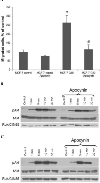
oxidases, which blocks the assembly of the active enzyme complex [21]. In accordance with our previ-ous results [16], we demonstrated that the motility of MCF-7 G10 cells with a high level of Ruk/CIN85
overexpression was signiicantly higher than that of
control cells (Fig. 1A). The treatment of G10 cells with apocynin resulted in the suppression of their
Fig. 1. Apocynin attenuates motility and induces the transition from sustained to transient EGF-dependent Akt activation in hu-man breast adenocarcinoma MCF-7 cells that overexpress adap-tor protein Ruk/CIN85. A. The transwell migration assay was used to study the motility of control (MCF-7 control) and Ruk/ CIN85-overexpressing (MCF-7 G10) cells. The number of mi-grated control cells was set equal to 100 %. Data are mean
± SEM of three independent experiments. *P ≤ 0.05 compared with control. #P ≤ 0.05 compared with MCF-7 G10. B, C. West-ern blotting of pAkt, tAkt and Ruk/CIN85 in lysates of control MCF-7 cells (B) and Ruk/CIN85-overexprerssing MCF-7 G10 cells (C) treated with EGF (100 ng/mL) for indicated periods of time. The representative blots of three independent experiments are shown.
A
B
migratory potential to the level of control cells. As can be seen from Fig.C, EGF induced more potent activation of Akt kinase up to 30 min of stimulation in comparison with the dynamics of Akt activity characteristic of control cells (Fig. 1B). The pretreat-ment of G10 cells with apocynin resulted in the dis-appearance of a signal for pAkt at 30 min while in control cells any changes in the Akt activity caused by apocynin were not observed (Fig. 1B, 1C). These data allow us to conclude that in Ruk/CIN85-overexpressing MCF-7 cells apocynin induces the transition from sustained to transient EGF-dependent Akt activation. Several studies demonstrated that ROS, such as H2O2, can modulate the Akt activity in various cell types through different mechanisms [for
review see 7] thereby inluencing the cell migration
and invasion [22]. To elucidate the molecular mech-anisms involved in the Ruk/CIN85-mediated regula-tion of NADPH oxidases and ROS producregula-tion, the control of Akt signaling and biological responses, further studies are necessary
Conclusions
The data obtained can suggest that ROS produced by NADPH oxidases are signaling components up-stream to Akt kinase that possibly mediate the in-creased migratory potential of Ruk/CIN85-overex-pressing MCF-7 cells.
Funding
This work was partially supported by bilateral grant between National Academy of Sciences (NAS) of Ukraine and Russian Fund for Basic Research (RFBR) – 2012.
REFERENCES
1. Meitzler JL, Antony S, Wu Y, Juhasz A, Liu H, Jiang G, Lu J, Roy K, Doroshow JH. NADPH oxidases: a perspective on reactive oxygen species production in tumor biology. Anti-oxid Redox Signal. 2014;20(17):2873–89.
2. Díaz B, Courtneidge SA. Redox signaling at invasive microdo-mains in cancer cells. Free Radic Biol Med. 2012;52(2):247–56. 3. Roy K, Wu Y, Meitzler JL, Juhasz A, Liu H, Jiang G, Lu J,
Antony S, Doroshow JH. NADPH oxidases and cancer. Clin Sci (Lond). 2015;128(12):863–75.
4. Lambeth JD. NOX enzymes and the biology of reactive oxygen. Nat Rev Immunol. 2004;4(3):181–9.
5. Katsuyama M. NOX/NADPH oxidase, the superoxide-gen-erating enzyme: its transcriptional regulation and physio-logical roles. J Pharmacol Sci. 2010;114(2):134–46. 6. Finkel T. Reactive oxygen species and signal transduction.
IUBMB Life. 2001;52(1–2):3–6.
7. Drobot LB, Samoylenko AA, Vorotnikov AV, Tyurin-Kuzmin PA, Bazalii AV, Kietzmann T, Tkachuk VA, Komisarenko SV.
Reactive oxygen species in signal transduction. Ukr Biokh-im Zh. 2013;85(6):209–17.
8. Pawson T. Dynamic control of signaling by modular adap-tor proteins. Curr Opin Cell Biol. 2007;19(2):112–6. 9. Levchenko A, Bruck J, Sternberg PW. Scaffold proteins may
biphasically affect the levels of mitogen-activated protein kinase signaling and reduce its threshold properties. Proc Natl Acad Sci U S A. 2000;97(11):5818–23.
10. Havrylov S, Rzhepetskyy Y, Malinowska A, Drobot L, Redo-wicz MJ. Proteins recruited by SH3 domains of Ruk/CIN85
adaptor identiied by LC-MS/MS. Proteome Sci. 2009;7:21. 11. Bazalii AV, Samoylenko AA, Petukhov DM, Rynditch AV, Re-dowicz M-J, Drobot LB. Interaction between adaptor pro-teins Ruk/CIN85 and Tks4 in normal and tumor cells of dif-ferent tissue origins. Biopolym Cell. 2014; 30(1): 37–41. 12. Gianni D, Diaz B, Taulet N, Fowler B, Courtneidge SA,
Bo-koch GM. Novel p47(phox)-related organizers regulate lo-calized NADPH oxidase 1 (Nox1) activity. Sci Signal.
2009;2(88):ra54.
13. Gianni D, DerMardirossian C, Bokoch GM. Direct interac-tion between Tks proteins and the N-terminal proline-rich region (PRR) of NoxA1 mediates Nox1-dependent ROS generation. Eur J Cell Biol. 2011;90(2–3):164–71.
14. Samoylenko AA, Byts NV, Pasichnyk GV, Kozlova NV, Baza-lii AV, Gerashchenko DS, Shandrenko SG, Vorotnikov AV, Kietzmann T, Komisarenko SV, Drobot LB. Recombinant lentivirus-mediated silencing of adaptor protein Ruk/CIN85
expression inluences biological responses of tumor cells.
Biotechnologia Acta. 2013; 6(4): 182–9.
15. Bazalii AV, Horak IR, Pasichnyk GV, Komisarenko SV, Drobot LB. Transcriptional regulation of NOX genes ex-pression in human breast adenocarcinoma MCF-7 cells is modulated by adaptor protein Ruk. CIN85. Ukr. Biochem. J. 2016; 88(1): 119–25.
16. Samoylenko A, Vynnytska-Myronovska B, Byts N, Kozlova N, Basaraba O, Pasichnyk G, Palyvoda K, Bobak Y, Barska M, Mayevska O, Rzhepetsky Y, Shuvayeva H, Lyzogubov V, Usenko V, Savran V, Volodko N, Buchman V, Kietzmann T, Drobot L. Increased levels of the HER1 adaptor protein Rukl/CIN85 contribute to breast cancer malignancy. Carci-nogenesis. 2012;33(10):1976–84.
CIN85: desining of expression vector and functional char-acterization. Biotechnologia Acta. 2015; 8(5): 19–26. 18. Ushio-Fukai M. Compartmentalization of redox signaling
through NADPH oxidase-derived ROS. Antioxid Redox Signal. 2009;11(6):1289–99.
19. Dikic I. CIN85/CMS family of adaptor molecules. FEBS Lett. 2002;529(1):110–5.
20. Havrylov S, Redowicz MJ, Buchman VL. Emerging roles of Ruk/CIN85 in vesicle-mediated transport, adhesion, migra-tion and malignancy. Trafic. 2010;11(6):721–31.
21. Stefanska J, Pawliczak R. Apocynin: molecular aptitudes. Mediators Inlamm. 2008;2008:106507.
22. McAuliffe PF, Meric-Bernstam F, Mills GB, Gonzalez-An-gulo AM. Deciphering the role of PI3K/Akt/mTOR pathway in breast cancer biology and pathogenesis. Clin Breast Can-cer. 2010;10 Suppl 3:S59–65.
Апоцинін пригнічує рухливість і індукує перехід від тривалої до тимчасової EGF-індукованої активації Akt в клітинах MCF-7 з надекспресією адаптерного протеїну Ruk/CIN85
А. В. Базалій, Л. Б. Дробот, С. В. Комісаренко
Мета. З’ясувати можливе залучення NADPH оксидаз до контр
-олю рухливості клітин і Akt-залежного сигнального шляху в аденокарциномних клітинах грудної залози людини лінії MCF-7, що стабільно надекспресують адаптерний протеїн
Ruk/CIN85. Методи. Для аналізу рухливості клітин використо
-вували модифіковану камеру Бойдена. Динаміку EGF-індукованої активації Akt досліджували Вестерн-блот аналі
-зом. Результати. Було показано, що апоцинін, інгібітор зби
-рання мембранних NADPH оксидаз, значно пригнічує рухли
-вість клітин MCF-7, що надекспресують Ruk/CIN85 (субклон G10) у порівнянні з контрольними клітинами. Крім цього, апоцинін індукував перехід від тривалої до тимчасової EGF-індукованої активації Akt у G10 клітинах і не впливав на тим
-часову активацію Akt у контрольних клітинах. Висновки.
Отримані дані дозволяють припустити, що АФК, продукти NADPH оксидаз, є сигнальними компонентами, розташовани
-ми вище від кінази Akt, які опосередковують підвищений мі
-граційний потенціал клітин MCF-7 з надекспресією Ruk/
CIN85.
К л юч ов і с л ов а: NADPH оксидази, апоцинін, адаптерний протеїн Ruk/CIN85, рухливість, EGF, Akt-залежне сигналю
-вання.
Апоцинин угнетает подвижность и индуцирует переход от длительной к временной EGF-индуцированной активации Akt в клетках MCF-7 со сверхэкспрессией адаптерного белка Ruk/CIN85
А. В. Базалий, Л. Б. Дробот, С. В. Комисаренко
Цель. Выяснить возможное вовлечение NADPH оксидаз в контроль подвижности клеток и Akt-зависимого сигнального пути в аденокарциномных клетках грудной железы человека линии MCF-7, которые стабильно сверхэкспрессируют адап
-терный белок Ruk/CIN85. Методы. Для анализа подвижности клеток использовали модифицированную камеру Бойдена. Динамику EGF-индуцированной активации Akt исследовали Вестерн-блот анализом. Результаты. Было показано, что апо
-цинин, ингибитор сборки мембранных NADPH оксидаз, зна
-чительно угнетает подвижность клеток MCF-7, которые сверхэкспрессируют Ruk/CIN85 (субклон G10) по сравнению с контрольными клетками. Кроме этого, апоцинин индуцировал переход от длительной к временной EGF-индуцированной ак
-тивации Akt в G10 клетках и не влиял на временную актива
-цию Akt в контрольных клетках. Выводы. Полученные дан
-ные позволяют предположить, что АФК, продукты NADPH оксидаз, являются сигнальными компонентами, расположен
-ными выше от киназы Akt, которые опосредуют повышенный миграционный потенциал клеток MCF-7 со сверхэкспрессией
Ruk/CIN85.
К л юч е в ы е с л ов а: NADPH оксидазы, апоцинин, адаптер
-ный белок Ruk/CIN85, подвижность, EGF, Akt-зависимая сиг
-нализация.