Induced Erythrocyte Hemolysis and Tumoricidal Action
Mehboob Hoque
1, Sandeep Dave
2, Pawan Gupta
2, Mohammed Saleemuddin
1*1 Interdisciplinary Biotechnology Unit, Aligarh Muslim University, Aligarh, India, 2 Institute of Microbial Technology (CSIR), Chandigarh, India
Abstract
A chance discovery of the tumoricidal action of a human milk fraction led to the characterization of the active component as oleic acid complex of the α-lactalbumin, which was given the acronym HAMLET. We report in this study that the oleic acid complex of bovine α-lactalbumin (BAMLET) is hemolytic to human erythrocytes as well as to those derived from some other mammals. Indirect immunofluorescence analysis suggested binding of BAMLET to erythrocytes prior to induction of hemolysis. Free OA was hemolytic albeit at higher concentrations, while sodium oleate caused hemolysis at far lower concentrations. Amiloride and BaCl2 offered protection against
BAMLET-induced hemolysis suggesting the involvement of a cation leak channel in the process. BAMLET coupled to CNBr-activated Sepharose was not only hemolytic but also tumoricidal to Jurkat and MCF-7 cells in culture. The Sepharose-linked preparation was however not toxic to non-cancerous peritoneal macrophages and primary adipocytes. The tumoricidal action was studied using the MTT-assay while apoptosis induction measured by the annexin V-propidium iodide assay. Repeated incubation of the immobilized BAMLET with erythrocytes depleted oleic acid and decreased the hemolytic activity of the complex. Incubation of MCF-7 and Jurkat cells with OA, soluble or immobilized BAMLET resulted in increase in the uptake of Lyso Tracker Red and Nile red by the cells. The data presented support the contention that oleic acid plays the key role, both in BAMLET-induced hemolysis and tumoricidal action.
Citation: Hoque M, Dave S, Gupta P, Saleemuddin M (2013) Oleic Acid May Be the Key Contributor in the BAMLET-Induced Erythrocyte Hemolysis and Tumoricidal Action. PLoS ONE 8(9): e68390. doi:10.1371/journal.pone.0068390
Editor: Luis Eduardo Soares Netto, Instituto de Biociencias - Universidade de São Paulo, Brazil
Received February 27, 2013; Accepted June 3, 2013; Published September 11, 2013
Copyright: © 2013 Hoque et al. This is an open-access article distributed under the terms of the Creative Commons Attribution License, which permits unrestricted use, distribution, and reproduction in any medium, provided the original author and source are credited.
Funding: The authors are grateful to the Department of Biotechnology, Government of India for support. MS is a recipient of UGC-BSR faculty fellowship and MH UGC-Maulana Azad National Junior Research fellowship. The funders had no role in study design, data collection and analysis, decision to publish, or preparation of the manuscript.
Competing interests: The authors have declared that no competing interests exist. * E-mail: [email protected]
Introduction
A serendipitous observation in the year 1995 that a casein fraction from human milk blocked the binding of bacteria to epithelial cell and was selectively toxic to a lung cancer cell line [1], evoked tremendous interest in the characterization of the tumoricidal molecule. The active fraction was soon traced to the casein fraction of human milk, obtained by acid precipitation [2] and was subsequently identified as oleic acid (OA)-bound partially unfolded α-lactalbumin (α-LA). The complex was given the acronym HAMLET (Human α-lactalbumin Made Lethal to Tumor cells) [3]. HAMLET exhibited broad tumoricidal activity against carcinomas, melanomas, glioblastomas and leukemias derived from human and various non-human sources [4]. Other studies showed that HAMLET like complexes with OA can also be prepared from bovine α-LA, which was designated as BAMLET [5], as well as from the protein derived from the milk of several other mammals [6]. The tumoricidal effect of HAMLET and BAMLET has been observed
also in vivo, in animal models [4,7] and in a clinical investigation [8]. The exact mechanism by which HAMLET-like complexes exert tumoricidal action continues to remain elusive despite concerted global efforts. Available data however suggest that the OA complex of α-LA selectively enters tumor cells and induces apoptosis-like events, stimulates macroautophagy and inhibits proteasomes [9,10]. Storm et al. [11,12] using small-hairpin RNA inhibition, proteomic and metabolomics technologies showed that c-Myc oncogene is an important determinant of HAMLET sensitivity; cells expressing high levels of c-Myc were more sensitive, while knock down of the gene lowered HAMLET sensitivity. More recent studies showed that both prophylactic and therapeutic effects of HAMLET against colon cancer are mediated by β-catenin signalling [13].
β-lactoglobulin and parvalbumin also formed OA complexes that were selectively toxic to tumour cells [15]. A recombinant variant of human α-LA in which all cysteine residues were replaced with alanine and lacked tertiary structure was not tumoricidal on its own, but could be transformed into one after association with OA [16]. This suggested that both unfolding and OA binding are essential for the killer activity against tumor cells. Considering that wide variations in sequence and conformation of the α-LA do not negatively affect its transformation to an antitumor molecule and near specific requirement of OA, questions were raised on the seminal role of the protein in the HAMLET/BAMLET [14,15]. In a more recent study Brinkmann et al. [17] showed that some primary cells and even human erythrocytes are also sensitive to BAMLET and attributed the lytic activity primarily to the OA component. While other studies that support the contention that OA is the toxic principle and α-LA acts to facilitate its solubilisation appeared [18,19], these claims are contested by works which continue to attribute tumoricidal effect to the unfolded α-LA [11,12]. We have investigated in this study the effect of soluble and immobilized BAMLET on red blood cells as well as Jurkat and MCF-7 cell lines and observed that prevention of entry of α-LA component of the complex in the cells by coupling it to Sepharose support does not abolish hemolytic/tumoricidal action. The results support the contention that OA may be playing the seminal role in the toxic action of BAMLET on erythrocytes and at least some tumor cells.
Materials and Methods
Ethics statements
Blood was withdrawn by expert technician under the supervision of a senior physician in the University Medical College from volunteers. Goat and buffalo blood was collected from the local slaughterhouse, Frigerio Conserva Allana Ltd, Aligarh, from the routinely slaughtered animals after obtaining a written permission from the management. Blood from rats, mice and rabbits was collected from the institutional animal house (registration number 332) according to the recommendations of Committee for the Purpose of Control and Supervision of Experiments on Animals (CPCSEA), Government of India. The Aligarh Muslim University Bioethical Committee approved the use of blood from the slaughterhouse animals, animals maintained in the animal house and human blood from volunteers after obtaining their written consent on prescribed pro forma.
Materials
Bovine α-LA (type III, Ca2+ depleted), Sodium Oleate (Na
Oleate), Sepharose 4B and MTT assay reagent were obtained from Sigma Chemical Company, St. Louis, USA. OA was purchased from LOBA Chemie India. DMEM, RPMI, fetal bovine serum, penicillin–streptomycin and trypsin were purchased from Invitrogen. All chemicals not listed here were of analytical grade.
Preparation of BAMLET
BAMLET was prepared by the heat-treatment procedure described by Kamijima et al. [20]. Briefly, α-LA was dissolved in phosphate-buffered saline (PBS), pH 7.4 to a concentration of 700 µM. OA (120 molar equivalents) was added to the protein solution, mixed gently and the mixture was incubated at 50 °C for 10 min. The tubes were cooled to 25 °C and centrifuged. After removal of excess OA by aspiration the preparation was subjected to dialysis against PBS for 18 h.
Protein estimation
The concentration of α-LA was estimated spectrophotometrically. The molar extinction, A1%
1 cm value of
20.1 (for native α-LA) and 22.8 (for α-LA in BAMLET) at 280 nm were used for calculating the concentration of protein [21].
OA estimation
Concentration of OA was estimated by the procedure described by Wawrik and Harriman [22] with few modifications. Briefly, to Eppendorf tubes containing 400 µL of copper reagent (9 volumes (vol.) aq. 1.0 M triethanolamine, 1.0 vol. 1.0 N acetic acid, 10 vol. 6.45% (w/v) Cu(NO3)2.3H2O) was added
equal vol. of OA containing samples prepared in 100 mM Tris-HCl buffer, pH 8.0 and the tubes were mixed by Vortexing. Chloroform (500 µL) was added and the tubes were Vortexed again for another 1 min. The samples were centrifuged at 14,000 x g for 3 min and the organic phase was carefully transferred into a new tube. Upon addition of 50 µL of 1% (w/v) sodium diethyldithiocarbamate in 2-butanol, yellow colour was developed which was read at 440 nm. For the preparation of standard curve OA was dissolved in ethanol and diluted in 100 mM Tris-HCl buffer, pH 8.
Chemical cross-linking of α-LA and BAMLET
α-LA and BAMLET prepared as described earlier were chemically cross-linked following the method described by Spolaore et al. [19] with a few modifications. Briefly, 5 mg.mL-1
of protein in phosphate buffer (20 mM, pH 7.4) was incubated at 25 °C with 0.01% glutaraldehyde prepared freshly in the same buffer. The cross-linking reaction was stopped by adding 1 M Tris buffer, pH 7.4 at different time period and the samples were characterized by SDS-PAGE in presence of 1% β-mercaptoethanol. BAMLET was prepared from the cross-linked α-LA as described earlier.
Immobilization of α-LA/ BAMLET on Sepharose 4B matrix
α-LA was converted to BAMLET by the same procedure as described earlier for soluble preparation.
Isolation of erythrocytes
Nine vol. of blood was collected from healthy volunteers in the tubes containing 2.7% (w/v) EDTA to prevent coagulation and centrifuged at 650 x g for 5 min. Plasma was removed carefully and the white buffy layer was completely removed by aspiration with a pipette with utmost care. The erythrocytes were then washed for additional three times in wash buffer (10 mM Tris, 0.9% NaCl and 5.0 mM glucose, pH 7.4) and used for further experiments.
Effect of BAMLET on erythrocyte membrane
Erythrocyte membranes were prepared as described by Fairbanks et al. [24]. The cells were hemolyzed in 20 vol. of 5 mM Tris-HCl buffer, pH 8.0 at 4 °C. The membrane was then pelleted by centrifugation at 12,000 x g for 10 minutes at 4 °C and repeatedly washed with the buffer to prepare hemoglobin-free membranes. The membrane was treated with 70 µM BAMLET for 2 h. Membrane was also prepared from the BAMLET-treated erythrocytes and analysed by SDS-PAGE under reducing condition.
Hemolysis assay
Ten microliters of packed erythrocytes were incubated with various concentrations of BAMLET for 90 min at 25 °C in a total vol. of 100 µL. The cells were then diluted to 1.0 mL with normal saline, incubated at 25 °C for 10 min. After removal of intact cells by centrifugation at 650 x g, haemoglobin in the supernatant was measured spectrophotometrically at 410 nm. For total lysis the erythrocytes were lysed in distilled water instead of saline and the value was used as 100 for calculation of percent hemolysis. All the experiments were reformed in quadruplets and repeated for at least three times.
Indirect Immunofluorescence of BAMLET treated erythrocytes
Ten microliters of packed human erythrocytes were treated with 20 µM BAMLET for 10 min at 25 °C and washed with PBS, pH 7.4. The pelleted cells were incubated with polyclonal anti-BAMLET antibody raised in mice (1:100 dilution in PBS, pH 7.4) for 30 min at 37 °C. The cells were again washed with PBS, pH 7.4 and finally suspended in 100 µL of the buffer. FITC-conjugated rabbit anti-mouse IgG (Sigma) was added to the cell suspension at 1:100 dilution and incubated for 30 min at 37 °C in dark. The cells were washed thoroughly and visualized under fluorescence microscope using a 100X oil immersion objective (Zeiss M2 Imager; Zeiss, Göttingen, Germany).
Cell culture
Cell lines were procured from cell repository at National Centre For Cell Science, Pune, India. Jurkat were grown in culture medium composed of RPMI-1640 and MCF-7 in DMEM with glutamine supplemented with 10 mM HEPES, 23.8 mM NaHCO3, 10% heat-treated fetal bovine serum,
penicillin (60 µg.mL-1) and streptomycin (0.1 mg.mL-1). Cells
growing in suspension (Jurkat) were kept at between 1 × 10⁵ to
1.5 × 10⁶ cells/mL. Cells were switched to serum-free medium
6 h prior to different treatment.
MTT assay
MTT assay was performed as described previously [25]. Briefly, Cells were seeded at 5,000 cells/well on 96-well plates and incubated for 24 to 48 h before the treatment. Cells treatment was continued with or without stimuli for 24 h, a 20 µL aliquot of 3-(4,5-Dimethylthiazol-2-yl)-2,5-diphenyltetraolium bromide (MTT, a yellow tetrazole; 5 mg.mL-1 in PBS) was
added to the wells and incubated for 4 h at 37 °C. After removing supernatant carefully, 200 µL of DMSO was added and mixed, and the absorbance was read at 563 nm.
Staining with annexin-V and PI
Apoptosis studied by annexin V-FITC Apoptosis Detection Kit (Calbiochem) to stain cells in annexin binding buffer according to the manufacturer’s instructions. Cells were analyzed with a BD FACS Calibur flow cytometer (BD Biosciences).
LysoTracker Red-uptake
Cells were cultured in media at 37°C and after treatment with or without compound. The acidotropic dye LysoTracker Red DND-99 was diluted in DMEM, resuspended in prewarmed (37 °C) medium containing 50 nM LysoTracker Red for 30 min [26]. Cells were then resuspended in PBS and lysosomal fluorescence of 5,000 cells per sample was determined by flow cytometry using the FL3 channel. CELLQUEST-PRO/Flowjo software was used to analyze all of the data from flow cytometric experiments.
Nile Red Staining
Nile red staining was performed as described by Greenspan et al. [27]. Briefly, Nile red (1 mg.mL-1) in acetone was prepared
and stored protected from light. The excitation wavelength was set at 488 nm with a 2 nm slit width; the emission wavelength was set at 540 nm with a 20 nm slit width and the fluorescence spectra were recorded. Excitation and emission fluorescence spectra were determined with a Cary Eclipse spectrofluorimeter (Varian). The relative fluorescence intensity of Nile Red in the presence of the various samples was obtained after subtraction of both the autofluorescence of the samples and the fluorescence intensity of Nile red alone in the buffer.
Isolation of peritoneal macrophages and primary adipocytes
9-week-old male C57BL/6J mice as described previously [29,30]. Briefly, the epididymal fat pads were removed, minced, and digested using collagenase at 37 °C for 2 h. The primary adipose cells were then washed extensively and incubated at 37 °C in Dulbecco’s modified Eagle’s medium containing 5% bovine calf serum.
Statistical analysis
Results are expressed as the mean ± SD unless otherwise mentioned. SigmaPlot (SyStat Software) and SPSS (IBM) were used for statistical analysis. All statistical data were from averages of three or more independent experiments. Two-tailed Student t test was performed to obtain P values. Statistical significance was established at * P<0.01.
Results and Discussion
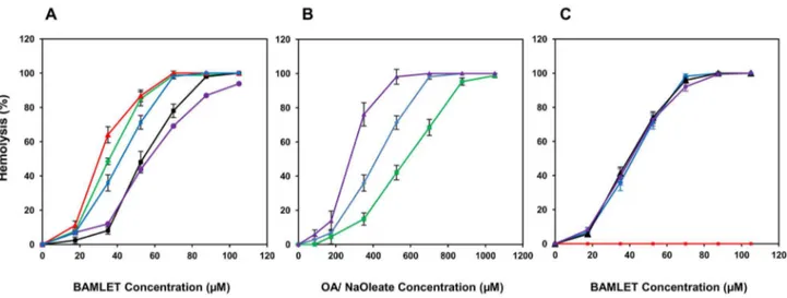
BAMLET used in the study was prepared following the procedure described by Kamijima et al. [20], in which heat-treated α-LA is exposed to OA. As also reported by the authors, BAMLET prepared thus exhibited altered spectroscopic properties, compared to α-LA, suggesting a molten globule like structure (Figure 1). While UV-absorption spectra indicate the exposure of side chains of aromatic residues to the solvent, loss of tertiary structure and acquisition of additional secondary structure are evident from the far and near UV-CD spectra. Intrinsic fluorescence measurements also suggest exposure of tryptophan(s) to the solvent and loss of tertiary structure. The structure of the complex bears striking similarity with that of BAMLET/HAMLET prepared by the ion exchange procedure [20]. The OA/protein molar ratio of the preparation was 10 (Table 1), which is close to that reported by Kamijima et al. [20], but an order of magnitude higher than the value reported by Svensson et al. [5] for the complex originally prepared by the ion-exchange procedure. More recent studies however suggest that in HAMLET-like complexes, obtained using various procedures including that based on ion-exchange, each molecule of α-LA binds to multiple molecules of OA [31]. In a very recent study Nakamura et al. [32] compared stoichiometry of protein and OA in HAMLET prepared in the authors’s laboratory by the ion-exchange procedure [3] and the heat-treatment procedure [20], also used in this study. Both the preparations contained 8.5 molecules of OA per protein molecule, a value close to that observed by us in this study.
While studies showing selective killing of cancer cells by BAMLET are numerous [1,4,33,34], recent reports suggest that the OA complex of α-LA may also be toxic to some non-cancerous cell, human erythrocytes [17] as well as to antibiotic sensitive and resistant strains of Streptococcus pnuemoniae
[35,36]. Figure 2A shows that in addition to human erythrocytes, those derived from several other mammals were readily hemolyzed by BAMLET, although their sensitivity to the α-LA-OA complex varied. While buffalo erythrocytes were most susceptible, those from mouse exhibited maximum recalcitrance, requiring 100 µM BAMLET for causing complete hemolysis. Ruminant erythrocytes lack phosphatidyl choline, the prominent phospholipid of most mammalian red cells [37],
yet of the two ruminant erythrocytes investigated buffalo erythrocytes were highly susceptible and goat erythrocytes more resistant to BAMLET-induced hemolysis. This suggests that hemolytic action of BAMLET may not necessitate specific composition of the membrane lipid(s). Toxic action of BAMLET against some bacteria [35,36] with membranes differing remarkably in lipid composition also argues against specific role of membrane lipids. Some studies have however provided evidence that nature of membrane lipids may determine the mode of action of HAMLET/BAMLET. HAMLET binds uniformly to the vesicles prepared from egg yolk and soybean phospholipids but when exposed to those prepared from tumor cell membranes reveal punctate binding pattern [38] and influence the permeability of tumor cell membranes to ions [1,39]. In a recent study it was shown that S. pneumoniae D39 cell membrane damage caused by BAMLET was mediated through Na+/Ca+2 exchange activity, calcium entry and
membrane depolarization [15]. The indirect immunofluorescent images of BAMLET treated erythrocytes provided evidence for the binding of BAMLET to erythrocyte surface (Figure 3). The BAMLET binding was detectable after incubation of the human erythrocytes with mouse anti-BAMLET antibody followed by FITC conjugated secondary antibody. The fluorescence was however lost on longer incubation due to hemolysis of the cells. We have however noticed no significant binding of BAMLET to human erythrocyte membrane proteins by SDS-PAGE after incubation of the intact cells or hemoglobin-free ghosts with the complex (Figure S1).
A recent report suggests that HAMLET forms annular oligomers when deposited on phospholipid monolayers, a property shared by α-LA and the peptide C derived from the protein but only at higher concentrations [40]. While studies that provide evidence supporting the formation of annular oligomers in intact cells exposed to HAMLET/BAMLET are lacking, the authors envisage a role of the annular oligomers in the permeability alterations and accumulation of the complex observed cytoplasm and subcellular organelles of the HAMLET exposed tumor cells.
Free OA was hemolytic but caused the lysis of human erythrocytes at concentrations higher than those required when present in the form of complex with α-LA (Figure 2B). Brinkmann et al. [17], however observed OA to be equally hemolytic in free form or when associated with the protein. While we have no definite explanation for the discrepancy, poor solubility of OA, its tendency to stick to the walls of plastic tubes under the conditions of the study and consequent low availability [17] for interacting with the cells apparently
Table 1. α-LA and OA molar ratios in various BAMLET preparations.
BAMLET Preparation α-LA:OA molar ratio*
Uncrosslinked 1: 10 (±1.4)
Crosslinked prior to OA binding 1: 11 (±1.52)
Crosslinked after OA binding 1: 10 (±1.4)
necessitated high concentrations of the fatty acid to induce hemolysis. Oleate, that forms HAMLET-like complexes [41] and its relevant constituent [42], was however more hemolytic than OA and moderately more hemolytic than even α-LA associated OA (Figure 2B). BAMLET prepared by the procedure of Kamijima et al. [20], when subjected to crosslinking with 0.01% glutaraldehyde resulted in the formation of adducts that moved in SDS-PAGE as polypeptides ranging from 50 kDa to those that were too large to enter the gel (Figure S2) as also reported by Spolaore et al. [19]. The amount of OA associated with the crosslinked preparations containing mostly protein oligomers
was comparable with that of the uncrosslinked BAMLET (Table 1). Both crosslinked BAMLET or α-LA that was heat-treated and crosslinked with glutaraldehyde prior to binding of OA were also hemolytic (Figure 2C) suggesting a mechanism of hemolytic action that may not require the entry of the BAMLET as entry of the covalently linked protein aggregate in the erythrocytes is unlikely, due to the inability of the erythrocytes to take up large molecules by phagocytosis/ endocytosis.
In order to further investigate the relative roles of α-LA and OA in inducing the hemolysis, BAMLET was immobilized on Sepharose 4B and hemolytic property of the immobilized
Figure 1. Spectrometric analysis of BAMLET (red line) native α-LA (black line). UV absorption spectra were recorded on a PerkinElmer Lambda 25 UV/VIS spectrophotometer in a quartz cuvette with 1.0 cm path length. Protein concentration used was 1.0 mg.mL-1 (panel A). Intrinsic fluorescence spectra were recorded at 25 °C between 300 to 500 nm by exciting Trp at 295 nm in a
Shimadzu RF-5301 PC spectrofluorophotometer using a quartz cuvette with 1.0 cm excitation path length. Protein concentration used was 0.1 mg.mL-1 (Panel B). Near-UV CD spectra were recorded between 320 and 250 nm using quartz cuvette with 1.0 cm
path length. Samples were prepared with a protein concentration 1.0 mg.mL-1 (panel C). Far-UV CD spectra were recorded between
250 and 195 nm at the scan rate of 200 nm/min using a quartz cuvette with 0.1 cm path length. Protein concentration used was 0.3 mg.mL-1 (panel D). The CD spectra were obtained using a JASCO J-815.
preparation on human erythrocytes investigated. The idea was to prevent the entry of the protein component of the complex into the erythrocytes and restrict its action to the cell surface. OA binding was not affected when BAMLET was coupled onto Sepharose and the matrix bound α-LA was equally effective in binding the fatty acid when carried through the procedure of
BAMLET formation (Table 2). As shown in Figure 4A, both the immobilized preparations were hemolytic towards human erythrocytes, albeit to a lower extent than the soluble BAMLET. While conformational alterations in α-LA resulting from the immobilization were not studied, the ability of the immobilized α-LA to bind OA and the resulting complex to hemolyze
Figure 2. BAMLET induced hemolysis of erythrocytes. Erythrocytes isolated from the blood of mouse (purple line), goat (black line), human (blue line), rabbit (green line) and buffalo (red line) were incubated with BAMLET and the resulting hemolysis determined as described under methods (Panel A). Human erythrocytes were treated either with free OA (green line), BAMLET (blue line), and free Na oleate (purple line) and the resulting hemolysis determined (panel B). Human erythrocytes were incubated with BAMLET (blue line), glutaraldehyde-treated BAMLET (purple line), and glutaraldehyde-treated α-LA converted to BAMLET (black line) and crosslinked α-LA (red line) (Panel C). Data are expressed as mean ± SD.
doi: 10.1371/journal.pone.0068390.g002
Figure 3. Interaction of BAMLET with erythrocytes. Human erythrocytes were incubated with 20 µM BAMLET in PBS for 10 min at 25 °C and washed thrice with PBS, pH 7.4. Anti-BAMLET polyclonal antibodies raised in mouse were added and the cells were further incubated at 37 °C and again washed thoroughly. The interaction was then probed with FITC conjugated secondary rabbit anti-mouse IgG (Sigma Chemical Co.) and the cells visualized under a fluorescence microscope at 100X magnification. The images shown are those observed under fluorescence (Panel A), bright field (Panel B) as well as that merged of both fluorescence and bright filed (Panel C).
erythrocytes is indicative of conformational similarity between the BAMLET and the Sepharose-immobilized preparation. Since immobilized BAMLET preparations did not leach out any significant amount of the protein, when incubated in absence of erythrocytes for up to 12 h, as analysed by ELISA (detection limit 0.16 ng), using specific anti-α-LA polyclonal antibodies that also recognize BAMLET (data not included), it is unlikely that protein released from the Sepharose support contributed towards hemolysis.
Studies on the reuse of the immobilized BAMLET further supported the seminal role of OA (Figure 4B). In these studies, the immobilized BAMLET preparations, after incubation with erythrocytes, were collected, washed and reincubated with a fresh batch of erythrocytes. The hemolytic activity decreased remarkably after second incubation and disappeared completely after the third. Analysis of bound OA suggests that the OA content of the immobilized BAMLET preparations decreased remarkably after first exposure to erythrocytes and became almost undetectable after the second (Table 2).
Storm [12] observed that HAMLET induces activation of an ion channel involved in the transport of K+, Na+ and Ca2+ in the
membranes of tumor cells that explains the earlier observed intracellular calcium accumulation by the tumor cells exposed to the complex. Interestingly, neither OA nor native α-LA caused the ion channel activation. More recent studies from the group suggest that enhanced ion fluxes across the plasma membrane are essential for the HAMLET-induced initiation of the processes that eventually lead to tumor cell death [43]. Studies employing lymphomas and carcinomas also suggest the involvement of a K+, Na+ and Ca2+ permeable non-selective
cation channel, distinct from the classical TRP, ENaC- or CNG channels in HAMLET-mediated cell death. Fully differentiated HRTEC cells however do not reveal comparable ion channel activation in response to HAMLET. Our studies also suggest the possible involvement of a non-specific cation channel activation in the hemolysis induced both by soluble (Figure 5A & B) and Sepharose bound BAMLET (Figure 5C & D) in human as well as goat erythrocytes. The BAMLET-induced hemolysis was preceded by cation leakage and inhibited by BaCl2 and
amiloride, suggesting similarity with several non-specific ion channels and inhibitors. Also while inclusion of EGTA moderately decreased the susceptibility of erythrocytes to the BAMLET-induced lysis, indicating a role of Ca2+, inclusion of
CaCl2 in the medium was protective against hemolysis. Ca2+
release OA from BAMLET [19], form insoluble salt and lower
Table 2. α-LA/OA molar ratios in the immobilized α-LA complexes of OA after incubation with human erythrocytes*
. BAMLET Preparation No incubation Single incubation Two incubations Three incubations Sepharose-linked α-LA incubated with OA
1:10 (±1.6) 1: 5 (±0.81) 1: 2 (±0.82) 1:0
Sepharose-linked 1:10 (±1.53) 1: 6 (±0.78) 1: 3.5 (±0.58) 1:0 *. Each value represents the mean ±SD of four determinations doi: 10.1371/journal.pone.0068390.t002
the availability of OA for interaction with the membrane [44]. The sensitivity of soluble and Sepharose-bound BAMLET-induced hemolysis to the inhibitors suggests similarities in their mechanism of action. Similarly OA-induced hemolysis was also sensitive to BaCl2 (data not included). Evidence for the
existence of non-selective cation channels, by applying patch-clamp single chain recording have been provided in human erythrocytes [45] and these were further characterized with respect to the stimulants [46,47]. More recently Föller et al. [48] have shown that human and mouse erythrocytes contain a transient receptor potential channel that contributes towards cation leak. During their investigation of HAMLET-induced ion channels in A549 lung carcinoma cells, Storm et al. [43] observed that the increase in free intracellular calcium was not restricted by inclusion of EGTA in the medium but by the inhibition of INsP3-gated ER Ca2+ channel with U73122,
implying that it originated mainly from intracellular stores. In another recent study [42] it was however shown that HAMLET and oleate trigger overlapping but fundamentally different responses on the ion channels of the lung carcinoma and Jurkat cells.
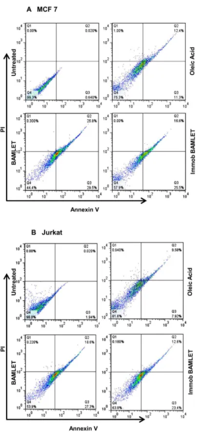
Immobilized BAMLET was also tumoricidal against MCF-7 and Jurkat cells. The MTT assay indicates that, unlike the soluble and immobilized α-LA which were barely toxic, BAMLET markedly lowered the number of surviving cells when added either as soluble or Sepharose bound preparations (Figure 6A & B). Neither decrease in cell viability nor apoptosis induction was however evident when non-cancerous mouse primary adipocytes or peritoneal macrophages were incubated with the soluble or immobilized BAMLET preparations (Figure 6C & D). The annexin-V staining assay, that detects apoptotic cell memebrane phosphatidyl serine (PS) externalization [49], was employed to further examine if the observed decrease in cell viability is related to induction of apoptosis-like processes [50]. Figure 7 reveals that the decrease in the viability of Jurkat and MCF-7 cells is indeed related to induction of apoptosis-like process by BAMLET with large fraction of cells exposed either to soluble or immobilized complex exhibiting enhanced PS exposure.
[51]. This suggests that lysosomal acidification precedes
permeabilization of lysosomes especially in MCF7 cells. Lysosomal acidification has earlier been correlated to leakymonovalent permeable ion channel TRP-ML1 which is involved
Figure 4. Hemolysis of human erythrocytes incubated with soluble and immobilized BAMLET. Human erythrocytes were
incubated with soluble BAMLET, immobilized α-LA, immobilized BAMLET and immobilized α-LA converted to BAMLET and the resulting hemolysis quantitated (panel A). Erythrocytes were incubated with immobilized BAMLET or immobilized α-LA converted to BAMLET and hemolysis quantitated. The immobilized preparations were collected, washed and incubated with a fresh batch of erythrocytes. The immobilized preparations were reused after the third cycle (panel B). Data are expressed as mean ± SD.
in control of pH in the organalle [54]. Also Cadmium induced tandem acidification and permeabilization of lysososmes has been reported [55]. It is tempting to suggest of a possible role of the cation channel in acidification and permeabilization of lysosomes in the BAMLET/OA-treated cells. Soluble and immobilized BAMLET-mediated increase in Nile Red uptake (Figure 8B) is suggestive of oleic acid access to the cells. HAMLET induced uptake of OA by cancer cells has also been demonstrated in an earlier study as well [11]. A corollary question is whether BAMLET has cell specific attribute and mechanism to induce cell death remain unanswered. There has been a spectrum of multipronged reports implicating the cytotoxicity of the BAMLET to tumor cells by different
mechanisms including apoptosis, necrosis, autophagy, perturbance in the chromatin structure, proteosome degradation, mitochondrial membrane depolarization, cytochrome C release, PS exposure, and a low caspase response [52]. In its simplest rendition, this complexity might underlie as few of these mechanisms associated with specific cell lines, although different mechanisms operative in a single cell lineage to variable parameters such as concentrations and different time points have also been suggested.
The ability of OA to exert tumoricidal activity on its own has been documented in some early and more recent studies [56-59]. Antitumor potential of OA was recognized several decades earlier [60,61] and its protective role against various
Figure 5. Effect of calcium, EGTA and ion-channel inhibitors on BAMLET-induced hemolysis of erythrocytes. Human (Panel A & C) and goat (Panel B & D) erythrocytes were incubated with 1 mM of each the test substances for 30 min at 25 °C and 50 µM soluble (Panel A & B) or 70 µM immobilized BAMLET (Panel C & D) were added. The samples were further incubated for additional 90 min at 25 °C and the resulting hemolysis measured. The results shown are average of three independent experiments, which were each conducted in triplicate. Data are expressed as mean ± SD.
forms of cancer has been supported by epidemiological studies [62,63]. The central role of OA in killing of the tumor cells was also envisaged from the observations that α-LA isolated from milk of several mammals and differing to varying extent in amino acid sequence [6], mutants of the protein [16] and even those structural unrelated [15], could be converted to tumoricidal complexes, while OA requirement was almost exclusive. More direct evidence in favour of the seminal role of OA in the toxic action of HAMLET/BAMLET was provided by Permyakov et al. [15], on the basis of their studies on OA complexes of few proteins structurally and functionally different from α-LA, both on Hep-2 tumor cell cells and S. pneumoniae.
Also considering the observed decrease in thermal stabilization that accompanied complex formation of β-lactoglobulin with OA after exposure to high temperature, even the requirement for molten globule like conformation of the protein in the complexes to exert killer action against tumor cells was questioned [15].
Menendez et al. [56] observed that OA suppresses the Her-2/neu overexpression and in turn interacts synergistically with anti-Her-2/neu breast cancer immunotherapy by promoting apoptotic death of breast cancer cells exhibiting amplification of the Her-2/neu gene. Khamaisie et al. [59] reported that OA inhibited Bcr-Abl kinase autophosphorylation in Ba/F3 cells and
Figure 6. Cytotoxicity of immobilized BAMLET. The cytotoxicity of free OA, soluble αLA, immobilized αLA, soluble BAMLET, immobilized BAMLET and immobilized αLA converted to BAMLET was evaluated on tumor cell line (A) MCF-7 and (B) Jurkat cells and primary cells (C) Mouse Peritoneal Macrophage and (D) Primary Mouse adipocytes, with the MTT assay after incubation for 24 h with mild shaking. The results were confirmed by three independent experiments, which were each conducted in triplicate. Data are expressed as the mean ± SD.
Figure 7. A comparison of BAMLET-induced apoptosis and necrosis in MCF-7 (A) and Jurkat cells (B). Cells were incubated with immobilized-BAMLET, soluble BAMLET or OA for 24 h with mild shaking before flow cytometric analysis. OA was dissolved in ethanol, diluted in media and was then sonicated before treatment to the cells. The results were confirmed by three independent experiments, which were each conducted in triplicate. Data are expressed as the mean ± SD. *P<0.01 vs. controls.
showed anti-CML activity in a BCR/ABL-positive mouse model. Interestingly the Gas Chromatography-Mass Spectrometry (GC-MS) profiles of cellular metabolites of A549 lung carcinoma cells incubated with HAMLET revealed rapid uptake of OA from the complex [11].
It is now recognized that unfolded α-LA facilitates the solubilisation/dissociation of large aggregates of ionized fatty acid that are formed above the critical micellar concentration [14,17,19]. The complex prepared using different procedures comprise between 1 to 20 molecules of OA per α-LA
depending on the procedure used and the fatty acid may bind at multiple sites [64]. Binding to the protein is likely to facilitate availability of higher and toxic concentration of OA for erythrocytes and tumor cells. The studies of Permyakov et al. [15] have also showed a direct correlation between the fatty acid concentration in protein complexes and toxicity.
A large number of studies have however shown that the partially unfolded α-LA present in HAMLET/BAMLET plays important role in its toxicity against the tumor cells. Evidence for the penetration of HAMLET/BAMLET into tumor cells and
Figure 8. Effect of soluble and immobilized BAMLET on the uptake of Lyso Tracker and Nile Red. (A) LysoTracker signal in the MCF-7 (Top panel) and Jurkat (Bottom panel) cells after 4 h of incubation with OA, BAMLET or immobilized BAMLET. Cells were labelled with LysoTracker and the signal was analyzed by flow cytometry as described in Methods. (B) Nile red uptake by MCF-7 (left panel) and Jurkat (Right panel) cells after 4 h incubation with OA, BAMLET, and immobilized BAMLET. The results were confirmed by three independent experiments, which were each conducted in triplicate. Data are expressed as the mean ± SD. *P<0.05 vs. Controls.
attack on several critical organalles including mitochondria, endoplasmic reticulum, proteasomes, lysosomes and trigger cell death pathways are available [9,34,53]. Storm et al. [11] observed that that c-Myc oncogene is an important determinant of HAMLET sensitivity; cells expressing high levels of the c-Myc gene were more sensitive while knock down of the gene lowered HAMLET sensitivity and modified the glycolytic state of the cells. The in vivo interaction of Alexa Fluor 568-labelled HAMLET with hexokinase1 using confocal microscopy as well as direct binding to the enzyme in vitro with the help of a high functional protein array has also been taken as evidence in support of the tumoricidal role of α-LA in the HAMLET [11]. A recent review has also summarized the potential role of the partially unfolded protein in HAMLET/BAMLET-like complexes [34]. Puthia et al. [13], in a more recent study have shown that HAMLET acts by triggering a Wnt-independent and ion channel dependent mechanism controlling β-catenin level in addition to the effects on β-catenin signalling.
Nakamura et al. [32] in recent study characterized that OA binding site in HAMLET and a similar complex prepared using goat α-LA (GAMLET) by 2D NMR spectroscopy and observed remarkable differences in OA binding sites; in HAMLET most of the residues affected by OA binding are within the A- and B-helices while those of GAMLET are located between the α and β subdomains. This suggested little role of α-LA, as no unique OA binding structure may be essential in the protein for expression of antitumor activity. In contrast, another very recent report concluded, based on a comparative analysis of ion fluxes, gene expression and tumoricidal activity that oleate and HAMLET produce overlapping yet clearly distinct effects [42].
Most studies describing tumoricidal action of HAMLET observed little or no toxicity of native α-LA against the tumor cells. Our studies on human erythrocytes and MCF-7 and Jurkat cells substantiate these observations (Figure 6A and B). Some earlier studies however show that native α-LA may induce apoptosis at least in some cells. For example native α-LA has been shown to cause apoptosis of mouse, cape fur seal and human mammary and cell lines as well as fibroblasts and myoepithelial cells and the activity was lost on heat treatment and pepsinization [65]. Apoptosis induction by native α-LA in human colon adenocarcinoma [66] and RAW 264.7 cells [67] has also been documented.
Erythrocyte hemolysis and tumoricidal action of HAMLET/ BAMLET may involve different mechanisms of action. Unlike in case of the tumor cells, erythrocyte hemolysis as shown by experiments with immobilized preparations, may be mediated through the action of BAMLET or the bound OA principally on the cell surface molecules. Cytosolic events requiring genomic involvements are essential for causing apoptosis-like cell death in case of the tumor cells. Although BAMLET binds to erythrocytes (Figure 3), its possible entry in to the cells could not be studied by ELISA of the protein due to ensuring hemolysis. HAMLET has been shown to lose its tumoricidal activity in presence of serum albumin [68]. Characterization of an equine lysozyme multimeric complex with OA, a preparation resembling HAMLET but with higher OA: protein ratio, subsequent to interaction with membranes reveals conversion
to nearly native like state, apparently after offloading of the bound fatty acid [69]. It is therefore likely that HAMLET/ BAMLET may be stripped of the bound OA during its interaction with the target cells. Unfortunately no study describing the nature of α-LA after internalization of the complex is available.
In a recent study Permyakov et al. [15], assuming the low contribution of the protein component in HAMLET towards its tumoricidal action and ability of proteins widely differing in structure to form BAMLET-like complexes suggested that association of OA with established antitumor proteins may generate superior tumoricidal molecules. Native α-LA can also be tumoricidal at least against some cells as discussed earlier and possible presence of even OA-depleted BAMLET in the cells may also enhance the killer action of OA. The observed moderately low effect of immobilized BAMLET, as compared to the soluble complex (Figure 6A & B) may indicate such supplementation of the OA-induced tumoricidal action by the protein. To the best of our knowledge the exact nature of structure assumed by α-LA after the entry of BAMLET/ HAMLET in the tumor cells is not known. It is however not likely that it will remain in the form of complex, because interaction with the membrane and cytoplasmic proteins that may deplete the molecule of its OA. Characterization of the structure of BAMLET internalized by the tumor cells will certainly be interesting.
In conclusion, this study provides evidence that OA component of BAMLET may play a key role both in causing hemolysis of mammalian erythrocytes as well as in the killing of Jurkat and MCF-7 cells. This was apparent from the experiments which showed that coupling of BAMLET to Sepharose support did not abolish the hemolytic and tumoricidal activities. Immobilized BAMLET exposed to erythrocytes was depleted both of bound OA and the ability to hemolyze erythrocytes. A recent exhaustive review by Fontana et al. [31] provides several arguments to show that OA can exert nearly all cytotoxic effects reported for HAMLET-like complexes and tends to conclude that the protein moiety does not possess toxic effect on its own. It is however likely that OA additionally facilitates the entry of α-LA in to the cells which after attaining a specific conformation may act to enhance the tumoricidal action.
Supporting Information
Figure S1. SDS-PAGE of the membranes prepared from
erythrocytes incubated with BAMLET. Membranes were
prepared from control and BAMLET-treated human erythrocytes. The lane M contained molecular weight markers, Lane 1, membrane prepared from control erythrocytes; Lane 2 and 3, membranes prepared from hemolyzed and unhemolyzed erythrocytes after incubation with BAMLET respectively. Lane 4 contained human erythrocyte membranes incubated with BAMLET. 20 µg of membrane protein was applied in each lane.
Figure S2. SDS-PAGE of glutaraldehyde treated α-LA (A) and BAMLET (B). α-LA and BAMLET were subjected to glutaraldehyde treatment as described under methods. Lane M: protein molecular weight marker; Lane 1: uncrosslinked α-LA/BAMLET; Lane 2, 3, 4, 5 and 6 contain the preparations crosslinked for 1, 5, 30, 60 and 360 min, respectively.
(TIF)
Acknowledgements
Facilities provided by the Interdisciplinary Biotechnology Unit (IBU), Aligarh Muslim University are gratefully acknowledged.
The authors are also grateful to Dr. M. Owais of the IBU for some useful suggestions.
Author Contributions
Conceived and designed the experiments: MS PG. Performed the experiments: MH SD. Analyzed the data: MS MH PG. Contributed reagents/materials/analysis tools: MS MH SD PG. Wrote the manuscript: MS PG.
References
1. Håkansson A, Zhivotovsky B, Orrenius S, Sabharwal H, Svanborg C (1995) Apotosis induced by a human milk protein. Proc Natl Acad Sci U S A 92: 8064-8068. doi:10.1073/pnas.92.17.8064. PubMed: 7644538. 2. Svensson M, Sabharwal H, Håkansson A, Mossberg AK, Lipniunas P
et al. (1999) Molecular characterization of alpha-lactalbumin folding variants that induce apoptosis in tumor cells. J Biol Chem 274: 6388-6396. doi:10.1074/jbc.274.10.6388. PubMed: 10037730. 3. Svensson M, Håkansson A, Mossberg AK, Linse S, Svanborg C (2000)
Conversion of alpha-lactalbumin to a protein inducing apoptosis. Proc Natl Acad Sci U S A 97: 4221-4226. doi:10.1073/pnas.97.8.4221. PubMed: 10760289.
4. Svanborg C, Agerstam H, Aronson A, Bjerkvig R, Düringer C et al. (2003) HAMLET kills tumor cells by apoptosis-like mechanism- cellular, molecular, and therapeutic aspects. Adv Cancer Res 88: 1-29. doi: 10.1016/S0065-230X(03)88302-1. PubMed: 12665051.
5. Svensson M, Fast J, Mossberg AK, Duringer C, Gustafsson L et al. (2003) α-Lactalbumin unfolding is not sufficient to cause apoptosis but is required for conversion to HAMLET (Human α-lactalbumin Made Lethal to Tumor cells). Prot Sci 12: 2794-2804. doi:10.1110/ps. 0231003.
6. Pettersson J, Mossberg AK, Svanborg C (2006) Alpha-Lactalbumin species variation, HAMLET formation, and tumor cell death. Biochem Biophys Res Commun 345: 260-270. doi:10.1016/j.bbrc.2006.04.081. PubMed: 16678133.
7. Xiao Z, Mak A, Koch K, Moore RB (2013) A molecular complex of bovine milk protein and oleic acid selectively kills cancer cells invitro and inhibits tumour growth in an orthotopic rat bladder tumour model. BJU Int. doi:10.1111/j.1464-410X.2012.11737.
8. Gustafsson L, Leijonhufvud I, Aronsson A, Mossberg AK, Svanborg C (2004) Treatment of skin papillomas with topical alpha-lactalbumin-oleic acid. N Engl J Med 350: 2663–2672. doi:10.1056/NEJMoa032454. PubMed: 15215482.
9. Mossberg AK, Mok KH, Morozova-Roche LA, Svanborg C (2010) Structure and function of human α-lactalbumin made lethal to tumor cells (HAMLET)-type complexes. FEBS J 277: 4614-4625. doi: 10.1111/j.1742-4658.2010.07890.x. PubMed: 20977665.
10. Ho CSJ, Rydström A, Trulsson M, Bålfors J, Storm P et al. (2012) HAMLET: functional properties and therapeutic potential. Future Oncology 8: 1301-1313. doi:10.2217/fon.12.122. PubMed: 23130929. 11. Storm P, Aits S, Puthia MK, Urbano A, Northen T et al. (2011)
Conserved features of cancer cells define their sensitivity to HAMLET-induced death; cMyc and glycolysis. Oncogene 30: 4765-4779. doi: 10.1038/onc.2011.196. PubMed: 21643007.
12. Storm P (2012) The mechanism of HAMLET-induced cell death: cellular signalling, oncogenes and clinical perspectives. Ph.D. thesis, LundsUniversitet, Sweden.
13. Puthia M, Storm P, Nadeem A, Hsiung S, Svanborg C (2013) Prevention and treatment of colon cancer by peroral administration of HAMLET (human α-lactalbumin made lethal to tumour cells). Gut 0: 1– 12. doi:10.1136/gutjnl-2012-303715.
14. Tolin S, De Franceschi G, Spolaore B, Frare E, Canton M et al. (2010) The oleic acid complexes of proteolytic fragments of alpha-lactalbumin display apoptotic activity. FEBS J 277: 163-173. doi:10.1111/j. 1742-4658.2009.07466.x. PubMed: 19968717.
15. Permyakov SE, Knyazeva EL, Khasanova LM, Fadeev RS, Zhadan AP et al. (2012) Oleic Acid is a Key Cytotoxic Component of HAMLET-like Complexes. Biol Chem 393: 85-92. PubMed: 22628302.
16. Pettersson-Kastberg J, Mossberg AK, Trulsson M, Yong YJ, Min S et al. (2009) Alpha-Lactalbumin engineered to be non-native and inactive, kills tumor cells when in complex with oleic acid: a new biological function resulting from partial unfolding. J Mol Biol 394: 994-1010. doi: 10.1016/j.jmb.2009.09.026. PubMed: 19766653.
17. Brinkmann CR, Heegaard CW, Petersen TE, Jensenius JC, Thiel S (2011) The toxicity of BAMLET is highly dependent on oleic acid and induces killing in cancer cell lines and non-cancer derived primary cells. FEBS J 278: 1955-1967. doi:10.1111/j.1742-4658.2011.08112.x. PubMed: 21457462.
18. Permyakov SE, Knyazeva EL, Leonteva MV, Fadeev RS, Chekanov AV et al. (2011) A novel method for preparation of HAMLET-like protein complexes. Biochemie 93: 1495-1501. doi:10.1016/j.biochi. 2011.05.002.
19. Spolaore B, Pinato O, Canton M, Zambonin M, Polverino de laureto P et al. (2010) Alpha-Lactalbumin forms with oleic acid a high molecular weight complex displaying cytotoxic activity. Biochemistry 49: 88658-88667.
20. Kamijima T, Ohmura A, Sato T, Akimoto K, Itabashi M et al. (2008) Heat-treatment method for producing fatty acid-bound alpha-lactalbumin that induces tumor cell death. Biochem Biophys Res Commun 376: 211-214. doi:10.1016/j.bbrc.2008.08.127. PubMed: 18774773.
21. Fast J, Mossberg AK, Svanborg C, Linse S (2005) Stability of HAMLET: A kinetically trapped α-lactalbumin oleic acid complex. Protein Sci 14: 329–340. doi:10.1110/ps.04982905. PubMed: 15659367.
22. Wawrik B, Harriman BH (2010) Rapid, colorimetric quantification of lipid from algal cultures. J Microbiol Methods 80: 262-266. doi:10.1016/ j.mimet.2010.01.016. PubMed: 20093146.
23. Porath J, Axen R, Ernback S (1967) Chemical coupling of proteins to agarose. Nature 215(5109): 1491-1492. doi:10.1038/2151491a0. PubMed: 6059570.
24. Fairbanks G, Steck TL, Wallach DFH (1971) Electrophoretic analysis of the major polypeptides of the human erythrocyte membrane. Biochemistry 10: 2606-2617. doi:10.1021/bi00789a030. PubMed: 4326772.
25. Dave S, Kaur NJ, Nanduri R, Dkhar HK, Kumar A et al. (2012) Inhibition of adipogenesis and induction of apoptosis and lipolysis by stem bromelain in 3T3-L1 adipocytes. PLOS ONE 7: e30831. doi: 10.1371/journal.pone.0030831. PubMed: 22292054.
26. Yuan XM, Li W, Dalen H, Lotem J, Kama R et al. (2002) Lysosomal destabilization in p53-induced apoptosis. Proc Natl Acad Sci U S A 99: 6286–6291. doi:10.1073/pnas.092135599. PubMed: 11959917. 27. Greenspan P, Mayer EP, Fowler SD (1985) Nile Red: A Selective
Fluorescent Stain for Intracellular Lipid Droplets. J Cell Biol 100: 965-973. doi:10.1083/jcb.100.3.965. PubMed: 3972906.
28. Mahajan S, Chandra V, Dave S, Nanduri R, Gupta P (2012) Stem bromelain-induced macrophage apoptosis and activation curtail Mycobacterium tuberculosis persistence. J Infect Dis 206: 366-376. doi: 10.1093/infdis/jis354. PubMed: 22615313.
29. Rodbell M (1964) Metabolism of isolated fat cells. i. Effects of hormones on glucose metabolism and lipolysis. J Biol Chem 239: 375-380..
31. Fontana A, Spolaore B, Polverino de Laureto P (2013) The biological activities of protein/oleic acid complexes reside in the fatty acid. Biochim Biophys Acta. Available: http://dx.doi.org/10.1016/j.bbapap. 2013.02.041.
32. Nakamura T, Aizawa T, Kariya R, Okada S, Demura M et al. (2013) Molecular mechanisms of the cytotoxicity of human α-lactalbumin made lethal to tumor cells (HAMLET) and other protein-oleic acid complexes. J Biol Chem. Available: http://www.jbc.org/cgi/doi/10.1074/ jbc.M112.437889. PubMed: 23580643.
33. Casbarra A, Birolo L, Infusini G, Dal Piaz F, Svensson M et al. (2004) Conformational analysis of HAMLET, the folding variant of human alpha-lactalbumin associated with apoptosis. Protein Sci 13: 1322-1330. doi:10.1110/ps.03474704. PubMed: 15075403.
34. Jøhnke M, Petersen TE (2012) The alpha-lactalbumin/oleic acid complex and its cytotoxic activity. In: WI (Ed.). Milk Protein. ISBN: 97-953-51-0743-9
35. Håkansson A, Svensson M, Mossberg AK, Sabharwal H, Linse S et al. (2000) A folding variant of α-lactalbumin with bactericidal activity against Streptococcus pneumoniae. Mol Microbiol 35: 589–600. PubMed: 10672181.
36. Hakansson AP, Roche-Hakansson H, Mossberg AK, Svanborg C (2011) Apoptosis-like death in bacteria induced by HAMLET, a human milk lipid-protein complex. PLOS ONE 6: e17717. doi:10.1371/ journal.pone.0017717. PubMed: 21423701.
37. White DA (1973) The phospholipid composition of mammalian tissues. in GB AnsellJN HawthorneRMC Dawson, Form and Function of Phospholipids. Amsterdam: Elsevier Scientific Publishing Co. 38. Mossberg A-K, Puchades M, Halasku O, Baumann A, Lanekoff I et al.
(2010) HAMLET interacts with lipid membranes and and perturbs their structure and intergrity. PLOS ONE, 5: e9384. doi:10.1371/ journal.pone.0009384. PubMed: 20186341.
39. Zherelova OM, Kataev AA, Grishchenko VM, Knyazeva EL, Permyakov SE et al. (2009) Interaction of antitumor alpha-lactalbumin-oleic acid complexes with artificial and natural membranes. J Bioenerg Biomembr 41: 229-237. doi:10.1007/s10863-009-9222-x. PubMed: 19588235.
40. Baumann A, Gjerde AU, Ying M, Svanborg C, Holmsen H et al. (2012) HAMLET forms annual oligomers when deposited with phospholipid monolayers. J Mol Biol 418: 90-102. doi:10.1016/j.jmb.2012.02.006. PubMed: 22343047.
41. Liskova K, Auty MAE, Chaurin V, Min S, Mok KH et al. (2011) Cytotoxic complexes of sodium oleate with β-lactoglobulin. Eur J Lipid Sci Technol 113: 1207–1218. doi:10.1002/ejlt.201100109.
42. Ho CSJ, Storm P, Rydström A, Bowen Ben et al Alsin F. (2013) Lipids as Tumoricidal Components of Human Alpha-lactalbumin Made Lethal to Tumor Cells (HAMLET); Unique and Shared Effects on Signaling and Death. J Biol Chem. Available: http://www.jbc.org/cgi/doi/10.1074/ jbc.M113.468405.
43. Storm P, Kjaer Klausen T, Trulsson M, Ho CSJ, Dosnon M et al. (2013) A Unifying Mechanism for Cancer Cell Death through Ion Channel Activation by HAMLET. PLOS ONE 8(3): e58578. doi:10.1371/ journal.pone.0058578. PubMed: 23505537.
44. Goré J, Hoinard C, Antoine JM, Couet C (1994) Effect of calcium and mineral waters on oleic-acid uptake by isolated hamster enterocytes. Reprod Nutr Dev 34: 439-448. doi:10.1051/rnd:19940505. PubMed: 7802936.
45. Christophersen P, Bennekou P (1991) Evidence for a voltage-gated, non-selective cation channel in the human red cell membrane. Biochim Biophys Acta 1065: 103-106. doi:10.1016/0005-2736(91)90017-3. PubMed: 1710495.
46. Kaestner L, Bernhardt I (2002) Ion channels in the human red blood cell membrane: their further investigation and physiological relevance. Bioelectrochemistry 55: 71-74. doi:10.1016/S1567-5394(01)00164-5. PubMed: 11786344.
47. Bennekou P (1993) The voltage-gated non-selective cation channel from human red cells is sensitive to acetylcholine. Biochim Biophys Acta 1147: 165-167. doi:10.1016/0005-2736(93)90328-W. PubMed: 7682111.
48. Foller M, Kasinathan RS, Koka S, Lang C, Shumilina E et al. (2008) TRPC6 Contributes to the Ca2+ Leak of Human Erythrocytes. Cell
Physiol Biochem 21: 183-192. doi:10.1159/000113760. PubMed: 18209485.
49. van Engeland M, Ramaekers FC, Schutte B, Reutelingsperger CP (1996) A novel assay to measure loss of plasma membrane asymmetry
during apoptosis of adherent cells in culture. Cytometry 24: 131-139. doi:10.1002/(SICI)1097-0320(19960601)24:2. PubMed: 8725662. 50. Vermes I, Haanen C, Steffens-Nakken H, Reutelingsperger C (1995) A
novel assay for apoptosis. Flow cytometric detection of phosphatidylserine expression on early apoptotic cells using fluorescein labelled Annexin V. J Immunol Methods 184: 39-51. doi: 10.1016/0022-1759(95)00072-I. PubMed: 7622868.
51. Rammer P, Groth-Pedersen L, Kirkegaard T, Daugaard M, Rytter A et al. (2010) BAMLET Activates a Lysosomal Cell Death Program in Cancer Cells. Mol Cancer Ther 9: 24-32. PubMed: 20053771. 52. Hallgren O, Aits S, Brest P, Gustafsson L, Mossberg AK et al. (2008)
Apoptosis and tumor cell death in response to HAMLET (human alpha-lactalbumin made lethal to tumor cells). Adv Exp Med Biol 606: 217-240. doi:10.1007/978-0-387-74087-4_8. PubMed: 18183931. 53. Mok KH, Pettersson J, Orrenius S, Svanborg C (2007) HAMLET,
protein folding and tumor cell death. Biochem Biophys Res Commun 354: 1-7. doi:10.1016/j.bbrc.2006.12.167. PubMed: 17223074. 54. Soyombo AA, Tjon-Kon-Sang S, Rbaibi Y, Bashllari E, Bisceglia J et al.
(2006)
TRP-L1 regulates lysosomal pH and acidic lysosomal lipid hydrolytic activity. J Biol Chem 281: 7294-7301.
55. Messner B, Ploner C, Laufer G, Bernhard D (2012) Cadmium activates a programmed, lysosomal membrane permeabilization-dependent necrosis pathway. Toxicol Lett 212: 268-275. doi:10.1016/j.toxlet. 2012.05.026. PubMed: 22677345.
56. Menendez JA, Vellon L, Colomer R, Lupu R (2005) Oleic acid, the main monounsaturated fatty acid of olive oil suppresses Her-2/neu (erbB-2) expression and synergistically enhances growth inhibitory effect of trastuzumab (Herceptin TM in breast cancer cells with Her-2/neu
oncogene amplication. Ann Oncol 16: 359-371. doi:10.1093/annonc/ mdi090. PubMed: 15642702.
57. Menendez JA, Papadimitropoulou A, Vellon L, Lupu R (2006) A genome explanation connecting “Mediterranean diet,” olive oil and cancer: Oleic acid the main monounsaturated fatty acid of olive oil, induces formation of inhibitory. Pea 3 transcription factor-PEA3 DNA binding site” complexes at the Her-2/neu (erbB-2) oncogene promoter in breast, ovarian and stomach cancer cells. Eur J Cancer 42: 2425-2432.
58. Fauser JK, Prisciandaro LD, Cummins AG, Howarth GS (2011) Fatty acids as potential adjunctive chemotherapeutic agents. Cancer Biol Ther 11: 724-731. doi:10.4161/cbt.11.8.15281. PubMed: 21430438. 59. Khamaisie H, Sussan S, Tal M, Najajreh Y, Ruthardt M et al. (2011)
Oleic acid is the active component in the mushroom Daedalea Gibbosa inhibiting Bcr-Abl kinase autophosphorylation activity. Anticancer Res 31: 177-183. PubMed: 21273596.
60. Nakahara W (1924) Effect of fatty acids on the resistance of mice to transplanted cancer. J Exp Med XL: 363-373.
61. Nakahara W (1925) Resistance to spontaneous mouse cancer induced by injections of oleic acid. J Exp Med XLI: 347-356.
62. Lipworth L, Martinez ME, Angell J, Hsieh CC, Trichopoulos D (1997) Olive oil and human cancer: assessment of the evidence. Prev Med 26: 181-190. doi:10.1006/pmed.1996.9977.
63. Martin-Moreno JM, Willett WC, Gorgojo L, Banegas JR, Rodriguez-Artalejo F et al. (1994) Dietary fat, olive oil intake and breast cancer risk. Int J Cancer 58: 774-780. doi:10.1002/ijc.2910580604.
64. Kehoe JJ, Brokorb A (2012) Interaction between sodium oleate and α-lactalbumin: the effect of temperature and concentration of on complex formation. Foods Hydrcolloids. doi:10.1016/j.foodhyd.2012.09.009. 65. Sharp JA, Lefevre C, Nicholasa KR (2008) Lack of functional
alpha-lactalbumin prevents involution in Cape fur seals and identifies the protein as an apoptotic milk factor in mammary gland involution. BMC Biol 6: 48. doi:10.1186/1741-7007-6-48. PubMed: 18986549. 66. Sternhagen LG, Allen JC (2001) Growth rates of human colon
adenocarcinoma cell lines are regulated by the milk protein alpha-lactalbumin. Adv Exp Med Biol 501: 115-120. doi: 10.1007/978-1-4615-1371-1_14.
67. Lin IC, Su SL, Kuo CD (2008) Induction in cell death in RAW 264.7 cells by alpha-lactalbumin. Food Chem Toxicol 46: 42-53. doi:10.1016/ j.fct.2008.09.032.
68. Brinkmann CR, Thiel S, Larsen MK, Petersen TE, Jensenius JC et al. (2011) Preparation and comparison of cytotoxic complexes formed between oleic acid and either bovine or human α-lactalbumin. J Diary Sci 94: 2159-2170. doi:10.3168/jds.2010-3622.