Vol-7, Special Issue3-April, 2016, pp2046-2054 http://www.bipublication.com Research Article
Immunohistochemical Expression of
cyclooxygenase-2 in oral lichen planus
Maryam Seyedmajidi1, Shahriar Shafaee2,
Kambiz Naderi Karvandan*3 and Hemmat Gholinia4
1Dental materials Research Center, Dental Faculty
Babol University of Medical Sciences, Babol, Iran.
2Medical Faculty, Babol University of Medical Sciences, Babol, Iran. 3
Dental Student, Dental Materials Research Center, dental faculty, Babol University of Medical Sciences, Babol, Iran
4Health Research Institute, Babol University of Medical Sciences, Babol, Iran.
*Corresponding author: KambizNaderiKarvandan Postal Address: Cancer Research Center, Babol University of MedicalSciences, Babol, Iran, Email:[email protected]: 09394696443.
ABSTRACT:
Background and goal: (cox2) Cyclooxygenase -2 as an enzyme is not present in normal tissues and in fact is a type of inductive enzyme during pathological phenomena such as inflammation and cancer are significantly increased. Increased expression of cox-2 in gastric carcinoma, pancreatic, lung and oral squamous cell carcinoma has been shown and seems to be one of the performance mechanisms of the inhibition of apoptosis in tumor cells. Regarding the role of cox-2 in apoptosis mechanisms and the creation of dysplastic changes and malignant, the study aimed to evaluate the immunohistochemical expression of cox-2 in oral lichen planus and were compared with normal mucosa and gingivitis.
Methods: The study was performed on 30 paraffin blocks related to patients referred to oral and maxillofacial pathology department in Dental School of Babol with confirmed diagnosis of oral lichen planus was done. The same number of samples of normal mucosa and samples of gingivitis were studied. Slices prepared from the above blocks bycoloring immunostaining cox-2 were stained and after reviewing the slides obtained, data using statistical software spss20 and x2 tests, Mann-Whitney and parametric tests such as T test was analyzed and P
˂
0.05 was considered significant
Results: In this study, significantly percentage of stained cells and staining intensity of cells in the basal layer of lichen planus was higher than normal mucosa (p <0.001). In parabasal layer the results were similar (p <0.001). This results in comparison to lichen planus and gingivitis in basal layer (p <0.001) and parabasal was seen (p <10.0). On the other hand, significantly the percentage of stained cells and staining intensity of cells in the basal layer of gingivitis was more than normal mucosa (p <0.001). In above comparison, similar result in the parabasal layer was seen (p <0.001). The percentage of stained cells and staining intensity of cells in lymphocytic infiltration was significantly higher in oral lichen planus compared to gingivitis (p <0.001).
Conclusion: According to the results of this study may be differences in the expression of COX2 in oral lichen planus and normal mucosa attributed to malignant potential of the lesion. However, further studies in the field with larger sample size and painting method is necessary.
Keywords: COX2, oral lichen planus, gingivitis
[I] INTRUDUCTION
Lichen planus is a common chronic inflammatory disease and its reasons was unknown and affected
disease. (1, 3, 4 and 6). Although the cause of lichen planus remains unknown but attribute its make to immunological factors that through it the accumulation of inflammatory cells such as Tcell (especially CD8 +) that at the interface between epithelium and connective tissue accumulated,
created. Cyclooxygenase (COX) and
prostaglandin synthesis from arachidonic acid catalyzed. There are two isoforms of COX. One building can be expressed as (COX-1) and another induction (COX-2) (3,4).COX- 2 gene is a gene that early response by growth factors, oncogenes, carcinogens, esters creator of the tumor, various cytokines, hypoxia, UV radiations induced and in many neoplastic processes expressed, cell division and stimulates angiogenesis and inhibit apoptosis (7,8,9). Considering the fact that we are seeing an increase in the enzyme cox-2 in inflammatory conditions and in previous studies, a there is significant association between chronic inflammation in oral lichen planus and its progress to the scc seen, in this study, investigating the expression of cox-2 in oral lichen planus as a therapeutic target molecule analyzed.
II] MATERIALS AND METHODS
The study included 30 paraffin blocks obtained from the pathology department of Dental School, Babol University of Medical Sciences with a diagnosis of oral lichen planus (15 samples erosive, 15 samples reticular) (Based on similar studies) and 30 samples of oral normal mucosa of crown lengthening surgery in histopathologic had minimal inflammation and 30 samples gingivitis were conducted. Samples from patients enrolled in the study over the past month are not taking cox-2 inhibitors drugs. Each block of paraffin, 4-micron
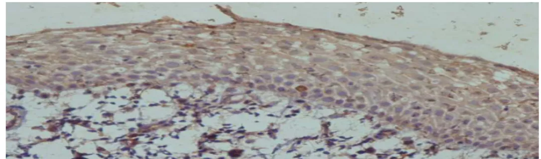
thick slices were prepared with H & A to confirm the diagnosis and sufficient sample size stained. Then another 4 microns were prepared for immunohistochemical studies. The slides for 24 hours at 37 ° C and one hour were kept at a temperature of 60 ° C to excess paraffin on a slide is removed, then Deparaffinization& Rehydration by placing the samples in xylen 100% and alcohol were graded. Antigens by placing slides in TBS buffer in a microwave (for 22 minutes) were recovered.Then slides with primary antibody (monoclonal mouse anti human-clone 4H12-UK-Nvocastra) cox-2 carefully 1.100 incubated for one hour at room temperature.Then, slide in the container include TBS with PH = 7.6 washed for 5 minutes, and for an hour inside en Vision tube is placed, TBS buffer Were washed and after putting on their DAB were washed with running water and after staining with hematoxylin Mayer were mounted.The percentage of cells stained for indicator COX-2 just counting in ten microscopic fields were calculated based on the following system: when the stained cells was no score 0, between 1 and 19% of cells were stained score1, between 20 and 49% of cells were stained score2 and if more than 50% of cells stained were score3 were considered. Positive control samples were used for ulcerative colitis.
Tonality vascular endothelium was considered as an internal control. The percentage of stained cells in the basal and suprabasal layer and lymphocyte infiltration was calculated separately. The staining intensity of staining cells on the basis of four degrees, mild, moderate and severe, respectively Score of 0,1,2 and 3 belong to them, it was evaluated. (01)
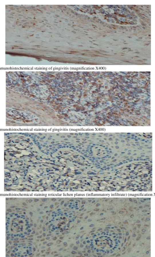
Figure 2: immunohistochemical staining of gingivitis (magnification X400)
Figure 3:Immunohistochemical staining of gingivitis (magnification X400)
Figure 4:Immunohistochemical staining reticular lichen planus (inflammatory infiltrate) (magnification X40)
[III] RESULTS
90 samples including 30 cases of normal mucosa of 18 men( 60%) and 12 women (40%) with a mean age of 10.35 ± 33.8 and 30 gingivitis of 17 men (54.6%) and 13 women (43.4%) with a mean age 9.94 ± 37.67 and 30 lichen Planus (15
reticular and 15 cases of erosive), on biopsies obtained from patients ranging in age from 25 to 62 years with an average age of 8.26 ± 45.57 of the 19 cases (63.4% ) were female and 11 cases (36.6%) were male, entered the study (table 1)
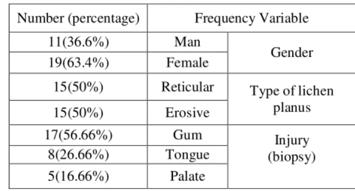
Frequency Variable Number (percentage) Gender Man 11(36.6%) Female 19(63.4%)
Type of lichen planus Reticular 15(50%) Erosive 15(50%) Injury (biopsy) Gum 17(56.66%) Tongue 8(26.66%) Palate 5(16.66%)
Table 1: Distribution of frequency lichen planus patients based on gender, location and type of lichen planus.
Percent and the number of samples stained and staining intensity in the basal layers, parabasal and infiltration of lymphocytes in lichen planus
patients and gingivitis, basal and parabasal for normal mucosa samples were analyzed for the results in Tables 2 and 3 below.
Layer The percentage of stained cell
score Lichen planus Gingivitis Normal mucosa Basal 0% 0 1(3.3%) 0(0%) 3(10%) 19-1% 1 3(10%) 6(20%) 14(46.7%) 49-20% 2 6(20%) 16(53.3%) 8(26.7%) >50% 3 20(66.7%) 8(26.7%) 5(16.7%) Parabasal 0% 0 1(3.3%) 0(0%) 3(10%) 19-1% 1 3(10%) 6(20%) 14(46.7%) 49-20% 2 6(20%) 16(53.3%) 8(26.7%) >50% 3 20(66.7%) 8(26.7%) 5(16.7%) Lymphocytic infiltration 0% 0 0(0%) 13(43.3%) _ 19-1% 1 1(3.3%) 10(33.3%) _ 49-20% 2 4(13.3%) 7(23.3%) _ >50% 3 25(83.3%) (0%) _
Table 2: Number and percentage of stained samples by staining cells in the basal layer, parabasal and lymphocyte infiltration in the three groups studied sample
Layer Staining intensity of cells
score Lichen planus
Gingivitis Normal mucosa
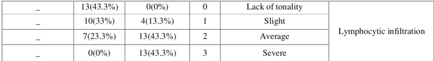
Basal Lack of tonality
0 1(3.3%) 0(0%) 3(10%) Slight 1 11(36.7%) 15(50%) 27(90%) Average 2 15(50%) 14(46.7%) 0(0%) Severe 3 3(10%) 1(3.3%) 0(0%) Parabasal Lack of tonality
Lymphocytic infiltration Lack of tonality
0 0(0%)
13(43.3%) _
Slight 1
4(13.3%) 10(33%)
_
Average 2
13(43.3%) 7(23.3%)
_
Severe 3
13(43.3%) 0(0%)
_
Table 3: Number and percentage of samples based on staining intensity of cells in the basal layer, parabasal and lymphocyte infiltration in three samples
Variable Layer
Normal mucosa
Mean Rank)*) Gingivitis
*(Mean Rank) Lichen Planus
Mean Rank)*) P-value
The percentage of stained cells Basal
31.38 45.6
59.52
0.001> 59.52 45.6 31.38 Parabasal
Staining intensity of cells Basal
28.15 52.05
56.30
0.001> 59.35 50.35 26.8 Parabasal
Table 4: Comparison of the percentage of stained cells and staining intensity of cells in the basal and parabasal layers of lichen planus patients, gingivitis and normal mucosa
Case
Variable Gingivitis
Mean Rank)*) Lichen Planus
Mean Rank)*) P-value
The percentage of stained cells in lymphocytic infiltration 16.37
44.63 0.001>
Staining intensity of cells in lymphocytic infiltration 18.62
42.38 0.001>
Table 5: Comparison of the percentage of stained cells and staining intensity of cells in the infiltration of lymphocytes in lichen planus and gingivitis.
For comparison, staining intensity and percentage of stained cells in the basal and parabasal layer in lichen planus and gingivitis for normal mucosa samples by Kruskal-wallis test were analyzed.Significantly the percentage of stained cells and staining intensity of cells in the basal layer of lichen planus was more than gingivitis (p <0.001). This result was seen in lichen planus and gingivitis in parabasal (p <0.001). Significantly the percentage of stained cells and staining intensity of cells in the basal layer of lichen planus was greater than normal mucosa (p <0.001). In comparison of lichen planus in normal mucosa and parabasal there similar results showed (p <0.001). On the other hand significantly the percentage of stained cells and staining intensity of cells in the basal layer of gingivitis was more than normal mucosa (p <0.001). In comparison, a similar result was seen above the parabasal layer (p <0.001). (Table 4)
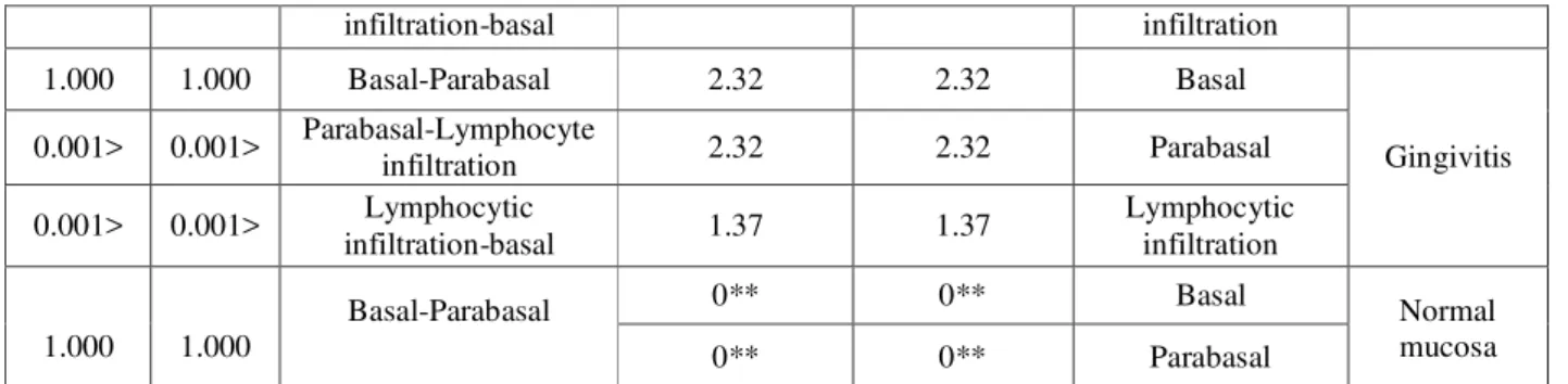
The percentage of stained cells and staining intensity in different layers separately for each of the groups were compared using wilcoxon and the following results were obtained (Table 6).
Sample Layer
Percent (mean rank)* Intensity (mean
rank)* Layers compared
P-value
Percent Intensity
Lichen Planus Basal
1.90 1.72
Basal-Parabasal 1.000
0.157
Parabasal 1.90
1.85 Parabasal-Lymphocyte
infiltration 0.38
0.001
Lymphocytic 2.2
2.43 Lymphocytic
infiltration infiltration-basal
Gingivitis Basal
2.32 2.32
Basal-Parabasal 1.000
1.000
Parabasal 2.32
2.32 Parabasal-Lymphocyte
infiltration 0.001>
0.001>
Lymphocytic infiltration 1.37
1.37 Lymphocytic
infiltration-basal 0.001>
0.001>
Normal mucosa Basal
0** 0**
Basal-Parabasal 1.000
1.000 0** 0** Parabasal
* Average grade (measure of comparison) **percentage of stained cells and staining intensity of
Table 6: Comparison of cells staining intensity and percentage of stained cells in the basal layer, Parabasal and lymphocytic infiltration of each of the samples
cells in the basal and parabasal layer of normal mucosa are similar in normal mucosa, intensity and percentage of stained cells in the basal and parabasal layer was equal (p =0.0001).In the case of gingivitis, degree and intensity of stained cells in the basal and parabasal layer was equal ( p =0.0001). However, staining intensity and percentage of stained cells in the basal and parabasal layers of lymphocytic infiltration was significantly higher (p <0.0001). The percentage of stained cells in lichen planus patients, basal and parabasal layers was equal (p =0.0001). It comes as the percentage of stained cells in lymphocytic infiltration significantly higher than basal and parabasal layers (p =0.038). Differences in staining intensity of cells in the basal layer and -para LP was not significant (p =0.157), staining intensity of cells in lymphocytic infiltration was significantly higher than the basal and parabasal layers (p =0.001). For comparison cells, staining intensity and percentage of stained cells in the basal and parabasal layer reticular and erosive lichen planus and infiltration of lymphocytes in the two groups Mann-Whitney test was used. As shown in Table 7 was no difference staining intensity and percentage of cells stained cells was not significant in any of the cases (p>0.05). The percentage of stained cells and staining intensity of cells in the infiltration of lymphocytes in oral lichen planus than the basal and parabasal layer.
Table 7: Comparison of the percentage of stained cells and staining intensity of cells in different layers in two groups of oral lichen planus and reticular
[IV] DISCUSSION
The study show increase in expression of cox-2 in oral lichen planus than in normal mucosa and gingivitis basal, parabasal levels and lymphocyte infiltration was in line with the results Neppelberg
and Renconen (11). However, piety and colleagues studied the expression of cox-2 in epithelial inflammatory infiltrate of lichen planus compared to the control group showed no significant difference while in the epithelium of
Case
Variable reticular
Mean rank* Erosive
Mean rank* P-value
The percentage of cells staining Basal
15.27 15.73
0.902
Parabasal 15.27
15.73 0.902
Lymphocytic infiltration 15.90
15.10 0.805
Staining intensity Basal
14.8 16.20
0.683
Parabasal 13.83
17.17 0.305
Lymphocytic infiltration 14.50
the difference in the expression of cox-2 in lichen planus and control group was not significant (12). In Thaneeyachankong et al demonstrated that increased expression cox2 significantly in normal epithelium lichen planus compared with oral mucosa (13). The results of this study showed that increased expression of cox2 in intensity is more inflamed oral lichen planus. However, the study stellalysitsa et al demonstrated that expression in oral lichen planus cox2 not associated with inflammation (14). Cox2 a stimulating enzymes in most cell types including keratinocytes, fibroblasts and Tcell is enabling the production of prostaglandins (15). Several processes including cell proliferation, apoptosis and angiogenesis in cancer cox2 is affected. cox2 in several ways, such as the proto-bcl2 adjustment and increase arachidonic acid inhibits apoptosis (16). Since cox2 factors such as cytokines and growth factors induce increase tissue growth factors seem to fit the role of erosive and atrophic because of the greater potential for malignant transformation are cox2 (17,18). Cox2 stable expression in the later stages of the disease can be a cause of the role of these factors in malignant (14).In the study, percentage difference cox2 expression and staining intensity of cells in the basal and parabasal layer of oral lichen planus not significant, difference between the percentage cox2 in the epithelium (basal and parabasal) by lymphocytic infiltration was observed. The intensity difference cox-2 expression in basal and parabasal layer of lymphocyt infiltration is significant lymphocytic infiltration, which is in line with the results of previous studies. The results Danillsson et al showed that cox2 mRNA and protein coc2 in the epithelium of oral lichen planus has greater extent than normal mucosal epithelium were expressed.The cox2 protein in the epithelium of oral lichen planus was observed only in the assessment by immunohistochemistry and Cox2 protein by western blot was observed in the epithelium of oral lichen planus. It seems that due to lack of western blot was observed in the levels of this protein in the epithelium is very low
(19). This study is a significant difference between the percentage of stained cells and staining intensity between OLP and reticular cells showed that contrary to the results Stella Lysitsa et al (14). Many studies show that only cox2 had no role in the pathogenesis of inflammatory diseases. Some tumors were such as colon tumors (20), bronchodilators (21), esophagus (22), head, and neck (23) of these cases. In addition, grade of tumor in the cox2 affect the change of state. During the dysplastic and hyperplastic lesions more aggressive rate increases will cox2 (14). Rankon et al undifferentiated carcinoma with high expression in language cox2 found (24). Recently Shibata et al cox1 and cox2 has high expression in oral dysplasia and oral scc observed. IHC showed that the expression of COX2 tissue dysplasia associated with severe dysplasia expression. For example, in high cox2 but the situation is different scc and the higher the grade, the difference between the two is realized in low cox2 which could be due to differences in materials and their methods (25). As noted in apoptosis and angiogenesis plays an important role in Cox2 however, according to study Shibata cox2 relationship don’t exist between apoptosis and angiogenesis in scc mouth (25). Now the question arises whether the cox2 inhibitor drugs could prevent progression of oral lichen planus to a dysplastic lesion? Recent studies show that drugs such as celecoxib reduced the growth of dysplastic lesions that inhibit the secretion is cox2. Cox2 resulting in high expression can become an indication of dysplastic epithelium of OLP to be and it is clear mechanism of action (26). So probably, the cox2 inhibitors can be used to reduce inflammation in the oral lichen planus, which is why it will be effective in the treatment of oral lichen planus, and future studies should address policies and treatment according to the conditions and services.
[V] CONCLUSION
Normal mucosa attributed to malignant potential of the lesion. However, cox2 expression in oral lichen planus can provide the groundwork for the transformation of this lesion to malignant tumors. More studies on the molecular level in the field can be helpful in prevention and treatment.
REFERENCE
1. Neville B W،Damm DD،Allen CM Bouquot ، JE.Oral and MaxillofacialPathology،3th ed.،Saunders Missouri، ،2009:741-815
2. Seyedmajidi M, shafaee s, Hejazi M, Hajiahmadi M, Siadati S. Expression of p53 and p63 in oral lichen planus and oral lichenoid reaction. JBUMS 2011;13(4):7-13.. 3. Tao X،Hunag Y،Li R،Qing R،Ma L،Rhodus
NL،et al.Assessment of local angiogenesis and vascular endothelial growth factor in the patients with atrophic-erosive and reticular oral lichen planes.Oral Surg،Oral Med،Oral Pathol ، Endod.2007:103(5):661-9.
4. Dan H،Liu W،Zhou Y،Wang J،Chen Q،Zeng X.Association of interleukin-8 gene polymorphisms and haplotypes with oral lichen
planes in a chinese
population.Inflammation.2010 : 33(2):76-81. 5. Mardani M،Ghabanchi J،Fattahi MJ،Tadbir
AA.Serum Level of Vascular Endothelial Growth Factor in Patient with Different Clinical Subtypes of Oral lichen planus.Iranian journal of medical sciences.2012:37(4):233-237
6. Kimkong I،Hirankarn N،Nakkuntod J،KitkumthornN.Tumour necrosis factor – alpha gene polymorphisms and susceptibility
to Oral lichen planes.Oral
Diseases.2011:17(2):206-9.
7. Herschman HR. prostaglandin synthase
2.Biochim BiophysActa. 1996 Jan 5;1299(1):125-40.
8. KarageceYalçin U, Seçkın S .The expression
of p53 and COX-2 in basal cell carcinoma, squamous cell carcinoma and actinic keratosis cases. Turk PatolojiDerg. 2012;28(2):119-27
9. Subbaramaiah K, Telang N, Ramonetti JT, Araki R, DeVito B, Weksler BB, et al. Transcription of cyclooxygenase-2 is enhanced in transformed mammary epithelial cells. Cancer Res. 1996 Oct 1;56(19):4424-9.
10.Pires I،Garcia A،Prada J،Queiroga F،Cox-1 and Cox-2 expression canine cutaneous.Oral andocular melanocytic tumors.J com Path.2010Oct؛143(2-3):142-9
11.Neppelberg E, Johannessen AC. DNA content, Cyclooxygenase-2 expression and loss of E-cadherin expression do not predict risk ofmalignant transformation in oral lichen planus. Eur Arch
12.Taghavi M ،Mahdavi N ،Shahla.evaluation of relationship expression two marker Bcl2 and Cox-2 in oral lichen planes.Community of Islamic dentists.2014؛26(1):51-58
13.ThaneeyaChankong, Pareena Chotjumlong, Thanapat Sastraruji, Surawut Pongsiriwet, AnakIamaroon, Suttichai Krisanaprakornkit. Increasedcyclooxygenase 2 expression inassociation with oral lichen planus severity.Journal of Dental Sciences (2016) xx, 1e7
14.Stella Lysitsa،Jacky Samson ،Christine
Gerber-Wicht ،Ursula Lang ،Tommaso
Lombardi .Cox-2 Expression in Oral Lichen Planus. Dermatology 2008;217:150–155. 15.Cortés-Ramírez DA, Rodríguez-Tojo MJ,
Gainza-Cirauqui ML, Martínez-Conde R, Aguirre-UrizarJM.Overexpression of cyclooxygenase-2 as a biomarker indifferent subtypes of the oral lichenoid disease. OralSurg Oral Med Oral Pathol Oral RadiolEndod 2010110(6):738-43.
16.Fernades AT, Armstrong RC, Krebs J, Srinivasula SM, Wang L, Bllrich F, et al. ProcNatlAcadSci U.S.A. 1996; 93: 7464-9.
17.Tsatsanis C, Androulidaki A,
VenihakiM,Margioris AN: Signalling networks
regulatingcyclooxygenase-2. Int J
BiochemCellBiol 2006; 38: 1654–1661.
18.Holmstrup P, Pindborg JJ: Erythroplakic
ActaDermVenereolSuppl (Stockh) 1979; 59: 77–84.
19.K. Danielsson , M. Ebrahimi, Y.B. Wahlin, K. Nylander, L. Boldrup .Increased levels of COX-2 in oral lichen planus supportsan autoimmune cause of the disease.DOI: 10.1111/j.1468-3083.2011.04306.x
20.Charles EE, Robert JC, Alamandla R,
FrancisMG, Suzanne F, Raymond D: Up-regulationof cyclooxygenase 2 gene expression
in humancolorectal adenomas and
adenocarcinomas.Gastroenterology 1994; 107: 1183–1188.
21.Wolff H, Saukkonen K, Anttila S, Karjalainen
A, Vainio H, RistimakiA:Expression
ofcyclooxygenase-2 in human lung
carcinoma.Cancer Res 1998; 58: 4997–5001.
22.Wolff H, Saukkonen K, Anttila S,
KarjalainenA, Vainio H, Ristimaki
A:Expression ofcyclooxygenase-2 in human
lung carcinoma.Cancer Res 1998; 58: 4997– 5001.
23.Sudbo J, Ristimäki A, Sondresen JE, et al:
Cyclooxygenase-2 (COX-2)expression in
highriskpremalignant oral lesions. Oral
Oncol2003; 39: 497–505.
24.Renkonen J, Wolff H, Paavonen T:
Expressionof cyclo-oxygenase-2 in human tonguecarcinoma and its precursor lesions. VirchowsArch 2002; 440: 594–597
25.Shibata M, Kodani I, Osaki M, et al:
Cyclooxygenase-1 and -2 expression in humanoral mucosa, dysplasias and squamous
cellcarcinomas and their pathological
significance.Oral Oncol 2005; 41: 304–312.
26.Feng L, Wang Z: Chemo preventive effect