Original Article
Evaluation of Personal Shields Used in Selected Radiology Departments
Mohsen Salmanvandi1, Mehdi Momennezhad2*, Shahrokh Naseri3, Reza Ghasemnezhad1,Hadi Didehvar1, Marym Salari4
Abstract
Introduction
The purpose of this study was to evaluate personal shields in radiation departments of hospitals affiliated to Mashhad University of Medical Sciences.
Materials and Methods
First, the information related to 109 personal shields was recorded and evaluated by imaging equipment. Afterwards, the equivalent lead thickness (ELT) of 62 personal shields was assessed, using dosimeter and standard lead layers at 100 kVp.
Results
In this study, 109 personal shields were assessed in terms of tears, holes and cracks. The results showed that 18 shields were damaged. Moreover, ELT was evaluated in 62 shields. As the results indicated, ELT was unacceptable in 8 personal shields and lower than expected in 9 shields.
Conclusion
According to the results, 16.5% of personal shields had defects (tears, holes and cracks) and 13% of them were unacceptable in terms of ELT and needed to be replaced. Therefore, regular quality control of personal shields and evaluation of new shields are necessary at any radiation department.
Keywords: Occupational Exposure; Radiography; Shield
1.Department of Radiology, School of Paramedical Sciences, Mashhad University of Medical Sciences (MUMS), Member of Student's Research Assembly of MUMS, Mashhad, Iran.
2. Nuclear Medicine Research Center, Mashhad University of Medical Sciences, Mashhad, Iran. *Corresponding author: Tel: +98 513 8511720, Fax: +98 513 8599359; E-mail: [email protected]
1. Introduction
Ionizing radiation is the largest source of artificial radiation [1]. Application of this type of radiation, which is important for the diagnosis and treatment of different diseases, is increasing annually [1]. However,the associated biological effects may lead to the damage of cells, tissues and organs. The stochastic effects of ionizing radiation have been reported at all radiation levels [2]. In fact, extensive research has demonstrated the incidence of cancer from medical exposure [3].
In order to reduce the adverse effects of ionizing radiation, use of personal shields such as apron, thyroid, gonad, skirt and vest shields has been proposed for the safety of systems. Personal shields are capable of reducing occupational exposure and increasing patient protection, according to As Low As Reasonably Achievable (ALARA) principle [4].
Personal shields significantly affect the absorption of scattered radiation during radiologic examinations. These shields can reduce the absorbed dose by critical organs such as bone marrow by approximately 75%. They can be either made of lead or lead-free.
Nevertheless, their thickness is expressed by equivalent lead thickness (ELT) [5]. The minimum ELT of personal shields in diagnostic imaging is 0.25 mm pb at 100 kV and 0.35 mm pb for more than 100 kV [6]. Considering the importance of personal shields for patients and staff, periodical quality control is necessary for ensuring the proper performance of these shields. Regarding the quality control of these shields, Michel and Zorn [7] suggested an annual fluoroscopy examination at 80 kV for new shields. The main purpose of the current study was to evaluate personal shields and compare different brands.[7]
2. Materials and Methods
The present descriptive study was performed at three hospitals affiliated to Mashhad University of Medical Sciences during 2011-2012. In this study, apron, gonad, thyroid, skirt and vest shields were evaluated. The sample size was determined by evaluating the number of personal shields used at each radiation department (table 1).
Table 1. The number of personal shields evaluated
Type Evaluation of tears and cracks Evaluation of ELT
Apron 38 26
Thyroid 32 15
Vest 14 9
Skirt 15 9
Gonad 10 3
Total 109 62
Table 2. Permissible defects for shields [9]
Lead equivalent (mm pb)
0.25 0.35 0.50
Type of apron The defined area Maximum length of defect (cm)
Double AP Whole body 13.5 17.5 27.0
Gonads 4.4 5.6 8.7
Single Whole body 5.9 5.6 5.4
Gonads 1.9 1.8 1.7
Thyroid 1.9 1.8 1.8
Double PA (10%) 17.0
LAT (30%) 9.8
First, all features of shields including the type, brand, lifetime, location, and
nominal ELT were recorded and
a specific code was allocated to each personal
shield. Afterwards, the shields
were scanned by C-Arm (Ziehm) or
fluoroscopy (Apelem, Baccara with 18-inch TV monitor) to find the defects (i.e., holes, cracks and tears). It should be mentioned that the scanned area was flat on the tabletop. Then, the location and size of defects were recorded. The size of defects was measured by 3D-Doctor 4.0 software(2012, Able Software Corporation; Lexington, USA). To calculate magnification factor and increase measurement accuracy, a marker with a known size was used. Finally, the total length of shield defects was measured in millimeter and the results were compared with acceptable standard values (table 2).
In addition, the ELT of personal shields was evaluated. For this purpose, a multi-o-meter dosimulti-o-meter (UNFORS Instruments, Billdal, Sweden) and a radiographic system (Semens with a 1mm AL filter) were applied. The dosimeter was placed on the tabletop at a distance of 100 cm from the focal spot of the x-ray tube. Then, the shields were placed at a 30-cm distance from the dosimeters by means of a suitable preservation. It is included light materials such as yonolit. This arrangement was designed to avoid scatter radiation to dosimeters.
The radiation field size was selected in a way to cover the whole dosimeter. This examination was performed to measure attenuation at primary radiation (100 kVp). Each measurement was repeated three times and the average output was calculated. The testwas performed under the same conditions with lead sheets of 0.2, 0.3, 0.4, and 0.6 mm thickness; then, the attenuation curve at 100 kVp was determined, based on the obtained results. To assess the actual ELT of the evaluated personal shields, the measured attenuation values were placed on the attenuation curve.
3. Results
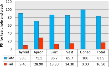
In this study, 109 personal shields were evaluated in terms of tears, cracks, and holes. Figure 1 indicates the percentage of intact and damaged personal shields. As to the obtained findings, 18 shields (16.5%) were considered damaged. Moreover, the results showed that the majority of damaged shields were apron shields (11 shields). Table 3 shows the number of intact and damaged personal shields with regard to their brand and lifetime.
Table 3. The evaluation results of personal shields with different lifetimes and brands in terms of tears, holes and cracks
Age (year) 1-3 4-6 6< Results
Brand
S* FΩ S F S F
Alikhani 1 4
Amray 6 2 1
Burkhart 2 1
Navidpartonama 26 2 3 2
Infab 5 1 3
Mavig 3 1
Medtronic 3 2
Dr Goos 2 1
Kelida 2
Cawo flex 1
X-Raynox 1
Sum 44 2 9 3 16 1
PSℓ: personal shields, ELT€: Equivalent Lead Thickness S*: Safe, Aπ: Acceptable, FΩ: Failed
Table 4. The results of the evaluation of personal shields with different lifetimes and brands in terms of ELT€
Age (year) 1-3 4-6 6<
Result Brand
S* A
π FΩ S A F S A F
Alikhani 1 1
Amray 2 1
Burkhart 2 1
Navidpartona ma
9 2 2 2 1
Infab 2 3 1 3
Medtronic 2
Dr Goos 1
Kelida 2
X-Raynox 1
Unknown 1 2 4 1 1
0
4 1
Sum 1
5
3 7 1 4
1 1
6
Figure 1: The results of the evaluation of personal shields in terms of tears, holes and cracks
Figure 2. Evaluation of personal shields in terms of ELT
The evaluation of 62 personal shields showed that ELT was unacceptable in 8 shields. In addition, 9 shields had less thickness,
compared to the value stated by
the manufacturer; however, the protection level was acceptable. Figure 2 shows the percentage of intact, damaged, and acceptable personal shields with respect to ELT. Table 4 shows the evaluation results of personal shields with different lifetimes and brands in terms of ELT.
4. Discussion
In total, to determine inefficient personal shields, rejection criteria are required. In a previous study, Lambert and McKeon
investigated the additional absorbed dose due to different types of defects (crack, tear and hole), with respect to the standard dosage (50 mSv per year). As they indicated, personal shields should be replaced in case the defect is larger than 15 mm² in the gonad area or larger than 11 mm² in the thyroid. If defects exist in areas that are not related to critical organs, the maximum permissible size is 670 mm². [8]
Moreover, as Duran and Phillips demonstrated, the maximum area of holes and cracks was 10 cm² in whole body, 0.2 cm² in the genital area, and 1cm² in the neck area, considering the maximum dose of 20 mSv per year for whole body scan and the cost of shields.[4]
Thyroid Apron Skirt Vest Gonad Total
Safe 90.6 71.1 86.7 85.7 100 83.5
Fail 9.40 28.90 13.30 14.30 0.00 16.50
0 20 40 60 80 100 120
PS
for
t
e
ar
, h
o
le
an
d
c
rac
k
Thyroid Apron Gonad Skirt Vest Total
Safe 87 69 100 67 56 73
Fail 0 4 0 33 44 13
Acceptable 13 27 0 0 0 14
0 20 40 60 80 100 120
Per
son
al
Sh
ie
ld
for
E
Stam and Pillary [9] determined permissible defects for shields, according to the maximum additional effective dose of 0.22 (table 2). [9]The introduced rejection criteria were different, mainly due to the relation between the rejection criteria and the permissible dosage. In the present study, the criteria established by Stam and Pillary were selected as the rejection criteria, since they were in accordance with the recent protection protocols [10]. Considering the rejection criteria, 16.5% of the evaluated shields (n=109) had defects more than the permissible amount and had to be replaced by new shields. Finnerty and Brennan assessed personal shields by fluoroscopy to find and measure the size of defects [5]; however, the results were not comparable with the rejection criteria. In addition, they determined ELT of lead-free and composite (lead with other elements) shields by an ionizing chamber.
In this study, we assessed ELT of personal shields (made of lead or lead-free) by a multi-o-meter dosimeter . The results indicated that 8 shields had an ELT less than 0.25 mm (the minimum permissible thickness in diagnostic application); thus, their application had to be limited. Nine shields had an ELT less than the amount permitted by the manufacturer, although they had the minimum permissible thickness. At this stage, the sample size reduced since some shields were found to be similar.
Defects in recently purchased shields are of significance at every radiation department. Therefore, it is recommend that diagnostic and therapeutic departments purchase new shields which do not rely on physical health.
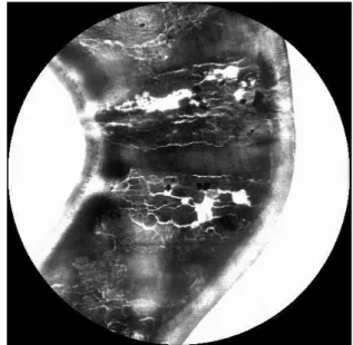
Moreover, these shields should be evaluated in terms of defects (tear, hole and crack) and ELT (Fig. 3 and 4). According to the findings (fig. 1 and 2), gonad shields were less damaged considering their small size and limited usage in the selected hospitals.
We faced some limitations during the study. First, some departments did not cooperate with us since the authorities in charge were sure about the reliability of the applied personal shields. Second, the exact production date of
the shields was not available. We hope that this article highlights the importance of protection issues.
Figure 3. Defects on a newly purchased shield
Figure 4. A seemingly intact thyroid shield, which appeared to be defected in the X-ray examination
5. Conclusion
shields. In addition, personal shields, which are not in use, should be hanged and not folded.
Acknowledgments
This study was funded by the Student’s Research Assembly of Mashhad University of Medical Sciences. The authors wish to thank
the radiology departments of Ghaem, Imam Reza, and Dr. Sheikh hospitals for their cooperation. We also express our gratitude to Mr. Behnam, Mr. Elhami, Mr. Jani, Mr. Ahmadi and Mrs. Sharghi.
References
1 Schauer DA, Linton OW. National Council on Radiation Protection and Measurements report shows substantial medical exposure increase. Radiology. 2009 Nov;253(2):293-6.
2 Shannoun F, Blettner M, Schmidberger H, Zeeb H. Radiation protection in diagnostic radiology. Dtsch Arztebl Int. 2008 Jan;105(3):41-6. 3 Berrington de Gonzalez A, Darby S. Risk of cancer from diagnostic X-rays: estimates for the UK and 14 other countries. Lancet. 2004 Jan 31;363(9406):345-51. 4 Duran E, Philips B. Rejection criteria for defects in lead apparel used for radiation protection of X-ray workers. Radiation Protection Sevices, British Columbia Centre for Disease Control. 2003;1.
5 Finnerty M, Brennan PC. Protective aprons in imaging departments: manufacturer stated lead equivalence values require validation. Eur Radiol. 2005 Jul;15(7):1477-84.
6 Medical and Dental Guidance Notes. A Good Practice Guide on all Aspects of Ionising Radiation Protection in the Clinical Environment. Journal of Radiological Protection. 2002;22(3):334.
7 Michel R, Zorn MJ. Implementation of an X-ray radiation protective equipment inspection program. Health Phys. 2002 Feb;82(2 Suppl):S51-3.
8 Lambert K, McKeon T. Inspection of lead aprons: criteria for rejection. Health Phys. 2001 May;80(5 Suppl):S67-9.
9 Stam W, Pillay M. Inspection of lead aprons: a practical rejection model. Health Phys. 2008 Aug;95 Suppl 2:S133-6.
10 The University of Texas Southwestern Medical Center (2010) Lead Apron Policy. The Environmental
Health and Safety, Dallas. Available via:
![Table 2. Permissible defects for shields [9]](https://thumb-eu.123doks.com/thumbv2/123dok_br/18171242.329968/2.892.183.716.707.904/table-permissible-defects-shields.webp)